Dentist in pune.(BDS. MDS) - Dr. Amit T. Suryawanshi..Zygomaticomaxillary complex fractures
•Download as PPTX, PDF•
42 likes•7,629 views

Dentist in pune. (BDS. MDS) - Dr. Amit T. Suryawanshi. Seminar-Canine Impaction. Email ID- amitsuryawanshi999@gmail.com Contact -Ph no.-9405622455 Subscribe our channel on youtube - https://www.youtube.com/channel/UC_gylEXTrjmEbbOTSXjuZ4Q/videos?view_as=public Follow us on slideshare

Recommended
More Related Content
What's hot
What's hot (20)
Viewers also liked
Similar to Dentist in pune.(BDS. MDS) - Dr. Amit T. Suryawanshi..Zygomaticomaxillary complex fractures
Similar to Dentist in pune.(BDS. MDS) - Dr. Amit T. Suryawanshi..Zygomaticomaxillary complex fractures (20)
More from All Good Things
More from All Good Things (20)
Recently uploaded
Recently uploaded (20)
Dentist in pune.(BDS. MDS) - Dr. Amit T. Suryawanshi..Zygomaticomaxillary complex fractures
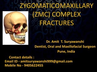
- 1. ZYGOMATICOMAXILLARY (ZMC) COMPLEX FRACTURES Dr. Amit T. Suryawanshi Dentist, Oral and Maxillofacial Surgeon Pune, India Contact details : Email ID - amitsuryawanshi999@gmail.com Mobile No - 9405622455
- 2. CONTENTS • INTRODUCTION • ANATOMY • CLASSIFICATION • SIGNS & SYMPTOMS • CLINICAL EXAMINATION • RADIOLOGIC EXAMINATION • MANAGEMENT • COMPLICATIONS
- 3. INTRODUCTION • Most common facial fractures, the second in frequency after nasal fractures. • The high incidence relates to the zygoma’s prominent position within the facial skeleton. • Male predilection, 4:1 over females. • Peak incidence- 2nd and 3rd decades of life.
- 4. INTRODUCTION • Zygomatic injury is mostly due to altercations followed by motor vehicle accidents. • In zygomatic fractures caused by altercations, the left zygoma is most commonly affected. • The zygoma plays an important role in facial contour. Disruption of the zygomatic position creates impairment of ocular and mandibular function.
- 5. INTRODUCTION • The zygoma or malar complex forms the central support of the cheek and is a strong buttress of the lateral and middle third of the facial skeleton. • It is for this reason, the zygoma is frequently fractured, either alone or in combination with other bony structures of the midface.
- 6. ANATOMY • The zygoma, a major buttress of the facial skeleton, is the principal structure of lateral midface.
- 7. ANATOMY • Zygoma is roughly quadrilateral in shape, with an outer convex (cheek) surface and an inner concave (temporal) surface. • It forms the point of greatest prominence of the cheek. • Resembles a four sided pyramid, which has temporal, orbital, maxillary and frontal processes.
- 8. ANATOMY • Zygoma articulates with four bones- the frontal, sphenoid, maxillary and temporal. • Body of the zygoma extensively articulates with the maxilla along the anterior maxilla and along the orbital floor. • It forms the superolateral aspect and part of the superoanterior aspect of the maxillary sinus.
- 9. ANATOMY
- 10. ANATOMY • The frontal process is thick and triangular in cross section, with facial, orbital and temporal surfaces. • The temporal process is flat and projects posteriorly to articulate with the zygomatic process of the temporal bone. • The combination of the two makes up the zygomatic arch.
- 11. ANATOMY • The zygomaticotemporal articulation is a very thin, delicate connection, which fractures frequently. • The zygoma provides origin to a major portion of the masseter muscle along the body and temporal surface. • The temporal fascia also attaches along the arch and the temporal process.
- 12. ANATOMY
- 13. SURGICAL ANATOMY ‘the malar bone represents a strong bone on fragile supports, and it is for this reason that, though the bone is rarely broken, the four processes – frontal, orbital, maxillary and zygomatic – are frequent sites of fracture.’ -- H.D.Gillies, T.P. Kilner and D. Stone, 1927
- 14. SURGICAL ANATOMY • 2 zygomatic bones & their temporal processes • Zygomatic processes of temporal bones Form part of the middle third of the facial skeleton which may be fractured following trauma.
- 15. SURGICAL ANATOMY • Fractures involving the orbit may give rise to alteration in the position of the globe of the eye. • The level of the globe is normally maintained by the suspensory ligament of Lockwood which passes from its medial attachment on the lacrimal bone to be inserted laterally into the Whitnall’s tubercle.
- 16. SURGICAL ANATOMY • Zygomatic and Le fort III fractures commonly result in drop in the level of the globe of the eye. • As the globe of the eye drops, the upper eyelid follows downwards giving rise to the physical sign ‘hooding of the eye’.
- 17. FRACTURE PATTERNS • The fracture pattern of any bone depends mainly on the direction and magnitude of the force. • The fracture lines pass through the areas of greatest weakness of a bone or between bones.
- 20. FRACTURE PATTERNS • The inferior orbital fissure is the key to remember the usual lines of ZMC fractures. • Three fracture lines extend from the inferior orbital fissure in an anteromedial, a superolateral, and an inferior direction.
- 21. FRACTURE PATTERNS 1. One fracture extends from the inferior orbital fissure anteromedially along the orbital floor mostly through the orbital process of the maxilla.
- 22. FRACTURE PATTERNS 2. Second line of fracture from the inferior orbital fissure runs inferiorly through the posterior(infratemporal) aspect of maxilla and joins the fracture from the anterior aspect of maxilla, under the zygomatico maxillary buttress.
- 23. FRACTURE PATTERNS 3. Third line of fracture extends superiorly from the inferior orbital fissure along the lateral orbital wall posterior to the rim, usually separating the zygomaticosphenoid suture.
- 24. CLASSIFICATION • Knight and North (1961) • Rowe and Killey (1968) • Yanagisawa ( 1973) • Larsen and Thomson (1978) • Rowe and Williams (1985) • Poswillo (1988)
- 25. Knight and North (1961) Based on the direction of displacement on a Water’s view radiograph, • Group I – Non displaced fractures • Group II – Arch fractures • Group III – Unrotated fractures • Group IV – Medially rotated fractures • Group V - Laterally rotated fractures • Group VI – Complex fractures
- 26. Knight and North (1961)
- 27. ROWE AND KILLEY (1968) • Type I – no significant displacement • Type II – fracture of the arch • Type III – rotation around a vertical axis • Type IV – rotation around a longitudinal axis • Type V – displacement of the complex en bloc • Type VI – displacement of orbito-antral partition • Type VII – displacement of the orbital rim segments • Type VIII – Complex comminuted fractures
- 28. Yanagisawa ( 1973) • GROUPS I & II - Unchanged • GROUP III - Medial or lateral rotation around a vertical axis • GROUP IV - Medial or lateral rotation around a longitudinal axis • GROUP V - Medial or lateral displacement without rotation • GROUP VI - Isolated arch fracture • GROUP VII - All complex fractures
- 29. Larsen and Thomson (1978) • GROUP I – Non displaced fractures requiring no treatment • GROUP II – All fractures requiring treatment
- 30. ROWE AND WILLIAMS (1985) • Fractures stable after elevation a) Arch only (medially displaced) b) Rotation around the vertical axis i) medially ii) laterally • Fractures unstable after elevation a) Arch only (inferiorly displaced) b) Rotation around horizontal axis i) medially ii) laterally c) Dislocations en bloc i) inferiorly ii) medially iii) postero - laterally d) Communited fractures
- 31. ROWE AND WILLIAMS (1985)
- 32. POSWILLO’S CLASIFICATION • Inward and downward displacement • Inward and posterior displacement • Outward displacement of the zygomatic complex • Communition • Fracture of the arch alone
- 33. SIGNS & SYMPTOMS • Periorbital Ecchymosis and Edema • Flattening of the malar prominence • Flattening over the zygomatic arch • Pain
- 34. SIGNS & SYMPTOMS • Ecchymosis of the maxillary buccal sulcus • Deformity at the zygomatic buttress of the maxilla • Deformity of the orbital margin • Trismus
- 35. SIGNS & SYMPTOMS • Abnormal nerve sensibility • Epistaxis • Subconjunctival Ecchymoses • Crepitation from air emphysema
- 36. SIGNS & SYMPTOMS • Displacement of the palpebral fissure • Unequal pupillary levels • Diplopia • Enophthalmos
- 37. RADIOLOGIC EXAMINATION • Plain films and Computed Tomography have their place in determining the type, location, magnitude, and direction of displacement of zygomatic fractures. • This includes, Water’s view, Submentovertex view, Computed Tomography.
- 38. RADIOLOGIC EXAMINATION • A single Water’s view is an important adjunct to clinical examination. • If fractures are noted, CT should be the procedure of choice. • Two dimensional CT is now considered the best and most useful means of radiologic assessment of the facial skeleton.
- 39. RADIOLOGIC EXAMINATION • CT scans allow complete assessment of the orbital floor and walls. • For ZMC injuries, it is optional to have both axial & coronal high resolution scans. • The axial scan is helpful in evaluating the medial and lateral orbital walls, and the coronal scan defines the extent of injury to the orbital floor.
- 47. END OF PART 1..
- 48. ZYGOMATICOMAXILLARY COMPLEX FRACTURES PART II PRESENTED BY, ASHWIN THAKARE(PART III)
- 49. IN THE LAST PART WE DISCUSSED…. • INTRODUCTION • ANATOMY (SURGICAL ANATOMY) • CLASSIFICATION • SIGNS & SYMPTOMS • CLINICAL EXAMINATION • RADIOLOGIC EXAMINATION
- 50. CONTENTS (PART 2) • TREATMENT.
- 51. TREATMENT • Historical review: • Various authors have given various treatment modalities and techniques for the management of ZMC fractures. • Dating back to 1751, when Duverney stressed the role of contraction of temporal muscle in realigning the medial displacement of the zygomatic arch.
- 52. HISTORICAL REVIEW • Ferrier in 1825, attempted to reduce fracture of zygomatic arch through an incision above the arch. • Dupuytren in 1847, discovered the important relationship of the temporal fascia and the muscle as a pathway to the zygomatic arch.
- 53. HISTORICAL REVIEW • Gillies in 1927 emphasised the cosmetic value of placing the incision within the hair line. • Stroymeyer in 1844 described the percuteneous hook technique. • Cheyne and Burghard in 1901 discussed the intraoral digital manipulation technique. • Smith and Yanagisawa in 1961 stressed the importance of cosmetic aspects of the treatment.
- 54. GENERAL PRINCIPLES OF TREATMENT • No treatment • Indirect reduction with, a. No fixation b. Temporary support c. Direct fixation d. Indirect fixation • Direct reduction and fixation
- 55. NO TREATMENT • Cases with a minimal degree of displacement, which following union, are considered unlikely to result any cosmetic deformity, disturbance of vision, persistent paraesthesia or impairment of mandibular movement.
- 56. INDIRECT REDUCTION • NO FIXATION: • Includes procedures which do not involve exposure of the fracture sites. • The principle is to disimpact and reduce the fracture by direct application of an instrument, through an indirect approach remote from the fracture line.
- 57. NO FIXATION • The techniques which have been developed for this operative approach, are based upon the introduction of an instrument through, a. the temporal fossa, b. the upper buccal sulcus (intraoral), c. the cheek (percutaneous), d. the nose (transantral) e. the eyebrow (lateral brow)
- 58. NO FIXATION • Temporal fossa approach: • This method was introduced by Gillies et al (1927) for elevation of the zygomatic arch. • Incision of about 2.5 cm long, made above and parallel to the anterior branch of the temporal artery.
- 61. NO FIXATION • Lateral brow approach: (Dingman & Natwig 1964) • The advantage of this technique is that the fracture at the orbital rim is visualized directly. • The frontozygomatic area of the lateral orbital rim is exposed by the eyebrow incision. • The instrument is inserted to lift the zygoma anteriorly, laterally and superiorly.
- 62. NO FIXATION • Lateral eyebrow approach:
- 63. NO FIXATION • Upper buccal sulcus: • The advantages of this technique have been discussed by Balasubramaniam (1967) who considers that “ less force is required by the intraoral approach than by the extraoral, because the force is exerted where it should be, i.e., more at the centre of the fractured fragment”.
- 64. NO FIXATION • Upper buccal sulcus: (Keen’s approach) • Access is gained by an incision of about 1cm in length at the reflection of the upper buccal sulcus immediately behind the zygomatic buttress.
- 65. NO FIXATION • Quinn in 1977 described a modification. • This employs a lateral coronoid approach through an incision situated over the anterior border of ramus.
- 67. NO FIXATION • Percutaneous approach: (Stroymeyer 1844) • This method consists of inserting a hook through the skin below and behind the zygomatic bone so that it engages the deep aspect and allows reduction by strong outward traction on the handle of the instrument.
- 68. NO FIXATION • Percutaneous approach: • Poswillo advises that the exact location of the initial stab wound for insertion is found at the intersection of a perpendicular line dropped from the outer canthus of the eye and a horizontal line extended posteriorly from the alar margin of the nostril.
- 70. NO FIXATION • Intranasal transantral approach: (Lothrop’s approach 1906) • Not common in use. • An opening is made into the antrum below the inferior meatus, and a curved urethral sound introduced and manipulated so that its tip lies on the antral aspect of the zygomatic bone. Firm outward and upward pressure is applied to reposition the bone.
- 71. TEMPORARY SUPPORT • This may be indicated, as a supplementary measure, under the following circumstances: • When the zygomatic complex is unstable following reduction, • When there is gross comminution of the zygomatic bone. • When there is comminution without bone loss of the orbital floor.
- 72. TEMPORARY SUPPORT • Instability following adequate reduction could be due to: • Rupture of the enveloping periosteum or attached temporal fascia. • Comminution of the zygomatic arch. • Loss of bone from around the zygomatic buttress. • Residual fibrosis when treatment is delayed.
- 73. TEMPORARY SUPPORT • Temporary support is a concept which is primarily based upon the introduction of a pack or other material into the antrum so as to exert counter-pressure against those forces which tend to bring about a relapse of the position achieved by indirect reduction.
- 74. TEMPORARY SUPPORT • Since the pack will bring about repositioning of fragments by pressure from their antral aspect it will be evident that this selective effect can only take place if there is absolute stability of the remainder of the antrum and the other elements of the zygomatic complex.
- 75. DIRECT FIXATION • Indirect reduction, combined with direct fixation following exposure of the fracture site, provides an excellent method of treatment. • Direct fixation is needed when the fractures remain unstable after indirect reduction.
- 76. DIRECT FIXATION • Transosseous wiring or osteosynthesis: • Separation at the frontozygomatic suture line with displacement in excess of 2-3 mm, is likely to cause detachment of the periosteal and fascial attachments, and hence the direct fixation becomes necessary.
- 77. DIRECT FIXATION • Incisions on the face should be placed parallel to or within the skin creases. • It is preferable to incise the skin through the outer end of the eyebrow. The incision should not be at right angles to the skin, but directed downwards at the same angle as the emerging hairs.
- 78. DIRECT FIXATION • The Gillies temporal approach is preferable if there is separation at the suture. • A percutaneous repositioning may be preferred. • Access to the temporal aspect of the zygomatic bone can be obtained through the frontozygomatic incision by passing a curved elevator supraperiosteally posterior to the frontal process of the zygomatic bone.
- 79. DIRECT FIXATION • Dingman and Natvig (1964) recommend that the holes are drilled in an anteroposterior direction and when the external angular process is well formed. It also enables the wires to be placed in a figure of eight pattern which provides better lateral stability.
- 80. DIRECT FIXATION • Elevation of the zygomatic bone and transosseous wiring or boneplating at the frontozygomatic suture will achieve a stable realignment of the orbital rim.
- 81. DIRECT FIXATION • A more severe displacement, a dislocation of the zygomatic complex en bloc, will result in separation of the fracture ends at the inferior orbital margin, with the fracture passing into the orbital floor. • In this type of unstable fracture it is essential to carry out an osteosynthesis (transosseous wiring) or microplating technique.
- 82. DIRECT FIXATION
- 83. DIRECT FIXATION • Micro-plates positioned in such a way that the screw holes are situated well away from the fracture sites provides a very useful alternative to wires. • The use of malleable micro-plating equipment has greatly improved the management of such cases.
- 84. DIRECT FIXATION • Application of a small plate, across the fronto-zygomatic suture will usually ensure that there is absolute immobility, so that union can take place. Precision alignment of the inferior orbital margin is essential, open reduction will be indicated.
- 85. DIRECT FIXATION • Inadequate immobilisation of the fronto-zygomatic suture where the lateral orbital wall is displaced may lead to a loss of malar prominence.
- 86. INDIRECT FIXATION • Indirect fixation implies that the zygomatic bone will be rigidly secured to some point elsewhere on the facial skeleton until union occurs. • The required degree of firmness can only be achieved by means of internal (intramedullary) pins or wires or external pins and rods which are linked together.
- 87. INDIRECT FIXATION • The indirect fixation can be achieved by the following methods: 1. Zygomatico-zygomatic (Trans-maxillary) 2. Naso-zygomatic 3. Zygomatico-palatal 4. Maxillo-zygomatic 5. Fronto-zygomatic 6. Cranio-zygomatic
- 88. INDIRECT FIXATION • Indirect fixation has only limited application at the present time in view of the greater efficiency and comfort obtained by internal fixation techniques.
- 89. COMPLICATIONS • Infraorbital nerve disorders • Implant extrusion, displacement and infection • Maxillary sinusitis • Persistent diplopia • Enophthalmos • Ankylosis of zygoma to coronoid process • Malunion of the zygoma
- 90. CONCLUSION • Thus, the zygomatic complex fractures are common injuries, second in frequency after the nasal bone fractures. • There being a wide range of treatment modalities and techniques for the management of zygomaticomaxillary complex fractures. • It is the apt judgement and knowledge of the surgical anatomy on the part of the surgeon enabling him to effectively manage the ZMC fractures with the desired outcome.
- 91. REFERENCES • Row and williams volume -1 • Fonseca trauma volume -2 • Peter wardbooth • Peterson’s oral and maxillofacial surgery.
- 92. Subscribe our channel on youtube (Copy and paste this url )- https://www.youtube.com/channel/UC_g ylEXTrjmEbbOTSXjuZ4Q/videos?view_as= public Follow us on slideshare Thank you
Editor's Notes
- Zygomatic or malar fractures are the terms commonly used to describe fractures that involve the lateral one third of the middle face. Because of the impure nature of zygomatic fractures, other terms like zygomaticomaxillary complex, zygomaticomaxillary compound, zygomaticoorbital, zygomatic complex, malar, trimalar and tripod fractures have been adopted.. The later two terms are misnomers since the zygoma has not three but four processes.
- The zygoma also has a narrow, weak articulation with the zygomatic crest of the greater wing of the sphenoid bone at the lateral aspect of the inf. Orbital fissure. It forms a major portion of the lateral aspect of the floor of the orbit.
- The anatomic position of the zygoma. Lateral skull demonstrating its articulation with the temporal, frontal and maxillary bones.
- Because of its thickness, the frontal process is a frequent site for wire or bone plate fixation following fracture.
- The zygoma also provides attachments for the temporal and zygomatic muscles. the strong infraorbital and lateral orbital rim provides protection to the orbital contents.
- Owing to the strong buttressing nature of the zygoma and the thin bones surrounding it, most injuries involving the zygoma are accompanied by disruption of the adjacent articulating bones. This disruption occurs because when a force is applied to the body of the zygoma, it is distributed through its four processes to the articulating adjacent bones, which are weaker than the zygoma.
- Common fracture pattern in zmc injury. Frontal view of skull showing fracture medial to zygomaticomaxillary suture and along the zygomaticosphenoid suture within the orbit. Oblique frontal view of skull showing fractures through frontozygomatic suture and posterior to zygomaticotemporal suture.
- C. Temporal view of skull showing fractures extending from the inferior orbital fissure both superiorly through the zygomaticosphenoid suture and inferiorly through the zygomatic buttress of the maxilla. D. Inferior view of the skull showing triple fracture of the zygomatic arch. To be noted, the orbital floor, medial orbital wall, and zygomaticomaxillary buttress are frequently comminuted in addition to all these fracture patterns.
- The orbital floor and medial wall are often comminuted, creating multiple lines of fracture within the internal orbit. The fracture frequently extends through the infraorbital rim to the facial surface of the maxilla above or even slightly medial to the infraorbital foramen. The fracture extends from the infraorbital rim in the maxilla laterally and inferiorly under the zygomatic buttress of the maxilla. Comminution of the infraorbital rim and bone along the anterior and lateral maxilla is common, with frequent involvement of the infraorbital foramen.
- Extending superiorly, laterally, and anteriorly toward the lateral orbital rim, the fracture frequently separates the FZ suture at the lateral orbital rim. A ZMC fracture which follows this pattern usually has one additional fracture line through the zygomatic arch. However the variability of these fractures is great, owing to the difference in magnitude and direction of force, the amount of soft tissue covering the zygoma, and the density of the adjacent bones.
- Knight and North in 1961 classified ZMC #, after observing fracture patterns on water’s view radiographs.
- Rowe and killey thought that it is insufficient to classify ZMC fractures on a Water’s view alone, and recognized that the displacement of zygomatic bone might be a consequence of axial rotation around a vertical axis or en bloc displacement.
- Yanagisawa in 1973 described the zmc fractures with respect to vertical and longitudinal axes, and they maintained that it is not enough to classify ZMC fractures around one axis, i.e., only vertical axis as described by Row and Killey.
- Larsen and Thompson in 1978 considered stability of the fractures as a criteria while classifying them into unstable and stable fractures. Stable or non displaced fractures requiring no treatment and Unstable or displaced fractures requiring treatment.
- The most commonly accepted classification is by Rowe and Williams. They considered stability of the fractures from the treatment point of view as the criteria for classification, and classified ZMC fractures as fractures stable after elevation and fractures unstable after elevation.
- Poswillo described zmc fractures as per their displacement in different directions. Today the most favored classifications for ZMC fractures are Rowe and Williams classification and Knight & North classification.
- Edema and bleeding into the loose connective tissue of the eyelids and periorbital areas. Swelling may be present in the periorbital tissue, where the eyelids may be swollen closed. Flattening of the normal prominence in the malar area is striking. It is characteristic when there’s distraction of the frontozygomatic suture and medial rotation and/or comminution. A characteristic indentation or loss of the normal convex curvature in the temporal area accompanies fractures of the zygomatic arch. Severe pain is generally not a feature unless the fractured segment is mobile. Patient complains of discomfort. Palpation of the fracture site elicits a painful response.
- Ecchymosis is an important sign and may occur even with a small disruption of the anterior or lateral maxilla. Intraoral palpation of the anterior and lateral aspect of the maxilla reveals irregularities of the normally smooth contour, in the zygomatic buttress area. Crepitation from comminuted fragments of bone can also be elicited. Fractures running through the orbital rim often result in a gap or step deformity, frequently noted in the infraorbital and lateral orbital rims when ZMC # are present. Trismus is especially higher in isolated fractures of the zygomatic arch. Postfracture trismus is due to impingement of the translating coronoid process of the mandible on the displaced zygomatic fragments. Also due to muscle spasm secondary to impingement by the displaced fragments, esp on temporal muscle.
- Mostly due to impaired sensation of the infraorbital nerve. Infraorbital nerve paresthesia is more common in fractures that are displaced. Infraorbital anesthesia occurs when the fracture through the orbital floor/ or the anterior maxilla causes tearing, shearing or compression of the infraorbital nerve. Whenever sinus mucosa is disrupted, hemorrhage into the sinus is possible. The maxillary sinus drains into the nose via middle meatus, unilateral hemorrhage from the nose is possible. Subconjuctival ecchymoses may accompany even a hairline crack through the orbital rim if the periosteum has been torn. Subconjunctival ecchymoses usually have no posterior limit and will be bright red owing to the ability of the oxygen to diffuse through the conjunctiva to the collection of blood. Fracture through a sinus wall with tearing of the lining mucosa allows air to escape into the facial soft tissue if the pressure within the sinus is greater than that within the tissue. When inflation of air occurs, one can palpate crepitation, indicating subcutaneous emphysema. It produces characteristic crackling sound. Patients with a ZMC fracture must be advised to avoid nose blowing or holding the nose when sneezing, as surgical emphysema may result.
- The lateral palpebral ligament is attached to the zygomatic portion of the orbital rim. Displacement of the zygoma carries the palpebral ligament with it. When the zygoma is displaced in an inferior direction, the lateral palpebral ligament is also depressed, causing a downward slope of the fissure (antimongoloid slant). The loss of osseous support for the orbital contents and displacement of Tenon’s capsule and suspensory ligaments of the globe permit depression of the globe, leading to unequal pupillary levels, with the involved pupil at a lower level than that of the normal side. Diplopia means blurred vision. Monocular diplopia is blurring of vision through one eye with the other closed. Binocular diplopia is blurring of vision through both eyes. If the zygomatic injury has produced an increase in orbital volume, by lateral and inferior displacement of the zygoma, or disruption of the medial, lateral and inferior orbital walls, or has resulted in a decrease in orbital soft tissue volume by herniation of orbital soft tissues, enophthalmos can result.
- PA water’s view, with head 27 degree angle to the vertical plane. PA Caldwell view, the face is at a 15 degree angle to cassette. Submentovertex OR Jug Handle view also shows the position of the zygomatic arch clearly and its displacement can be well perceived on it. For more details, CT scan is obtained with axial and coronal 3-5 mm cuts.
- Or if there is involvement of the orbital floor and walls, which cannot be perceived well on a water’s view alone, a CT scan should be considered.
- Three dimensional CT scans offer no additional information beyond what is already present in two dimensional scans.
- Right sided depressed zygomatic arch fracture. Submentovertex view
- Left-sided ZMC fracture (yellow arrows) with fluid level (green arrow) in the maxillary sinus
- Axial CT scan showing zygomatic arch fracture.
- Axial CT scan demonstrating zygomaticomaxillary complex fracture on right with severe displacement
- A high resoluton 3d ct scan showing zmc fracture on the left side with comminution.
- USUAALLY By direct application of an instrument to the deep (temporal) aspect of the zygomatic bone.
- It still remains one of the best techniques for applying a powerful and controlled force to the zygomatic bone and arch. The temporal fascia is attached to the outer aspect of the zygomatic bone and superior border of the zygomatic arch., and beneath this layer and superficial to the temporal muscle lies a potential space or tissue plane into which a long, flat and narrow instrument can be introduced.
- Advantage of rowe’s pattern elevator over bristow’s elevator. 2.5 cm long incision above and parallel to the anterior branch of the temporal artery. The fascia is identified and incised, and a periosteal elevator is passed downwards and forwards as far as the temporal aspect of the zygomatic bone. After entering the space between the fascia and muscle, the periosteal elevator is withdrawn until the tip comes to the fascial incision, to act as a guide for the introduction of a rows pattern zygomatic elevator. The reduction is often accompanied by an audible click.
- At the same time during elevation, the other hand palpates along the infraorbital rim and body of the zygoma. Instruments used are dingman’s zygomatic elevator, urethral sound or a large kelly hemostat.
- Elevation of the zygoma through the lateral eyebrow approach. Disadvantage is that, it is difficult to generate a large amount of force especially in the superior direction.
- A pointed curved elevator (monks pattern) can be passed upwards supraperiosteally to contact the deep or infratemporal surface of the zygomatic bone and thus enable upward, forward and outward pressure to be exerted.
- This is deepened by blunt dissection in a supraperiosteal plane, following the lateral aspect of the coronoid process and the tendon of the temporal muscle until the medial aspect of the arch is reached. A suitable elevator is then placed in position and the arch is palpated extraorally while the instrument is moved antero-posteriorly to restore the original contour.
- The upper buccal sulcus approach and the position of the instrument before the application. ( taylor monk’s pattern elevator). A pointed curved elevator (monks pattern) can be passed upwards supraperiosteally to contact the deep or infratemporal surface of the zygomatic bone and thus enable upward, forward and outward pressure to be exerted.
- Various instruments have been designed for this purpose by crowe, ginestet and dautrey and poswillo.
- The percutaneous bone hook is rotated downwards through about 90 degree as the point is advanced until it engages the temporal aspect of the bone. Elevation of the ZMC can also be done with a screw inserted percutaneously. A caroll girard bone screw.
- This method is applicable only to those cases where a rotation around the vertical axis has taken place in a medial direction causing a depression of the zygomatic bone in the antral cavity.
- It will be necessary therefore to ensure that the orbital margins, floor and medial wall are either intact or reduced and stabilised by some form of fixation before inserting a pack or other temporary support.
- i.e., in the cases of category 2 fractures.
- Before deciding that there is separation at the frontozygomatic suture line, a comparison should be made with the uninjured side.
- Along the langers lines. In older patients, so called crow’s foot wrinkles around the outer aspect of the eye, an incision through one of these lines about 1 cm above the outer canthus. An incision of 1.5cm is usually enough.
- This is an excellent technique which provides better lateral stability.
- A separation of the bone ends at the FZ suture doesn’t mean that there will be an associated separation at the fracture site in the inferior orbital margin, since a pivotal movement may have taken place along the horizontal axis between the zygomatic arch and the inferior orbital rim with depression of the zygomatic bone into the antrum.
- Unlike the relatively robust bone at the frontozygomatic suture, the bone of the inferior orbital margin is thin and the antrum is in close proximity so that the drill holes must be carefully placed in relation to the fracture sites. They should pass from about 5mm below the outer aspect of the rim obliquely upwards and backwards, whenever possible be situated behind the rim and also be situated 3-5mm away from the fracture margins. A direct wire may be used but a figure of 8 pattern provides better stability. Wire used is 0.35 mm fine stainless steel wire, away from fracture margins.
- The fragments are maintained in position by a direct and a figure of 8 wire suture.
- When there is loss of bone, it will be necessary to select a stable point at the extreme limits of the comminuted segment through which a hole may be safely drilled. After passing a 0.35mm diameter soft stainless steel wire through the hole on one side, the ends are carefully twisted to form a cable sufficient to bridge the gap.
- Postoperative instability is due to a hinge rotation around the FZ suture or due to the separation of the bone ends at this site. And it may not be sufficient to control the zygomatic bone, with transosseous wiring especially if there is also a tendency to rotation and displacement at the inferior orbital margin , even after insertion of wires, when there is loss of support from the zygomatic support.
- In this case, a combination of the frontozygomatic wire and an intraoral approach to the zygomatic buttress where mini plates may be attached, thereby reducing and aligning the buttress and hence the lateral orbital wall, may be of significant help. A plate may also be applied across the frontozygomatic suture, although it is preferable to use a wire initially since a degree of mobility remains, thereby facilitating correct alignment of the zygomatic buttress when placing a miniplate across it.
- However, the technique of indirect fixation does provide a means of fixation when there has been gross loss of bone in the region of the fronto-zygomatic suture and inferior orbital rim. In all those methods, Kirschner wires are used to fix the fractures zygomatic bone to some point else where on the facial skeleton, which is stable.