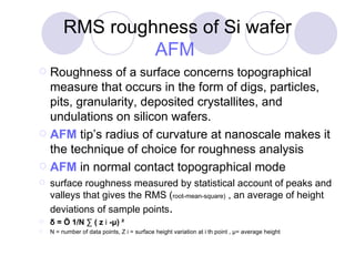
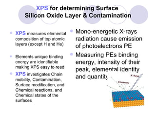
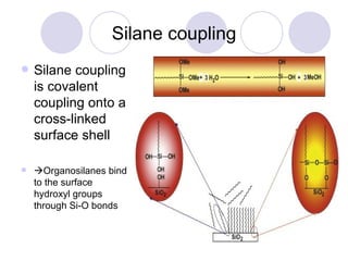
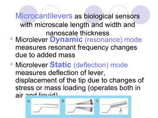
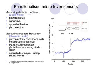
Characterisation of Biosensors  Oxford Workshop 4-5 July 2009 Post Grad Cert Nanotechnology   Nasrin Azadeh-McGuire University of Oxford Begbroke Science Park & Dept of Continuing Education
Bioaffinity Based Biosensor and immunosensor Biosensor is a detection device with high analytical specificity made of layers of biological elements with biorecognition quality that are immobilized on a substrate having reversible interactions with the analyte and a signal transducer. Immunosensors are biosensors that have antibodies as biological element.
Surface characterisation of biosensors Chemical analysis to confirm covalent Immobilization Surface density to detect distribution and location Structural analysis to inform spatial relation and anatomy Functional information about biorecognition
RMS roughness of Si wafer AFM   Roughness of a surface concerns topographical measure that occurs in the form of digs, particles, pits, granularity, deposited crystallites, and undulations on silicon wafers.  AFM  tip’s radius of curvature at nanoscale makes it the technique of choice for roughness analysis AFM  in normal contact topographical mode  surface roughness measured by statistical account of peaks and valleys that gives the RMS ( root-mean-square)  , an average of height deviations of sample points .  δ = Ö 1/N ∑ ( z  i  -μ) ²  N = number of data points, Z i = surface height variation at i th point , μ= average height
XPS  for determining Surface  Silicon Oxide Layer & Contamination XPS  measures elemental composition of top atomic layers (except H and He) Elements unique binding energy are identifiable making XPS easy to read XPS  investigates Chain mobility, Contamination, Surface modification, and Chemical reactions, and Chemical states of the surfaces Mono-energetic X-rays radiation cause emission of photoelectrons PE Measuring PEs binding energy, intensity of their peak, elemental identity and quantity
SAMs Self assembled monolayers SAMs are long chain monolayers of diverse functionalities organised on surface of noble metals or silane for use as sensor arrays Two main coupling schemes thiol coupling  silane coupling
Silane coupling Silane coupling is covalent coupling onto a cross-linked surface shell  Organosilanes bind to the surface hydroxyl groups through Si-O bonds
Thiolated Au film coupling First monolayer appears within few minutes after immersion in dilute solution of alkanothiols in ethanol After ~ 12 hours the film is highly packed by van der Waals forces , contaminations are replaced, defects are filled Chemisorption of thiols onto gold surfaces mostly has covalent character  Au oxide surface layer reduced by the thiol form covalent link to Thiol monolayers
Surface Characterisation of SAMs ToF SIMS   with elemental and molecular fingerprinting; detect distribution and location of biomolecules Tip enhanced Raman Spectroscopy ,  FT-Raman Spectroscopy for molecular recognition  AFM tapping mode for imaging biofunctionalised   surfaces, high resolution, ability to operate in liquid medium, topographical information, images of individual molecule measure molecular dimensions   AFM as force sensing tool ; AFM with functionalised tip, f-d mode probes functionality, elastic property, adhesion strength, and range of inter-atomic/molecular interactions for molecular recognition XPS complemented with Ellipsometric and contact angle   measurement can confirm the presence of immobilized molecules
ToF SIMS SIMS analyses mass of ions and molecular fragments ejected from surface (dept of 10-20  Å )  TOF-SIMS: pulse primary ion beam at low fluence  to ionize and desorbe the sample surface ToF analyzer detects time of flight of secondary ions to mass spectrometer with their different velocities  Mass spectrum counts of secondary ions emitted to determine molecular species.  Highly focused primary ion beam (ca 1  μ  dia ) images visualising molecular distribution on the surface TOF-SIMS spectrum of (PET) (mass peaks for the fragments of a PET molecule)
ToF SIMS Advantageous: Mass spectra with high mass resolution and high mass range (several thousand Daltons) Molecular characterisation of surfaces, functionalities, and oxidation states Limitations:  requires substantial interpretations of information rich spectra Peak overlaps and interference  Larger molecules produce many smaller fragment ions
Microcantilevers  as biological sensors with microscale length and width and nanoscale thickness Microlever  Dynamic  (resonance) mode  measures resonant frequency changes due to added mass  Microlever  Static  (deflection) mode  measures deflection of lever, displacement of the tip due to changes of stress or mass loading (operates both in air and liquid)
Functionalised micro-lever sensors   Measuring deflection of lever  (Static Mode) piezoresistive capacitive optical reflection piezoelectric  Measuring resonant frequency  (Dynamic mode) piezoelectric - oscillations with measurable amplitude magnetically actuated photothermal – using diode laser  acoustic technique – using sound waves  bending responses due to various concentration of streptavidin
References   Bruno Pettinger, Tip-enhanced Raman scattering: Influence of the tip-surface geometry on optical resonance and enhancement, Surface Science 603 (2009) 1335–1341 Myhra S, A review of enabling technologies based on scanning probe microscopy relevant to bioanalysis, Biosensors and Bioelectronics, Volume 19, Issue 11, 15 June 2004, Pages 1345-1354 Matthias Rief, Single Molecule Force Spectroscopy on Polysaccharides by Atomic Force Microscopy, Science, Vol. 275. no. 5304, pp. 1295 - 129  Emanuele Ostuni, (Harvard Univ), The interaction of proteins and cells with self-assembled monolayers of alkanethiolates on gold and silver, Colloids and Surfaces B: Biointerfaces, Volume 15, Issue 1,  31 August 1999, Pages 3-30  Wagner P, (Stanford Uni), Immobilization strategies for biological scanning probe microscopy, Volume 430, Issues 1-2,  23 June 1998, Pages 112-115  Wang C et al, Ultrasensitive biochemical sensors based on microcantilevers of atomic force  microscope, Analytical Biochemistry, Volume 363, Issue 1, 1 April 2007, Pages 1-11  N Sandhyarani and T Pradeep, Characteristics of alkanethiol self assembled monolayers prepared on sputtered gold films: a surface enhanced Raman spectroscopic investigation, Vacuum, Volume 49, Issue 4, April 1998, Pages 279-284  Ijeoma M. Nnebe, Carnegie Univ, Dynamic Atomic Force Microscopy Studies to Characterize Heterogeneous Surfaces, http://www.andrew.cmu.edu/user/jamess3/inENN03.pdf Henke L et al, An AFM determination of the effects on surface roughness caused by cleaning of fused silica and glass substrates in the process of optical biosensor preparation, Biosensors and Bioelectronics 17 (2002) 547_/555 Urban G, Micro- and nanobiosensors—state of the art and trends, Measurement Science and Technology 20 (2009) 012001 (18pp) Louis Tiefenauer, Biointerface analysis on a molecular level: New tools for biosensor research, Colloids and Surfaces B: Biointerfaces,, Volume 23, Issues 2-3, February 2002, Pages 95-114 Yam Chi Ming et al, Preparation, characterization, resistance to protein adsorption, and specific avidin–biotin binding of poly(amidoamine) dendrimers functionalized with oligo(ethylene glycol) on gold, Journal of Colloid and Interface Science, Volume 296, Issue 1, April 2006, Pages 118-130 http://www.nb.engr.washington.edu/research/instrumentation/tofsims/7200.shtml http://www.iontof.com/products-tofsims5-IONTOF-TOF-SIMS-TIME-OF-FLIGHT-SURFACE-ANALYSIS.htm http://www.phi.com/techniques/xps.html http://www.uwo.ca/ssw/services/tof-sims.html

Workshop Biosensor Brief

  • 1.
    Characterisation of Biosensors Oxford Workshop 4-5 July 2009 Post Grad Cert Nanotechnology Nasrin Azadeh-McGuire University of Oxford Begbroke Science Park & Dept of Continuing Education
  • 2.
    Bioaffinity Based Biosensorand immunosensor Biosensor is a detection device with high analytical specificity made of layers of biological elements with biorecognition quality that are immobilized on a substrate having reversible interactions with the analyte and a signal transducer. Immunosensors are biosensors that have antibodies as biological element.
  • 3.
    Surface characterisation ofbiosensors Chemical analysis to confirm covalent Immobilization Surface density to detect distribution and location Structural analysis to inform spatial relation and anatomy Functional information about biorecognition
  • 4.
    RMS roughness ofSi wafer AFM Roughness of a surface concerns topographical measure that occurs in the form of digs, particles, pits, granularity, deposited crystallites, and undulations on silicon wafers. AFM tip’s radius of curvature at nanoscale makes it the technique of choice for roughness analysis AFM in normal contact topographical mode surface roughness measured by statistical account of peaks and valleys that gives the RMS ( root-mean-square) , an average of height deviations of sample points . δ = Ö 1/N ∑ ( z i -μ) ² N = number of data points, Z i = surface height variation at i th point , μ= average height
  • 5.
    XPS fordetermining Surface Silicon Oxide Layer & Contamination XPS measures elemental composition of top atomic layers (except H and He) Elements unique binding energy are identifiable making XPS easy to read XPS investigates Chain mobility, Contamination, Surface modification, and Chemical reactions, and Chemical states of the surfaces Mono-energetic X-rays radiation cause emission of photoelectrons PE Measuring PEs binding energy, intensity of their peak, elemental identity and quantity
  • 6.
    SAMs Self assembledmonolayers SAMs are long chain monolayers of diverse functionalities organised on surface of noble metals or silane for use as sensor arrays Two main coupling schemes thiol coupling silane coupling
  • 7.
    Silane coupling Silanecoupling is covalent coupling onto a cross-linked surface shell  Organosilanes bind to the surface hydroxyl groups through Si-O bonds
  • 8.
    Thiolated Au filmcoupling First monolayer appears within few minutes after immersion in dilute solution of alkanothiols in ethanol After ~ 12 hours the film is highly packed by van der Waals forces , contaminations are replaced, defects are filled Chemisorption of thiols onto gold surfaces mostly has covalent character Au oxide surface layer reduced by the thiol form covalent link to Thiol monolayers
  • 9.
    Surface Characterisation ofSAMs ToF SIMS with elemental and molecular fingerprinting; detect distribution and location of biomolecules Tip enhanced Raman Spectroscopy , FT-Raman Spectroscopy for molecular recognition AFM tapping mode for imaging biofunctionalised surfaces, high resolution, ability to operate in liquid medium, topographical information, images of individual molecule measure molecular dimensions AFM as force sensing tool ; AFM with functionalised tip, f-d mode probes functionality, elastic property, adhesion strength, and range of inter-atomic/molecular interactions for molecular recognition XPS complemented with Ellipsometric and contact angle measurement can confirm the presence of immobilized molecules
  • 10.
    ToF SIMS SIMSanalyses mass of ions and molecular fragments ejected from surface (dept of 10-20 Å ) TOF-SIMS: pulse primary ion beam at low fluence to ionize and desorbe the sample surface ToF analyzer detects time of flight of secondary ions to mass spectrometer with their different velocities Mass spectrum counts of secondary ions emitted to determine molecular species. Highly focused primary ion beam (ca 1 μ dia ) images visualising molecular distribution on the surface TOF-SIMS spectrum of (PET) (mass peaks for the fragments of a PET molecule)
  • 11.
    ToF SIMS Advantageous:Mass spectra with high mass resolution and high mass range (several thousand Daltons) Molecular characterisation of surfaces, functionalities, and oxidation states Limitations: requires substantial interpretations of information rich spectra Peak overlaps and interference Larger molecules produce many smaller fragment ions
  • 12.
    Microcantilevers asbiological sensors with microscale length and width and nanoscale thickness Microlever Dynamic (resonance) mode measures resonant frequency changes due to added mass Microlever Static (deflection) mode measures deflection of lever, displacement of the tip due to changes of stress or mass loading (operates both in air and liquid)
  • 13.
    Functionalised micro-lever sensors Measuring deflection of lever (Static Mode) piezoresistive capacitive optical reflection piezoelectric Measuring resonant frequency (Dynamic mode) piezoelectric - oscillations with measurable amplitude magnetically actuated photothermal – using diode laser acoustic technique – using sound waves  bending responses due to various concentration of streptavidin
  • 14.
    References Bruno Pettinger, Tip-enhanced Raman scattering: Influence of the tip-surface geometry on optical resonance and enhancement, Surface Science 603 (2009) 1335–1341 Myhra S, A review of enabling technologies based on scanning probe microscopy relevant to bioanalysis, Biosensors and Bioelectronics, Volume 19, Issue 11, 15 June 2004, Pages 1345-1354 Matthias Rief, Single Molecule Force Spectroscopy on Polysaccharides by Atomic Force Microscopy, Science, Vol. 275. no. 5304, pp. 1295 - 129 Emanuele Ostuni, (Harvard Univ), The interaction of proteins and cells with self-assembled monolayers of alkanethiolates on gold and silver, Colloids and Surfaces B: Biointerfaces, Volume 15, Issue 1, 31 August 1999, Pages 3-30 Wagner P, (Stanford Uni), Immobilization strategies for biological scanning probe microscopy, Volume 430, Issues 1-2, 23 June 1998, Pages 112-115 Wang C et al, Ultrasensitive biochemical sensors based on microcantilevers of atomic force microscope, Analytical Biochemistry, Volume 363, Issue 1, 1 April 2007, Pages 1-11 N Sandhyarani and T Pradeep, Characteristics of alkanethiol self assembled monolayers prepared on sputtered gold films: a surface enhanced Raman spectroscopic investigation, Vacuum, Volume 49, Issue 4, April 1998, Pages 279-284 Ijeoma M. Nnebe, Carnegie Univ, Dynamic Atomic Force Microscopy Studies to Characterize Heterogeneous Surfaces, http://www.andrew.cmu.edu/user/jamess3/inENN03.pdf Henke L et al, An AFM determination of the effects on surface roughness caused by cleaning of fused silica and glass substrates in the process of optical biosensor preparation, Biosensors and Bioelectronics 17 (2002) 547_/555 Urban G, Micro- and nanobiosensors—state of the art and trends, Measurement Science and Technology 20 (2009) 012001 (18pp) Louis Tiefenauer, Biointerface analysis on a molecular level: New tools for biosensor research, Colloids and Surfaces B: Biointerfaces,, Volume 23, Issues 2-3, February 2002, Pages 95-114 Yam Chi Ming et al, Preparation, characterization, resistance to protein adsorption, and specific avidin–biotin binding of poly(amidoamine) dendrimers functionalized with oligo(ethylene glycol) on gold, Journal of Colloid and Interface Science, Volume 296, Issue 1, April 2006, Pages 118-130 http://www.nb.engr.washington.edu/research/instrumentation/tofsims/7200.shtml http://www.iontof.com/products-tofsims5-IONTOF-TOF-SIMS-TIME-OF-FLIGHT-SURFACE-ANALYSIS.htm http://www.phi.com/techniques/xps.html http://www.uwo.ca/ssw/services/tof-sims.html