

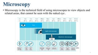
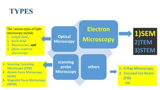
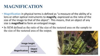
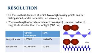
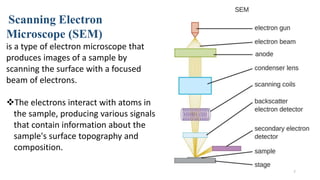
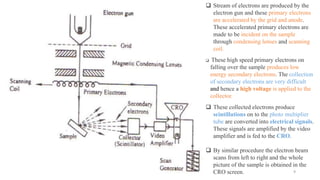
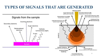
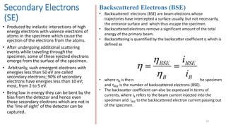
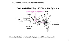
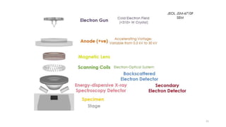
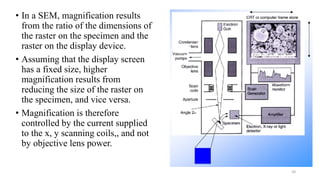
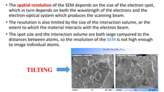


The document discusses the working principle of scanning electron microscopy (SEM). It describes how SEM uses a focused beam of electrons to scan the surface of a sample to produce images. SEM provides higher resolution than light microscopes due to the much shorter wavelength of electrons. The document outlines the various components of an SEM, including the electron gun, electromagnetic lenses, detectors for secondary electrons and backscattered electrons, and how these are used to control magnification and resolution. It also discusses some imaging parameters and artifacts that can influence SEM results.


