Download to read offline


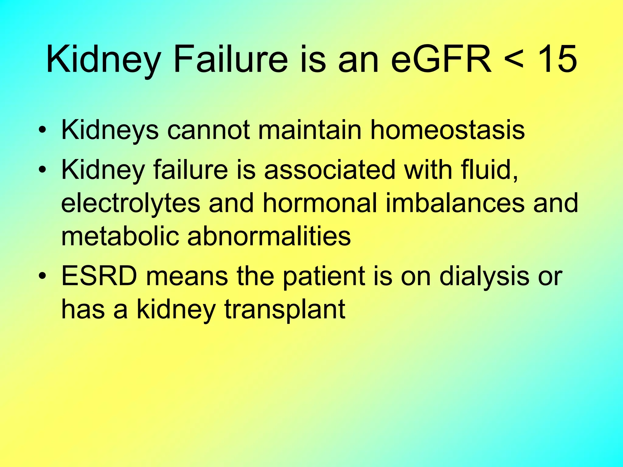

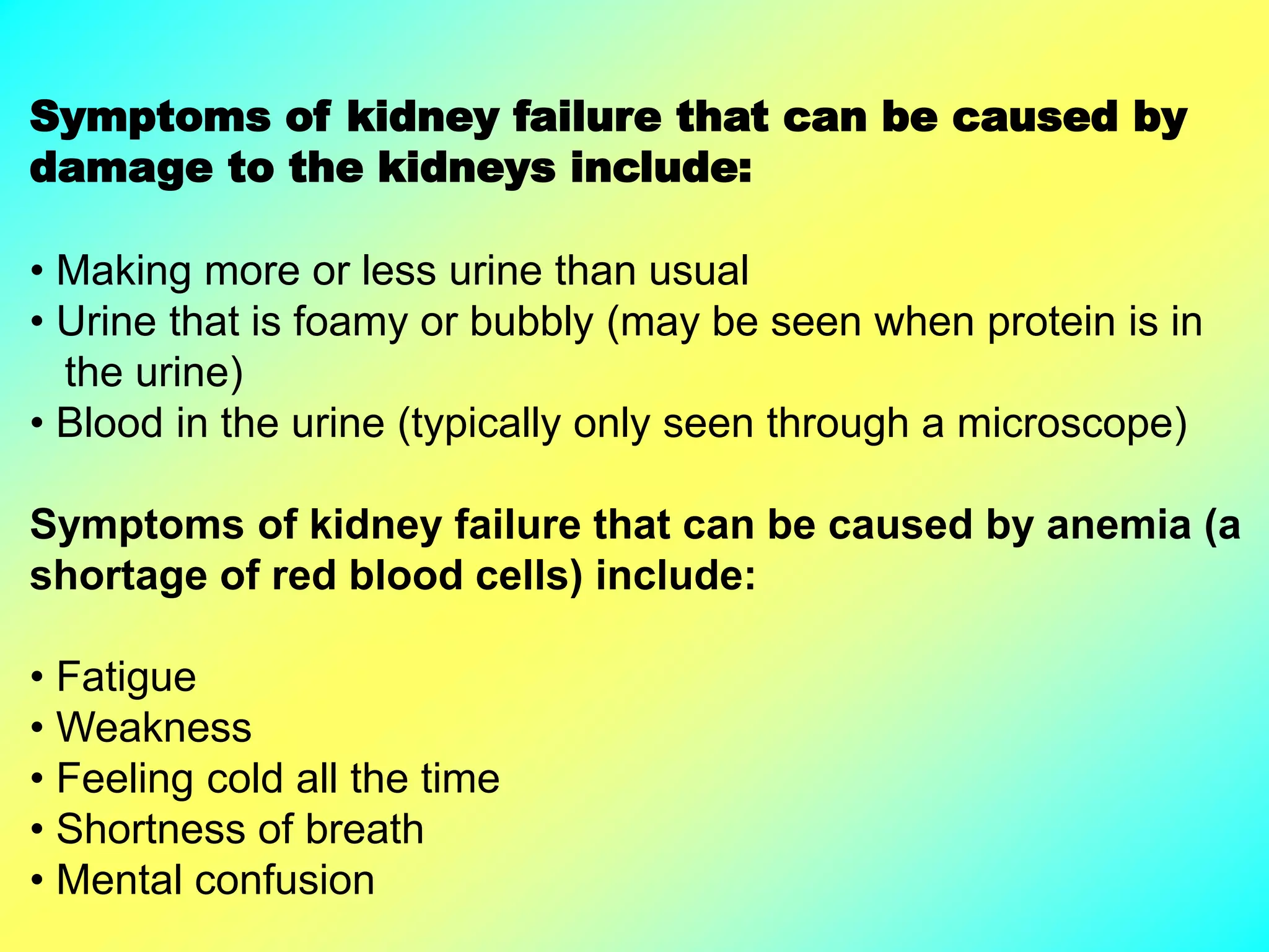


The document provides information about kidney structure and function, as well as kidney diseases. It discusses that the kidneys contain nephrons and filter blood to remove waste and regulate fluid levels. It describes acute kidney injury (AKI) as a sudden decline in kidney function, and chronic kidney disease (CKD) as long-term decreased function. For kidney failure, dialysis or transplantation is needed to replace lost kidney function.


![KIDNEY FAILURE MD5 [Autosaved].pptx](https://cdn.slidesharecdn.com/ss_thumbnails/kidneyfailuremd5autosaved-221027143100-3940946c-thumbnail.jpg?width=640&height=640&fit=bounds)


