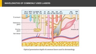
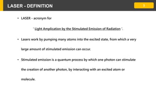
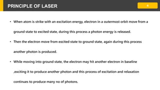
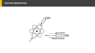
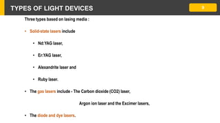
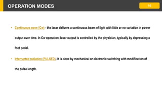
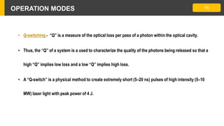
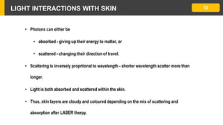
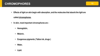
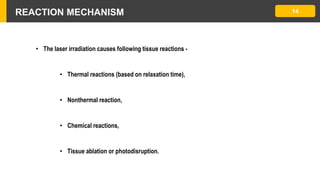
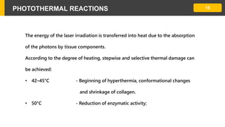
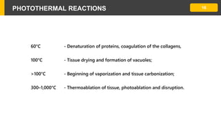
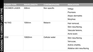
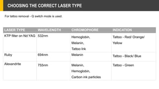
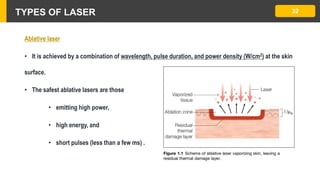
LASER THERAPY
BASICS OF
Dr.SRINIVASAN.G,
Post Graduate in DVL
MADURAI MEDICAL COLLEGE
1LIGHT SPECTRUM
• Light is a complex system of radiant energy that is composed of waves and energy
packets known as photons. It is arranged into the electromagnetic spectrum (EMS)
according to the wavelength.
• The distance between two successive troughs or crests of the waves, measured in
meters, is the wavelength.
• The number of wave crests (or troughs) in given point in a second is called frequency.
2LIGHT SPECTRUM
Light spectrum contains -
• Ultra Violet Rays (200–400 nm),
• Visible Spectrum (400–700 nm),
• Near Infra Red “I” (755–810 nm),
• Near Infra Red “II” (940–1,064 nm),
• Mid Infra Red (1.3–3 mm),
• Far Infra Red (3 mm and beyond)
WAVELENGTHS OF COMMONLY USED LASERS
3LASER - DEFINITION
• LASER - acronym for
' Light Amplication by the Stimulated Emission of Radiation '.
• Lasers work by pumping many atoms into the excited state, from which a very
large amount of stimulated emission can occur.
• Stimulated emission is a quantum process by which one photon can stimulate
the creation of another photon, by interacting with an excited atom or
molecule.
4PRINCIPLE OF LASER
• When atom is strike with an excitation energy, electron in a outermost orbit move from a
ground state to excited state, during this process a photon energy is released.
• Then the electron move from excited state to ground state, again during this process
another photon is produced.
• While moving into ground state, the electron may hit another electron in baseline
,exciting it to produce another photon and this process of excitation and relaxation
continues to produce many no of photons.
PHOTON GENERATION
DEFINITION
• Laser light -
• monochromic - output with single wavelength of light ,
• has cohorence - all waves of light travel in phase spacially and temporally ,
• highly collimated - travel long distance without divergence.
• LASER stimulate tissue remodelling by a complex process that mimics large wound healing
epidermal regeneration, induction of metalloproteinases, and formation of new dermal
elastin fibrils and collagen types I and III.
• Compared with gross wound healing, there is minimal inflammation and no scarring.
7
5PRINCIPLE OF LASER APPARATUS
• A laser apparatus is shaped like a long, narrow tube or cylinder to harness laser energy,
• The cylinder has mirrors at either end so that photons are reflected back and forth and are
constantly renewing the process of stimulated emission as they strike more of the excited
atoms in the laser chamber.
• The mirrors also align the photons so that they are traveling parallel.
LASER APPARATUS 6
• One of the two mirrors is only partially silvered (reflective) so that it allows some
transmission of the laser light.
• The transmitted light becomes the laser beam.
COMPONENTS OF LASER DEVICE
• Lasing medium - Amplifier of laser, which can be
• Solid , Gas or Liquid.
• Excitation source,
• Feedback apparatus - feedback mechanism is produced by the resonator, where the light is
reflected by two mirrors so that the photons pass several times through the laser material.
• Output coupler
8
TYPES OF LIGHT DEVICES
Three types based on lasing media :
• Solid-state lasers include
• Nd:YAG laser,
• Er:YAG laser,
• Alexandrite laser and
• Ruby laser.
• The gas lasers include - The Carbon dioxide (CO2) laser,
Argon ion laser and the Excimer lasers,
• The diode and dye lasers.
9
OPERATION MODES
• Continuous wave (Cw) - the laser delivers a continuous beam of light with little or no variation in power
output over time. In Cw operation, laser output is controlled by the physician, typically by depressing a
foot pedal.
• Interrupted radiation (PULSED)- It is done by mechanical or electronic switching with modification of
the pulse length.
10
OPERATION MODES
• Q-switching - “Q” is a measure of the optical loss per pass of a photon within the optical cavity.
• Thus, the “Q” of a system is a used to characterize the quality of the photons being released so that a
high “Q” implies low loss and a low “Q” implies high loss.
• A “Q-switch” is a physical method to create extremely short (5–20 ns) pulses of high intensity (5–10
MW) laser light with peak power of 4 J.
11
LIGHT INTERACTIONS WITH SKIN
• Photons can either be
• absorbed - giving up their energy to matter, or
• scattered - changing their direction of travel.
• Scattering is inversely proprtional to wavelength - shorter wavelength scatter more than
longer.
• Light is both absorbed and scattered within the skin.
• Thus, skin layers are cloudy and coloured depending on the mix of scattering and
absorption after LASER therpy.
12
CHROMOPHORES 13
• Effects of light on skin begin with absorption, and the molecules that absorb the light are
called chromophores.
• In skin, most important chromophores are -
• Hemoglobin,
• Melanin,
• Exogenous pigments ( Tattoo ink, drugs )
• Water,
• Lipid.
REACTION MECHANISM 14
• The laser irradiation causes following tissue reactions -
• Thermal reactions (based on relaxation time),
• Nonthermal reaction,
• Chemical reactions,
• Tissue ablation or photodisruption.
PHOTOTHERMAL REACTIONS 15
The energy of the laser irradiation is transferred into heat due to the absorption
of the photons by tissue components.
According to the degree of heating, stepwise and selective thermal damage can
be achieved:
• 42–45°C - Beginning of hyperthermia, conformational changes
and shrinkage of collagen.
• 50°C - Reduction of enzymatic activity;
PHOTOTHERMAL REACTIONS 16
60°C - Denaturation of proteins, coagulation of the collagens,
100°C - Tissue drying and formation of vacuoles;
>100°C - Beginning of vaporization and tissue carbonization;
300–1,000°C - Thermoablation of tissue, photoablation and disruption.
SELECTIVE PHOTOTHERMOLYSIS 17
• It causes Selective histological damage, which requires heat confinement to desirable target
structures.
• Selective photothermolysis combines appropriate wavelength (‘color’ of light), fluence
(‘dose’ of light), pulse duration, and protective skin cooling for the treatment of a variety of
diseases
• Selective Photothermolysis ( SP ) operate in the visible and Near Infrared (NIR) spectrum.
• In the visible light spectrum, a target chromophore is treated using wavelengths of light of a
complimentary color.
SELECTIVE PHOTOTHERMOLYSIS 18
• SP involves in extreme localized heating, which relies on -
1. Wavelength (COLOUR OF LIGHT)- havre to select a wavelength that reaches and is
preferentially absorbed by the desired target structures;
2. Pulse duration ,
3. Fluence (DOSE OF LIGHT) - It denote the amount of energy needed to damage the the
tissue. It is inversely proprtional to absorption by the target tissue– stronger
absorption requires lower fluence, and vice versa.
PULSE DURATION 19
• An exposure duration less than or equal to the time necessary for cooling of the target
structures;
• It is directly proportional to the square of the target size in millimeters.
• It allows heat to be confined during the laser pulse in or near the target structures. The
moment the heat is formed , the target begins to cool by conduction .
PULSE DURATION 20
Thermal Relaxation Time ( TRT )
• TRT is defined as the time required for substantial ( half the time ) cooling of the target
structure.
• TRT is strongly related to target size,
TRT ≈ d2,
• TRT is in units of seconds, and 'd' is the target size in millimeters.
SKIN COOLING 23
• Sparing of the epidermis and superficial dermis is important for selective destruction of deeper
structures and can be improved by the use of appropriate skin cooling.
• Cooling can be applied
• before (pre-cooling),
• during (parallel cooling), and
• after the laser pulse (post-cooling).
SKIN COOLING 24
• The greater the depth of an anatomical structure, the longer the cooling should be applied.
• 20–50 milliseconds is enough for epidermal protection.
• 5–10 seconds is needed to protect both epidermis and dermis for lasers targeting
subcutaneous fat.
SKIN COOLING 25
• Cooling can be applied using
• Direct solid contact cooling (e.g. cold sapphire window),
• Automated cryogen spray (direct cooling devices) or
• By blowing direct cold air.
FRACTIONAL THERMOLYSIS 21
• It uses microbeams of laser to target the tissue, inducing microthermal zones (MTZ) of
injury.
• Each MTZ is typically 100–300 µm in diameter.
• The depth and density (number per unit area) of the microlaser beams applied to the tissue
can be adjusted depending on the clinical indication.
FRACTIONAL THERMOLYSIS 22
• Advantage - it spares untreated skin surrounding each MTZ, allowing fast healing
and reducing the risk of side effects.
• Fractionated devices also play role in drug delivery into the tissue and for
extruding material out of the skin.
CHOOSING CORRECT LIGHT SOURCE 26
1. Define clinical indication
2. Choose correct light wavelength (nm) based on histological target chromophore
3. Observe whether continuous wave (CW) or pulsed source is required
4. Choose right pulse width if necessary (seconds)
5. Choose pulse frequency (Hz), if necessary
CHOOSING CORRECT LIGHT SOURCE 27
6. Set skin cooling parameters
7. Choose appropriate light dose
8. Test laser to check whether it is working properly
9. Trigger single pulse on target skin and observe clinical end point
10. Adjust dosimetry if necessary
11. If no unwanted sign is observed, continue with treatment
LASER TYPE WAVELENGTH CHROMOPHORE INDICATION
EXCIMER LASER 308nm Non specific Vitiligo
Psoriasis
Atopic dermatitis
Morphea
Nd:YAG 1064nm Melanin Hair removal,
Skin resurfacing,
Vascular lesions,
Acne scars
CO2 10600nm Cellular water Skin resurfacing,
Verrucae,
Actinic cheilitis,
Ablative skin resurfacing
CHOOSINGTHECORRECTLASERTYPE
CHOOSING THE CORRECT LASER TYPE
LASER TYPE WAVELENGTH CHROMOPHORE INDICATION
KTP filter on Nd:YAG 532nm Hemoglobin,
Melanin,
Tattoo Ink
Tattoo - Red/ Orange/
Yellow
Ruby 694nm Melanin Tattoo - Black/ Blue
Alexandrite 755nm Melanin,
Hemoglobin,
Carbon ink particles
Tattoo - Green
For tattoo removal - Q switch mode is used.
PRECAUTIONS 28
• Anesthetize area if necessary (e.g. tattoos, ablative lasers)
• Wear appropriate personal protection (e.g. wavelength specific eye goggles/glasses, fume-
resistant mask, gloves)
• Turn smoke evacuator on if performing ablative procedures
• Dress the area with petrolatum ointment and protect from sun exposure until treated area is
healed.
TOPICAL ANAESTHESIA
TOPICAL ANAESTHESIA
• Clean the area with either mild cleanser or with water.
• Use a gloved finger or a tongue depressor
• Apply a uniform layer of the product: approximately 1/8” thick
• If the product is applied with a bare finger (not recommended), wash the finger
right after the application.
• Topical anesthetic is left on skin for at least 30 min, depending upon the choice
of product
TOPICAL ANAESTHESIA
• Usually, the agent is left on skin for 30–60 min.
• Superficial penetration - 30 mins ( hair removal )
• For deeper penetration - ablative skin resurfacing treatments - 60 min.
• Immediately preceding the treatment:
• Remove topical anesthetic with dry gauze
• Clean the skin by wiping it with a water-moistened gauze.
TYPES OF LASER 29
LASER treatment is grossly divided into -
• Ablative laser
• those that vaporize tissue by boiling water inside the tissue.
• Non ablative laser
• those that do not vaporize the tissue.
TYPES OF LASER 30
Ablative laser
• To remove tissue, ablative lasers must raise local tissue temperature beyond the boiling point of
100oC, plus add much more energy needed for changing water into steam.
• To vaporize 1 cm3
of water, more than 2000 J are required. An ablative laser must deliver about 2500
J of energy per cm3
to vaporize tissue.
• The energy must be delivered quickly to remove the hot tissue before heat is conducted deeply into
the skin, causing a burn.
TYPES OF LASER 31
Ablative laser
• The standard ablative lasers in dermatology are
• Erbium - 2940nm ,
• CO2 - 10600nm .
• They are used to precisely remove a thin layer for resurfacing or narrow column for fractional
treatment of skin, leaving behind minimal residual thermal damage.
• Minimum residual thermal injury is usually 0.1mm to achieve hemostasis.
TYPES OF LASER 32
Ablative laser
• It is achieved by a combination of wavelength, pulse duration, and power density (W/cm2) at the skin
surface.
• The safest ablative lasers are those
• emitting high power,
• high energy, and
• short pulses (less than a few ms) .
TYPES OF LASER 33
Non Ablative laser
• It improves skin quality without physical removal or vaporization of the skin.
• Non-ablative skin resurfacing is a safe and effective means of improving many aspects of
photoaged skin.
• It alters cellular and non-cellular components of the skin without causing an open wound.
• Non-ablative modalities can be done with local anaesthesia alone.
Laser basics -  srinivasan - final

Laser basics - srinivasan - final

  • 1.
    LASER THERAPY BASICS OF Dr.SRINIVASAN.G, PostGraduate in DVL MADURAI MEDICAL COLLEGE
  • 2.
    1LIGHT SPECTRUM • Lightis a complex system of radiant energy that is composed of waves and energy packets known as photons. It is arranged into the electromagnetic spectrum (EMS) according to the wavelength. • The distance between two successive troughs or crests of the waves, measured in meters, is the wavelength. • The number of wave crests (or troughs) in given point in a second is called frequency.
  • 3.
    2LIGHT SPECTRUM Light spectrumcontains - • Ultra Violet Rays (200–400 nm), • Visible Spectrum (400–700 nm), • Near Infra Red “I” (755–810 nm), • Near Infra Red “II” (940–1,064 nm), • Mid Infra Red (1.3–3 mm), • Far Infra Red (3 mm and beyond)
  • 4.
  • 5.
    3LASER - DEFINITION •LASER - acronym for ' Light Amplication by the Stimulated Emission of Radiation '. • Lasers work by pumping many atoms into the excited state, from which a very large amount of stimulated emission can occur. • Stimulated emission is a quantum process by which one photon can stimulate the creation of another photon, by interacting with an excited atom or molecule.
  • 6.
    4PRINCIPLE OF LASER •When atom is strike with an excitation energy, electron in a outermost orbit move from a ground state to excited state, during this process a photon energy is released. • Then the electron move from excited state to ground state, again during this process another photon is produced. • While moving into ground state, the electron may hit another electron in baseline ,exciting it to produce another photon and this process of excitation and relaxation continues to produce many no of photons.
  • 7.
  • 8.
    DEFINITION • Laser light- • monochromic - output with single wavelength of light , • has cohorence - all waves of light travel in phase spacially and temporally , • highly collimated - travel long distance without divergence. • LASER stimulate tissue remodelling by a complex process that mimics large wound healing epidermal regeneration, induction of metalloproteinases, and formation of new dermal elastin fibrils and collagen types I and III. • Compared with gross wound healing, there is minimal inflammation and no scarring. 7
  • 9.
    5PRINCIPLE OF LASERAPPARATUS • A laser apparatus is shaped like a long, narrow tube or cylinder to harness laser energy, • The cylinder has mirrors at either end so that photons are reflected back and forth and are constantly renewing the process of stimulated emission as they strike more of the excited atoms in the laser chamber. • The mirrors also align the photons so that they are traveling parallel.
  • 10.
    LASER APPARATUS 6 •One of the two mirrors is only partially silvered (reflective) so that it allows some transmission of the laser light. • The transmitted light becomes the laser beam.
  • 11.
    COMPONENTS OF LASERDEVICE • Lasing medium - Amplifier of laser, which can be • Solid , Gas or Liquid. • Excitation source, • Feedback apparatus - feedback mechanism is produced by the resonator, where the light is reflected by two mirrors so that the photons pass several times through the laser material. • Output coupler 8
  • 12.
    TYPES OF LIGHTDEVICES Three types based on lasing media : • Solid-state lasers include • Nd:YAG laser, • Er:YAG laser, • Alexandrite laser and • Ruby laser. • The gas lasers include - The Carbon dioxide (CO2) laser, Argon ion laser and the Excimer lasers, • The diode and dye lasers. 9
  • 13.
    OPERATION MODES • Continuouswave (Cw) - the laser delivers a continuous beam of light with little or no variation in power output over time. In Cw operation, laser output is controlled by the physician, typically by depressing a foot pedal. • Interrupted radiation (PULSED)- It is done by mechanical or electronic switching with modification of the pulse length. 10
  • 14.
    OPERATION MODES • Q-switching- “Q” is a measure of the optical loss per pass of a photon within the optical cavity. • Thus, the “Q” of a system is a used to characterize the quality of the photons being released so that a high “Q” implies low loss and a low “Q” implies high loss. • A “Q-switch” is a physical method to create extremely short (5–20 ns) pulses of high intensity (5–10 MW) laser light with peak power of 4 J. 11
  • 15.
    LIGHT INTERACTIONS WITHSKIN • Photons can either be • absorbed - giving up their energy to matter, or • scattered - changing their direction of travel. • Scattering is inversely proprtional to wavelength - shorter wavelength scatter more than longer. • Light is both absorbed and scattered within the skin. • Thus, skin layers are cloudy and coloured depending on the mix of scattering and absorption after LASER therpy. 12
  • 16.
    CHROMOPHORES 13 • Effectsof light on skin begin with absorption, and the molecules that absorb the light are called chromophores. • In skin, most important chromophores are - • Hemoglobin, • Melanin, • Exogenous pigments ( Tattoo ink, drugs ) • Water, • Lipid.
  • 17.
    REACTION MECHANISM 14 •The laser irradiation causes following tissue reactions - • Thermal reactions (based on relaxation time), • Nonthermal reaction, • Chemical reactions, • Tissue ablation or photodisruption.
  • 18.
    PHOTOTHERMAL REACTIONS 15 Theenergy of the laser irradiation is transferred into heat due to the absorption of the photons by tissue components. According to the degree of heating, stepwise and selective thermal damage can be achieved: • 42–45°C - Beginning of hyperthermia, conformational changes and shrinkage of collagen. • 50°C - Reduction of enzymatic activity;
  • 19.
    PHOTOTHERMAL REACTIONS 16 60°C- Denaturation of proteins, coagulation of the collagens, 100°C - Tissue drying and formation of vacuoles; >100°C - Beginning of vaporization and tissue carbonization; 300–1,000°C - Thermoablation of tissue, photoablation and disruption.
  • 20.
    SELECTIVE PHOTOTHERMOLYSIS 17 •It causes Selective histological damage, which requires heat confinement to desirable target structures. • Selective photothermolysis combines appropriate wavelength (‘color’ of light), fluence (‘dose’ of light), pulse duration, and protective skin cooling for the treatment of a variety of diseases • Selective Photothermolysis ( SP ) operate in the visible and Near Infrared (NIR) spectrum. • In the visible light spectrum, a target chromophore is treated using wavelengths of light of a complimentary color.
  • 21.
    SELECTIVE PHOTOTHERMOLYSIS 18 •SP involves in extreme localized heating, which relies on - 1. Wavelength (COLOUR OF LIGHT)- havre to select a wavelength that reaches and is preferentially absorbed by the desired target structures; 2. Pulse duration , 3. Fluence (DOSE OF LIGHT) - It denote the amount of energy needed to damage the the tissue. It is inversely proprtional to absorption by the target tissue– stronger absorption requires lower fluence, and vice versa.
  • 22.
    PULSE DURATION 19 •An exposure duration less than or equal to the time necessary for cooling of the target structures; • It is directly proportional to the square of the target size in millimeters. • It allows heat to be confined during the laser pulse in or near the target structures. The moment the heat is formed , the target begins to cool by conduction .
  • 23.
    PULSE DURATION 20 ThermalRelaxation Time ( TRT ) • TRT is defined as the time required for substantial ( half the time ) cooling of the target structure. • TRT is strongly related to target size, TRT ≈ d2, • TRT is in units of seconds, and 'd' is the target size in millimeters.
  • 24.
    SKIN COOLING 23 •Sparing of the epidermis and superficial dermis is important for selective destruction of deeper structures and can be improved by the use of appropriate skin cooling. • Cooling can be applied • before (pre-cooling), • during (parallel cooling), and • after the laser pulse (post-cooling).
  • 25.
    SKIN COOLING 24 •The greater the depth of an anatomical structure, the longer the cooling should be applied. • 20–50 milliseconds is enough for epidermal protection. • 5–10 seconds is needed to protect both epidermis and dermis for lasers targeting subcutaneous fat.
  • 26.
    SKIN COOLING 25 •Cooling can be applied using • Direct solid contact cooling (e.g. cold sapphire window), • Automated cryogen spray (direct cooling devices) or • By blowing direct cold air.
  • 27.
    FRACTIONAL THERMOLYSIS 21 •It uses microbeams of laser to target the tissue, inducing microthermal zones (MTZ) of injury. • Each MTZ is typically 100–300 µm in diameter. • The depth and density (number per unit area) of the microlaser beams applied to the tissue can be adjusted depending on the clinical indication.
  • 28.
    FRACTIONAL THERMOLYSIS 22 •Advantage - it spares untreated skin surrounding each MTZ, allowing fast healing and reducing the risk of side effects. • Fractionated devices also play role in drug delivery into the tissue and for extruding material out of the skin.
  • 29.
    CHOOSING CORRECT LIGHTSOURCE 26 1. Define clinical indication 2. Choose correct light wavelength (nm) based on histological target chromophore 3. Observe whether continuous wave (CW) or pulsed source is required 4. Choose right pulse width if necessary (seconds) 5. Choose pulse frequency (Hz), if necessary
  • 30.
    CHOOSING CORRECT LIGHTSOURCE 27 6. Set skin cooling parameters 7. Choose appropriate light dose 8. Test laser to check whether it is working properly 9. Trigger single pulse on target skin and observe clinical end point 10. Adjust dosimetry if necessary 11. If no unwanted sign is observed, continue with treatment
  • 31.
    LASER TYPE WAVELENGTHCHROMOPHORE INDICATION EXCIMER LASER 308nm Non specific Vitiligo Psoriasis Atopic dermatitis Morphea Nd:YAG 1064nm Melanin Hair removal, Skin resurfacing, Vascular lesions, Acne scars CO2 10600nm Cellular water Skin resurfacing, Verrucae, Actinic cheilitis, Ablative skin resurfacing CHOOSINGTHECORRECTLASERTYPE
  • 32.
    CHOOSING THE CORRECTLASER TYPE LASER TYPE WAVELENGTH CHROMOPHORE INDICATION KTP filter on Nd:YAG 532nm Hemoglobin, Melanin, Tattoo Ink Tattoo - Red/ Orange/ Yellow Ruby 694nm Melanin Tattoo - Black/ Blue Alexandrite 755nm Melanin, Hemoglobin, Carbon ink particles Tattoo - Green For tattoo removal - Q switch mode is used.
  • 33.
    PRECAUTIONS 28 • Anesthetizearea if necessary (e.g. tattoos, ablative lasers) • Wear appropriate personal protection (e.g. wavelength specific eye goggles/glasses, fume- resistant mask, gloves) • Turn smoke evacuator on if performing ablative procedures • Dress the area with petrolatum ointment and protect from sun exposure until treated area is healed.
  • 34.
  • 35.
    TOPICAL ANAESTHESIA • Cleanthe area with either mild cleanser or with water. • Use a gloved finger or a tongue depressor • Apply a uniform layer of the product: approximately 1/8” thick • If the product is applied with a bare finger (not recommended), wash the finger right after the application. • Topical anesthetic is left on skin for at least 30 min, depending upon the choice of product
  • 36.
    TOPICAL ANAESTHESIA • Usually,the agent is left on skin for 30–60 min. • Superficial penetration - 30 mins ( hair removal ) • For deeper penetration - ablative skin resurfacing treatments - 60 min. • Immediately preceding the treatment: • Remove topical anesthetic with dry gauze • Clean the skin by wiping it with a water-moistened gauze.
  • 37.
    TYPES OF LASER29 LASER treatment is grossly divided into - • Ablative laser • those that vaporize tissue by boiling water inside the tissue. • Non ablative laser • those that do not vaporize the tissue.
  • 38.
    TYPES OF LASER30 Ablative laser • To remove tissue, ablative lasers must raise local tissue temperature beyond the boiling point of 100oC, plus add much more energy needed for changing water into steam. • To vaporize 1 cm3 of water, more than 2000 J are required. An ablative laser must deliver about 2500 J of energy per cm3 to vaporize tissue. • The energy must be delivered quickly to remove the hot tissue before heat is conducted deeply into the skin, causing a burn.
  • 39.
    TYPES OF LASER31 Ablative laser • The standard ablative lasers in dermatology are • Erbium - 2940nm , • CO2 - 10600nm . • They are used to precisely remove a thin layer for resurfacing or narrow column for fractional treatment of skin, leaving behind minimal residual thermal damage. • Minimum residual thermal injury is usually 0.1mm to achieve hemostasis.
  • 40.
    TYPES OF LASER32 Ablative laser • It is achieved by a combination of wavelength, pulse duration, and power density (W/cm2) at the skin surface. • The safest ablative lasers are those • emitting high power, • high energy, and • short pulses (less than a few ms) .
  • 41.
    TYPES OF LASER33 Non Ablative laser • It improves skin quality without physical removal or vaporization of the skin. • Non-ablative skin resurfacing is a safe and effective means of improving many aspects of photoaged skin. • It alters cellular and non-cellular components of the skin without causing an open wound. • Non-ablative modalities can be done with local anaesthesia alone.