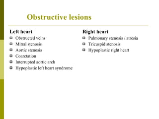
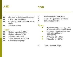
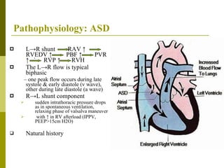
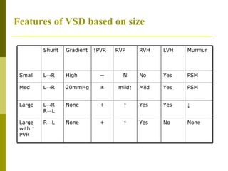
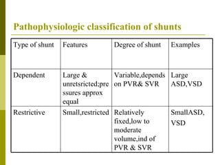
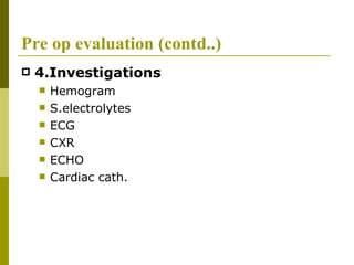
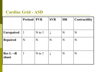
Classification of CHD ASD & VSD: Pathophysiology, preopevaluation, Anaesthetic management for incidental surgery Dr.Nishad.P.K AIIMS,  NewDelhi
Classification of CHD Left to right shunts Right to left shunts(cyanotic lesions) Obstructive lesions Single ventricle   Others  : Vascular rings, Venous anomalies,  Arteriovenous fistulae
Left to right shunts Atrial level :  ASD, PAPVC Ventricular level :  VSD Endocardial cushion defects Great artery level : PDA, AP window, Coronary level :  ALCAPA, coronary fistula
Right to left shunts With pulm. stenosis TOF TA Ebstein’s anomaly   With pulm HTN Eisenmenger’s syndrome With ↑ PBF TGA TAPVC SV W/O obst: to PBF PTA TA  DORV
Obstructive lesions   Left heart Obstructed veins Mitral stenosis Aortic stenosis Coarctation Interrupted aortic arch Hypoplastic left heart syndrome Right heart Pulmonary stenosis / atresia Tricuspid stenosis Hypoplastic right heart
ASD Opening in the interatrial septum i: 1 in 1500 live births 6- 10% of CHD.  30% of CHD in adults Females >males Types : Ostium secondum(75%) Ostium primum(15%) Sinus venosis(10%) Patent foramen ovale(5%) Coronary sinus(rare) VSD Most common CHD(20%) i: 2.6 – 5.7  per 1000 live births  10% of adult CHD Types : Subpulmonary (5 – 7 %)  , ass. With aortic valve insufficiency Perimembranous (80% ) , ass with tricuspid valve abnormality AV canal  (5 – 8 %) Muscular (5 – 20%) Small, medium, large
 
Pathophysiology
Pathophysiology: ASD L->R shunt  RAV ↑  RVEDV ↑  PBF ↑  PVR ↑  `  RVP ↑  RVH The L->R flow is typical biphasic –  one peak flow occurs during late systole & early diastole (v wave), other during late diastole (a wave) R->L shunt component sudden intrathoracic pressure drops as in spontaneous ventilation, relaxing phase of valsalva maneuver  with ↑ in RV afterload (IPPV, PEEP>15cm H2O) Natural history
Pathophysiology :VSD During systole. Bypasses the RV cavity.  L->R shunt  ↑in PBF,LAV,LVEDV  LVH Cause : CHF at 6-10 wks rec: RTI pulm HTN  Eisenmenger’s syndrome Adaptive mechanisms : ↑SV,contractility,HR& myocardial mass
Features of VSD based on size ↓ Yes Yes ↑ + None L->R R->L Large None No Yes ↑ + None R->L Large with  ↑  PVR  PSM Yes Mild mild↑ ± 20mmHg L->R Med PSM Yes No N ─ High L->R Small Murmur LVH RVH RVP ↑ PVR Gradient Shunt
Pathophysiologic classification of shunts SmallASD, VSD Relatively fixed,low to moderate volume,ind of PVR & SVR Small,restricted Restrictive Large ASD,VSD Variable,depends on PVR& SVR Large & unretsricted;pressures approx equal Dependent Examples Degree of shunt Features Type of shunt
Pre op evaluation 1.Review underlying anatomy & physiology of cardiac lesion Previous cardiac surgeries – palliative vs. reparative Evaluate existing residua or sequelae 2.Assess other pre existing or congenital anomalies Extra cardiac malformation in 20-45% Known chromosomal anomaly in 5 - 10% ASD  --  Trisomy 21,18,13,XXXY,Del 4p,5p,CHARGE,Fetal Hydantoin Syndrome,Fetal alcohol syndrome,Holt Oram ,Treacher collins,Noonan’s VSD  --   Trisomy 21,18,13,XXXY,Del 4p,5p,CHARGE,Fetal Hydantoin Syndrome,Fetal alcohol syndrome, Del 10q, 13q, 18q, Fetal valproate syndrome, Elis van creveld syndrome, Apert…
Pre op evaluation(contd..) Examination   General Airway  Cardiac Respiratory  3.History functional status CHF Cyanotic spells Recent RTI Coexisting illness:GERD, reactive airway d/s, seizure d/r
Pre op evaluation (contd..) 4.Investigations  Hemogram  S.electrolytes ECG CXR ECHO Cardiac cath.
ECG
CXR ASD  VSD
Pre op evaluation(contd..) 5.Review information from last cardiological examination Recent cardiac cath, echo  Functional status 6.Assess risk  factors Pulmonary HTN Cyanosis Reoperation Arrhythmias Vent dysfunction 7.Review changes since last cardiac examination History & physical examination Lab data Current medication
Pre op evaluation(contd..) 8.  Review proposed surgical procedure Elective vs. emergent Expected length& invasiveness 9.  Plan treatment of potential complications Dysrhythmias Pulmonary hypertension Ventricular dysfunction 10.  Plan post op care Monitoring Pain management Cardiology follow up 11.  Discuss anaesthetic plan & risk with patient/parent
Preop preparation   Fasting Guidelines: Avoid dehydration/hypovolemia Premedication: Benefits: Anxiolysis ↑ cooperation ↓ separation anxiety ↓ cardiovascular liability Detrimental effects: Hypoventilation Hypotension Pain on administration
IE Prophylaxis  (AHA 2007 guidelines) Unrepaired cynotic CHD (Eisenmenger’s) Completely repaired CHD with prosthetic material or device during 1 st  6 months Repaired CHD with residual defects at/near the patch or device which inhibit endotheliazation Previous IE
IE PROPHYLAXIS IS NO LONGER RECOMMENDED FOR ANY OTHER FORM OF CHD Procedures All dental procedures that involve manipulation of gingival tissue,or periapical region of teeth or perforation of oral mucosa Procedures on RT, infected skin, skin structures, MST NOT recommended for GI or GU tract procedures IE Prophylaxis (contd…)
IE Prophylaxis (contd…)
Monitoring Pulse Oximetry NIBP ECG Capnography Urine Output Temperature CVP IBP TEE
Anaesthetic goals Bubble avoidance Optimizing O2 delivery & ventilatory function Avoid hypovolemia ↓  L->R shunt
Bubble Avoidance: Remove all bubbles from iv tubing Connect iv tubing to the venous cannula while there is free flow of iv fluid and blood Eject small amount of solution from syringe to clear air from needle to hub before injection Aspirate injection port of 3 way before injection to clear air Hold the syringe upright to keep bubbles at the plunger end Do not inject the last ml from the syringe Do not leave a central line open to air
 
Hypovolemia is poorly tolerated in these patients. The low resistance pulmonary circulation tends to  steal   volume from the high resistance systemic circulation.  This is further increased by the systemic arterial vasoconstriction of hypovolemia
Cardiac Grid - ASD N N ↓ N to ↑ ↑ Res L->R shunt N N N N N Repaired N N ↓ N to ↑ ↑ Unrepaired Contractility HR SVR PVR Preload
Cardiac Grid - VSD N N N N ↑ Repaired N N ↓ N to ↑ ↑ Unrepaired Contractilit y HR SVR PVR Preload
 
Controlled ventilation is optimal as : Many patients with CHD cannot tolerate the high conc. Of inhalation agents required during SV Appropriate ventilation helps prevent atelectasis & hypercarbia Ventilation is a compromise between active hyperventilation & preservation of mean intrathoracic pressure Ideal tidal volume corresponds to FRC to obtain PaCO2 & lowest mean intrathoracic pressure
At  low Tidal volume: hypercarbia & atelectasis  ↑  PVR At high tidal volume: large extra alveolar vesels are much compressed&  ↑  PVR
Inhalation : In L->R shunts with ↑ pulmonary blood flow, speed of inhalation induction is unchanged. We found more episodes of severe hypotension & an increased incidence of  bradycardia and emergent drug use in the patients that received halothane than in patients who received sevoflurane (Anesth Analg 2001;92:1152–8) Intravenous: Time to appear   in systemic circulation is unchanged though peak plasma conc. are lower & effect is prolonged Thiopentone:↓ SVR, well tolerated in normovolemic, stable CHD Propofol: Significant ↓ in SVR & MAP  Ketamine: ↑ SVR, ↑ L->R shunt Etomidate: minimal cardiovascular effect  Induction:
Central Neuraxial Blockade Merit: ↓  SVR Demerits : Incremental IPPV represents a weak ↑ compared with already elevated pulmonary pressure & RV well adapted to ↑ afterload IPPV allows hypervent & ↓ PVR VD due to sudden profound fall in SVR can reverse shunt
Pregnancy & L -> R shunts Modest L->R shunts are well tolerated during pregnancy Anaesthetic management should pay attention to: Care to avoid bubble infusion LOR to saline rather than air during epidural catheterization Early administration of labor analgesia as pain ↑  SVR & catecholamine release worsening L->R shunt Slow onset of epidural anaesthesia is preferred Monitoring of SpO2 & provision of supplementary O2
Child with incidental murmer History, cardiac examination and ECG Echo and cardiac opinion prior to sx if: Younger than 1 yr Pathological murmurs Cardiac signs or symptoms Evidence of LVH or RVH Criteria for pathological murmurs All diastolic murmurs All PSM Late systolic Very loud Continuous murmurs With cardiac signs or symptoms
Eisenmenger’s syndrome Prevent further increase in Rt to Lt shunt Maintain COP Prevent arrhythmias Avoid hypovolemia,   PVR,   SVR  Marked increase in SVR should also be avoided as excessive systemic vasoconstriction can precipitate acute LVH Prevent thrombosis and optimize coagulation IE prophylaxis
Much more to come Are we all still awake?
THANK YOU

Vsd,Asd &Anaesthesia

  • 1.
    Classification of CHDASD & VSD: Pathophysiology, preopevaluation, Anaesthetic management for incidental surgery Dr.Nishad.P.K AIIMS, NewDelhi
  • 2.
    Classification of CHDLeft to right shunts Right to left shunts(cyanotic lesions) Obstructive lesions Single ventricle Others : Vascular rings, Venous anomalies, Arteriovenous fistulae
  • 3.
    Left to rightshunts Atrial level : ASD, PAPVC Ventricular level : VSD Endocardial cushion defects Great artery level : PDA, AP window, Coronary level : ALCAPA, coronary fistula
  • 4.
    Right to leftshunts With pulm. stenosis TOF TA Ebstein’s anomaly With pulm HTN Eisenmenger’s syndrome With ↑ PBF TGA TAPVC SV W/O obst: to PBF PTA TA DORV
  • 5.
    Obstructive lesions Left heart Obstructed veins Mitral stenosis Aortic stenosis Coarctation Interrupted aortic arch Hypoplastic left heart syndrome Right heart Pulmonary stenosis / atresia Tricuspid stenosis Hypoplastic right heart
  • 6.
    ASD Opening inthe interatrial septum i: 1 in 1500 live births 6- 10% of CHD. 30% of CHD in adults Females >males Types : Ostium secondum(75%) Ostium primum(15%) Sinus venosis(10%) Patent foramen ovale(5%) Coronary sinus(rare) VSD Most common CHD(20%) i: 2.6 – 5.7 per 1000 live births 10% of adult CHD Types : Subpulmonary (5 – 7 %) , ass. With aortic valve insufficiency Perimembranous (80% ) , ass with tricuspid valve abnormality AV canal (5 – 8 %) Muscular (5 – 20%) Small, medium, large
  • 7.
  • 8.
  • 9.
    Pathophysiology: ASD L->Rshunt RAV ↑ RVEDV ↑ PBF ↑ PVR ↑ ` RVP ↑ RVH The L->R flow is typical biphasic – one peak flow occurs during late systole & early diastole (v wave), other during late diastole (a wave) R->L shunt component sudden intrathoracic pressure drops as in spontaneous ventilation, relaxing phase of valsalva maneuver with ↑ in RV afterload (IPPV, PEEP>15cm H2O) Natural history
  • 10.
    Pathophysiology :VSD Duringsystole. Bypasses the RV cavity. L->R shunt ↑in PBF,LAV,LVEDV LVH Cause : CHF at 6-10 wks rec: RTI pulm HTN Eisenmenger’s syndrome Adaptive mechanisms : ↑SV,contractility,HR& myocardial mass
  • 11.
    Features of VSDbased on size ↓ Yes Yes ↑ + None L->R R->L Large None No Yes ↑ + None R->L Large with ↑ PVR PSM Yes Mild mild↑ ± 20mmHg L->R Med PSM Yes No N ─ High L->R Small Murmur LVH RVH RVP ↑ PVR Gradient Shunt
  • 12.
    Pathophysiologic classification ofshunts SmallASD, VSD Relatively fixed,low to moderate volume,ind of PVR & SVR Small,restricted Restrictive Large ASD,VSD Variable,depends on PVR& SVR Large & unretsricted;pressures approx equal Dependent Examples Degree of shunt Features Type of shunt
  • 13.
    Pre op evaluation1.Review underlying anatomy & physiology of cardiac lesion Previous cardiac surgeries – palliative vs. reparative Evaluate existing residua or sequelae 2.Assess other pre existing or congenital anomalies Extra cardiac malformation in 20-45% Known chromosomal anomaly in 5 - 10% ASD -- Trisomy 21,18,13,XXXY,Del 4p,5p,CHARGE,Fetal Hydantoin Syndrome,Fetal alcohol syndrome,Holt Oram ,Treacher collins,Noonan’s VSD -- Trisomy 21,18,13,XXXY,Del 4p,5p,CHARGE,Fetal Hydantoin Syndrome,Fetal alcohol syndrome, Del 10q, 13q, 18q, Fetal valproate syndrome, Elis van creveld syndrome, Apert…
  • 14.
    Pre op evaluation(contd..)Examination General Airway Cardiac Respiratory 3.History functional status CHF Cyanotic spells Recent RTI Coexisting illness:GERD, reactive airway d/s, seizure d/r
  • 15.
    Pre op evaluation(contd..) 4.Investigations Hemogram S.electrolytes ECG CXR ECHO Cardiac cath.
  • 16.
  • 17.
  • 18.
    Pre op evaluation(contd..)5.Review information from last cardiological examination Recent cardiac cath, echo Functional status 6.Assess risk factors Pulmonary HTN Cyanosis Reoperation Arrhythmias Vent dysfunction 7.Review changes since last cardiac examination History & physical examination Lab data Current medication
  • 19.
    Pre op evaluation(contd..)8. Review proposed surgical procedure Elective vs. emergent Expected length& invasiveness 9. Plan treatment of potential complications Dysrhythmias Pulmonary hypertension Ventricular dysfunction 10. Plan post op care Monitoring Pain management Cardiology follow up 11. Discuss anaesthetic plan & risk with patient/parent
  • 20.
    Preop preparation Fasting Guidelines: Avoid dehydration/hypovolemia Premedication: Benefits: Anxiolysis ↑ cooperation ↓ separation anxiety ↓ cardiovascular liability Detrimental effects: Hypoventilation Hypotension Pain on administration
  • 21.
    IE Prophylaxis (AHA 2007 guidelines) Unrepaired cynotic CHD (Eisenmenger’s) Completely repaired CHD with prosthetic material or device during 1 st 6 months Repaired CHD with residual defects at/near the patch or device which inhibit endotheliazation Previous IE
  • 22.
    IE PROPHYLAXIS ISNO LONGER RECOMMENDED FOR ANY OTHER FORM OF CHD Procedures All dental procedures that involve manipulation of gingival tissue,or periapical region of teeth or perforation of oral mucosa Procedures on RT, infected skin, skin structures, MST NOT recommended for GI or GU tract procedures IE Prophylaxis (contd…)
  • 23.
  • 24.
    Monitoring Pulse OximetryNIBP ECG Capnography Urine Output Temperature CVP IBP TEE
  • 25.
    Anaesthetic goals Bubbleavoidance Optimizing O2 delivery & ventilatory function Avoid hypovolemia ↓ L->R shunt
  • 26.
    Bubble Avoidance: Removeall bubbles from iv tubing Connect iv tubing to the venous cannula while there is free flow of iv fluid and blood Eject small amount of solution from syringe to clear air from needle to hub before injection Aspirate injection port of 3 way before injection to clear air Hold the syringe upright to keep bubbles at the plunger end Do not inject the last ml from the syringe Do not leave a central line open to air
  • 27.
  • 28.
    Hypovolemia is poorlytolerated in these patients. The low resistance pulmonary circulation tends to steal volume from the high resistance systemic circulation. This is further increased by the systemic arterial vasoconstriction of hypovolemia
  • 29.
    Cardiac Grid -ASD N N ↓ N to ↑ ↑ Res L->R shunt N N N N N Repaired N N ↓ N to ↑ ↑ Unrepaired Contractility HR SVR PVR Preload
  • 30.
    Cardiac Grid -VSD N N N N ↑ Repaired N N ↓ N to ↑ ↑ Unrepaired Contractilit y HR SVR PVR Preload
  • 31.
  • 32.
    Controlled ventilation isoptimal as : Many patients with CHD cannot tolerate the high conc. Of inhalation agents required during SV Appropriate ventilation helps prevent atelectasis & hypercarbia Ventilation is a compromise between active hyperventilation & preservation of mean intrathoracic pressure Ideal tidal volume corresponds to FRC to obtain PaCO2 & lowest mean intrathoracic pressure
  • 33.
    At lowTidal volume: hypercarbia & atelectasis ↑ PVR At high tidal volume: large extra alveolar vesels are much compressed& ↑ PVR
  • 34.
    Inhalation : InL->R shunts with ↑ pulmonary blood flow, speed of inhalation induction is unchanged. We found more episodes of severe hypotension & an increased incidence of bradycardia and emergent drug use in the patients that received halothane than in patients who received sevoflurane (Anesth Analg 2001;92:1152–8) Intravenous: Time to appear in systemic circulation is unchanged though peak plasma conc. are lower & effect is prolonged Thiopentone:↓ SVR, well tolerated in normovolemic, stable CHD Propofol: Significant ↓ in SVR & MAP Ketamine: ↑ SVR, ↑ L->R shunt Etomidate: minimal cardiovascular effect Induction:
  • 35.
    Central Neuraxial BlockadeMerit: ↓ SVR Demerits : Incremental IPPV represents a weak ↑ compared with already elevated pulmonary pressure & RV well adapted to ↑ afterload IPPV allows hypervent & ↓ PVR VD due to sudden profound fall in SVR can reverse shunt
  • 36.
    Pregnancy & L-> R shunts Modest L->R shunts are well tolerated during pregnancy Anaesthetic management should pay attention to: Care to avoid bubble infusion LOR to saline rather than air during epidural catheterization Early administration of labor analgesia as pain ↑ SVR & catecholamine release worsening L->R shunt Slow onset of epidural anaesthesia is preferred Monitoring of SpO2 & provision of supplementary O2
  • 37.
    Child with incidentalmurmer History, cardiac examination and ECG Echo and cardiac opinion prior to sx if: Younger than 1 yr Pathological murmurs Cardiac signs or symptoms Evidence of LVH or RVH Criteria for pathological murmurs All diastolic murmurs All PSM Late systolic Very loud Continuous murmurs With cardiac signs or symptoms
  • 38.
    Eisenmenger’s syndrome Preventfurther increase in Rt to Lt shunt Maintain COP Prevent arrhythmias Avoid hypovolemia,  PVR,  SVR Marked increase in SVR should also be avoided as excessive systemic vasoconstriction can precipitate acute LVH Prevent thrombosis and optimize coagulation IE prophylaxis
  • 39.
    Much more tocome Are we all still awake?
  • 40.