SlideShare a Scribd company logo
VASCULAR RINGS
& SLINGS
DR. NGUYEN HOANG LINH CHI
CARDIOVASCULAR DEPARTMENT
VIETNAM NATIONAL HOSPITAL OF PEDIATRICS
EMBRYOLOGY
 Develops during 3rd -8th
week of the fetal life
 Ventral aorta partly fuse 
Aortic sac
 Unfused parts remain as Rt
& Lt horns of the sac
 The first arteries to appear in
the embryo  Right & left
primitive aorta
 Continuous with endocardial
tube
 Each one – dorsal & ventral
portion
EMBRYOLOGY
 Six pairs of arterial arches appear,
connecting the dorsal & ventral
aorta
 All are not present at the same
time
 Selective regression & persistance
of these arch vessels forms the
major arteries of head, neck &
thorax
EMBRYOLOGY
 The 1st, 2nd, 5th arches disappear
- 1st arch : dissappeared, small
portion  maxillary artery
- 2nd arch : dissappeared, remain
portion  hyoid + stapedial
arteries
- 5th arch : never forms/form
incompletetly  regresses
EMBRYOLOGY
 3rd arch  Common carotid artery
 4th arch :
- Right : proximal part of RSA
- Left: part of aortic arch between LCA
& LSA
 6th arch  Pulmonary arteries &
Ductus arteriosus
- Right side:
+ Proximal part  proximal segment
of RPA
+ Distal portion  dissapears
- Left side:
+ Proximal part  proximal segment
of LPA
+ Distal part : forms ductus arterious
EMBRYOLOGY
 Aortic arch
+ Proximal segment from aortic
sac
+ Middle segment from the Lt 4th
arch arch
+ Distal segment from the Lt
dorsal aortic
EMBRYOLOGY
 Brachiocephalic A  Rt horn of
the aortic sac
 Rt subclavian :
+ Rt 4th arch artery
+ Rt dorsal aorta
+ 7th intersegmental A
 Lt subclavian :
7th intersegmental A
NORMAL ANATOMY
VASCULAR RING
DEFINITION
Vascular ring is a congenital
anomaly in which the aortic
arch and its branches
completely or incompletely
encircle and compress the
trachea or esophagus or both
INCIDENCE:
 Based on surgical case series
 True prevalence : ascertain ( ~ 1-
3% CHD)
- Double aortic arch : most
common
- Rt Aortic arch (RAA) & aberrant
LtSA
VASCULAR RING
CLASSIFICATION
 Complete Vascular Ring
- Double aortic ring
- Rt aortic arch (RAA) & retroesophageal component
+ Retroesophageal LSA & ligamentum/ductus arteriosum
+ Mirror-image branching & retroesophageal ligamentum arteriosum
+ Retroesophageal Lt brachiocephalic artery
- Lt aortic arch (LAA) & Rt DAo & Rt PDA/ligamenttum arteriosus
- Cervical aortic arch complex
 Incomplete Vascular Ring
- Lt aortic arch & Retroesophageal RSA
- Tracheal compression brachiocephalic or Lt common carotid artery
- Ductus arteriosus sling
- Malrotation of heart & PDA
 Pulmonary artery Sling
VASCULAR RING
KEY FEATURES
 Arch location
 Branching of aorta
 Compression of airway
 Diverticulum of Kommerel
 dEscending aorta
VASCULAR RING
VASCULAR RING
CLASSIFICATION
 Complete Vascular Ring
- Double aortic ring
- Rt aortic arch (RAA) & retroesophageal component
+ Retroesophageal LSA & ligamentum arteriosum
+ Mirror-image branching & retroesophageal ligamentum arteriosum
+ Retroesophageal Lt brachiocephalic artery
- Lt aortic arch (LAA) & Rt DAo & Rt PDA/ligamenttum arteriosus
- Cervical aortic arch complex
 Incomplete Vascular Ring
- Lt aortic arch & Retroesophageal RSA
- Tracheal compression b brachiocephalic or Lt common carotid
artery
- Ductus arteriosus sling
- Malrotation of heart & PDA
 Pulmonary artery Sling
DOUBLE AORTIC ARCH
•Most common vascular ring (40%)
• Both of embryonic right and left arches persist
•Ascending aorta arises normally  leaves the
pericardium  divides 2 branches (Lt & Rt arch)
 join posteriorly to Dao
• Lt arch :
-pass anterior, left of trachea  join by ductus
/ligamentum arteriosus
- gives 2 vessels: LCA , LSA
• Rt arch:
-pass posterior, right of esophageus  join
-left-side Dao
-Gives 2 vessels : RCA, RSA
• Right arch dominant is most common (75%) ,
•Left arch dominant (25%), 2 arch is equal (5%)
•Associated anomalies : uncommon (TOF,
DOUBLE AORTIC ARCH
CLINICAL PRESENTATIONS
RESPIRATORY SYMPTOMPSs FEEDING SYMPTOMS
- Inspiratory stridor ( onset <3 moth)
- “ Noisy breathing ” or wheezing
-Chronic cough
-Recurrent respiratory infections
-Hoarse cry
-ALTE/apnea
-Gagging or choking
-Recurrent emesis
-Dysphagia ( solid foods, in older
children )
-Failure to thrive
PHYSICAL EXAM
-Often normal
-Poor weight gain ( if severe compression)
-Pulmonary exam : wheezing, stridor, dyspnea, retraction
DOUBLE AORTIC ARCH
DIAGNOSIS
 Chest X-ray
 Barium constrat esophagrams
 Bronchography
 Echocardiography
 Magnetic resonance imaging (MRI)
 Computed tomography
 Angiography
Chest X-ray
Barium constrat esophagrams
Figure 51-7 , p1843,Kirklin, Barratt-Boyes-Cardiac Surgery 2013
Echocardiography
Computed tomography
Magnetic resonance imaging (MRI)
Angiography
Bronchography
STAGING
-Percentage is evaluated by using
ETT of different sizes the largest
ETT that can be place with 20cm
pressure is evaluated against a scale
developed by Myers and Cotton:
+ Grade 1: Obstruction of 0-50% of
the lumen
+ Grade 2: Obstruction of 51-70% of
the lumen
+ Grade 3: Obstruction of 71-99% of
the lumen
+ Grade 4: Obstruction of 100% ( no
visible lumen)
DOUBLE AORTIC ARCH
Figure 51-9 , p1846,Kirklin, Barratt-Boyes-Cardiac Surgery 2013
SURGICAL TREATMENT
VASCULAR RING
CLASSIFICATION
 Complete Vascular Ring
- Double aortic ring
- Rt aortic arch (RAA) & retroesophageal component :
+ Retroesophageal LSA & ligamentum/ductus arteriosum
+ Mirror-image branching & retroesophageal ligamentum arteriosum
+ Retroesophageal Lt brachiocephalic artery
+ RAA & Retroesophageal LSA
- Lt aortic arch (LAA) & Rt DAo & Rt PDA/ligamenttum arteriosus
- Cervical aortic arch complex
 Incomplete Vascular Ring
- Lt aortic arch & Retroesophageal RSA
- Tracheal compression brachiocephalic or Lt common carotid artery
- Ductus arteriosus sling
- Malrotation of heart & PDA
 Pulmonary artery Sling
RAA + ABERRANT LEFT SUBCLAVIAN ARTERY
 Second most common vascular ring
(30%)
 Regression of left aortic arch
segment between LCA & LSCA (4th
arch)  LSCA originates as last
branch from the aortic arch  LSA
pass posterior, left of esophagus.
 Left ductus ligament originates from
bulbous dilation at the base of LSA
(diverticulum of Kommerell) &
attaches to proximal LPA the ring
compresses esophagus + trachea
 DAo can be left/right side
 Usually an isolated anomaly
RAA+ ABERRANT LEFT SUBCLAVIAN ARTERY
BARIUM ESOPHAGRAM
Figure 51-7 , p1843,Kirklin, Barratt-Boyes-Cardiac Surgery 2013
RAA+ ABERRANT LEFT SUBCLAVIAN ARTERY
 AORTOGRAM:
Figure 51-8 , p1845,Kirklin, Barratt-Boyes-Cardiac Surgery 2013
CT/MRI
 SURGICAL TREATMENT
RAA+ ABERRANT LEFT SUBCLAVIAN ARTERY
Figure 51-10 , p1847,Kirklin, Barratt-Boyes-Cardiac Surgery 2013
VASCULAR RING
CLASSIFICATION
 Complete Vascular Ring
- Double aortic ring
- Rt aortic arch (RAA) & retroesophageal component
+ Retroesophageal LSA & ligamentum arteriosum
+ Mirror-image branching & retroesophageal ligamentum arteriosum
+ Retroesophageal Lt brachiocephalic artery
+ RAA & Retroesophageal LSA
- Lt aortic arch (LAA) & Rt DAo & Rt PDA/ligamenttum arteriosus
- Cervical aortic arch complex
 Incomplete Vascular Ring
- Lt aortic arch & Retroesophageal RSA
- Tracheal compression brachiocephalic or Lt common carotid artery
- Ductus arteriosus sling
- Malrotation of heart & PDA
 Pulmonary artery Sling
RAA with Mirror-image Branching &
Retroesophageal Ligamentum Arteriosus
 Regression of left dorsal aorta between the left 7th
intersegmental artery (LSA) & left 6th arch (ductus)
VASCULAR RING
CLASSIFICATION
 Complete Vascular Ring
- Double aortic ring
- Rt aortic arch (RAA) & retroesophageal component
+ Retroesophageal LSA & ligamentum arteriosum
+ Mirror-image branching & retroesophageal ligamentum arteriosum
+ Retroesophageal Lt brachiocephalic artery
- Lt aortic arch (LAA) & Rt DAo & Rt PDA/ligamenttum arteriosus
- Cervical aortic arch complex
 Incomplete Vascular Ring
- Lt aortic arch & Retroesophageal RSA
- Tracheal compression brachiocephalic or Lt common carotid artery
- Ductus arteriosus sling
- Malrotation of heart & PDA
 Pulmonary Vascular Sling
RAA with RETROESOPHAGEAL LEFT
INNOMINATE ARTERY
Left arch regresses between right arch and LCA
ANOMALOUS LEFT PULMONARY ARTERY
“PULMONARY VASCULAR SLING”
• Left PA arises extrapericardially from
posterosuperior wall of RPA & courses
behind the trachea , in front of the
esophagus to enter the hilum of the left
lung + compresses the right main bronchus
• RPA direct continuation of the
pulmonary trunk .
• Left lung hilum is lower than normal in
relation to the pulmonary trunk
•Ductus/ligamentum arteriosus follows a
normal course from pulmonary trunk RPA
to anomalous LPA  join the DAo 
Ring-Sling complex
PULMONARY ARTERY SLING
 DIAGNOSIS :
 CXR (AP and lateral)
- Tracheal narowing and/or displacement( lateral)
- Aortic arch location
- Tracheal position : abnormal deviation to the left (due
to the aorta coursin over the right main bronchus OR
Mildline position with double aortic arch
- Atelectasis, hyperinflation, or pneumonia may be present
PULMONARY ARTERY SLING
www.uhrad.com/pedsarc/peds056a.jpg
CXR
PULMONARY ARTERY SLING
BARIUM ESOPHAGRAM
-Most important and reliabe diagnostic tool
-Posterior (+ anterior) compression of sophagus on lateral
PULMONARY ARTERY SLING
 ECHOCARDIOGRAPHY
PULMONARY ARTERY SLING
 CT/MRI : identify vascular structures and anatomy of
tracheobronchial
PULMONARY ARTERY SLING
ANGIOGRAPHY : considered “gold standard”, but rarely
needed for diagnosis
PULMONARY ARTERY SLING
 BRONCHOSCOPY:
- Use as a diagnostic tool is controversial
- Recommended for diagnosis of a vascular sling rule out
concomitant tracheal rings ( and useful for abberant
innominate artery )
PULMONARY ARTERY SLING
 TREATMENT
- Asymptomatic patient need no surgical treatment,
even when anomalies are found incidentally
- Medical management is recommended for infants with
mild symptoms
- Respiratory distress, history of recurrent pulmonary
infections, apneic spells, FTT are indications for
surgical intervention
PULMONARY ARTERY SLING
p1853,Kirklin, Barratt-Boyes-Cardiac Surgery 2013
PULMONARY ARTERY SLING
p1855,Kirklin, Barratt-Boyes-Cardiac Surgery 2013
LAA + Retroesophageal RSCA
TRACHEAL COMPRESSION BY INNOMINATE
ARTERY
TRACHEAL COMPRESSION BY INNOMINATE
ARTERY
Figure 51-12 , p1849,Kirklin, Barratt-Boyes-Cardiac Surgery 2013
Vascular ring & Sling

More Related Content

What's hot

Pulmonary atresia with intact interventricular septum
Pulmonary atresia with intact interventricular septum Pulmonary atresia with intact interventricular septum
Pulmonary atresia with intact interventricular septum
Ramachandra Barik
 
An approach to cardiac xray Dr. Muhammad Bin Zulfiqar
An approach to cardiac xray Dr. Muhammad Bin ZulfiqarAn approach to cardiac xray Dr. Muhammad Bin Zulfiqar
An approach to cardiac xray Dr. Muhammad Bin Zulfiqar
Dr. Muhammad Bin Zulfiqar
 
Acquired valvular heart diseases with x ray findings
Acquired valvular heart diseases with x ray findingsAcquired valvular heart diseases with x ray findings
Acquired valvular heart diseases with x ray findingsairwave12
 
L-TGA or CCTGA
L-TGA or CCTGA L-TGA or CCTGA
L-TGA or CCTGA
Malleswara rao Dangeti
 
Aortic arch anomalies
Aortic arch anomaliesAortic arch anomalies
Aortic arch anomalies
Sanket Nale
 
Congenital heart disease and vascular abnormality(x-ray findings)
Congenital heart disease and vascular abnormality(x-ray findings)Congenital heart disease and vascular abnormality(x-ray findings)
Congenital heart disease and vascular abnormality(x-ray findings)
z2jeetendra
 
D TGA
D TGAD TGA
Av canal defect
Av canal defectAv canal defect
Av canal defect
drsrb
 
Cardiac chamber enlargement
Cardiac chamber enlargementCardiac chamber enlargement
Cardiac chamber enlargementairwave12
 
2 fetal echo part 1 Dr Ahmed Esawy
2 fetal echo part 1 Dr Ahmed Esawy2 fetal echo part 1 Dr Ahmed Esawy
2 fetal echo part 1 Dr Ahmed Esawy
AHMED ESAWY
 
Rib notching
Rib notching Rib notching
Rib notching
Madhu Reddy
 
Aortic arch final.ppt
Aortic arch final.pptAortic arch final.ppt
Aortic arch final.ppt
Hiralal Pawar
 
Tof physiology
Tof physiologyTof physiology
Tof physiology
Amit Verma
 
Fetal Echocardiography: Basics and Advanced
Fetal Echocardiography:  Basics and AdvancedFetal Echocardiography:  Basics and Advanced
Fetal Echocardiography: Basics and Advanced
Tarique Ajij
 
Cxr congenital
Cxr  congenitalCxr  congenital
Cxr congenital
Malleswara rao Dangeti
 
Pulmonary atresia with intact ventricular septum
Pulmonary atresia with intact ventricular septumPulmonary atresia with intact ventricular septum
Pulmonary atresia with intact ventricular septumRamachandra Barik
 
Approach to cyanotic congenital heart disease
Approach to cyanotic congenital heart diseaseApproach to cyanotic congenital heart disease
Approach to cyanotic congenital heart disease
ikramdr01
 
Congenitally corrected transposition of great arteries
Congenitally corrected transposition of great arteriesCongenitally corrected transposition of great arteries
Congenitally corrected transposition of great arteriesDheeraj Sharma
 

What's hot (20)

Pulmonary atresia with intact interventricular septum
Pulmonary atresia with intact interventricular septum Pulmonary atresia with intact interventricular septum
Pulmonary atresia with intact interventricular septum
 
An approach to cardiac xray Dr. Muhammad Bin Zulfiqar
An approach to cardiac xray Dr. Muhammad Bin ZulfiqarAn approach to cardiac xray Dr. Muhammad Bin Zulfiqar
An approach to cardiac xray Dr. Muhammad Bin Zulfiqar
 
Acquired valvular heart diseases with x ray findings
Acquired valvular heart diseases with x ray findingsAcquired valvular heart diseases with x ray findings
Acquired valvular heart diseases with x ray findings
 
L-TGA or CCTGA
L-TGA or CCTGA L-TGA or CCTGA
L-TGA or CCTGA
 
Aortic arch anomalies
Aortic arch anomaliesAortic arch anomalies
Aortic arch anomalies
 
Congenital heart disease and vascular abnormality(x-ray findings)
Congenital heart disease and vascular abnormality(x-ray findings)Congenital heart disease and vascular abnormality(x-ray findings)
Congenital heart disease and vascular abnormality(x-ray findings)
 
D TGA
D TGAD TGA
D TGA
 
Av canal defect
Av canal defectAv canal defect
Av canal defect
 
Cardiac chamber enlargement
Cardiac chamber enlargementCardiac chamber enlargement
Cardiac chamber enlargement
 
CONGENITAL CORONARY ANOMALIES AND VARIANTS, ALCAPA
CONGENITAL CORONARY ANOMALIES AND VARIANTS, ALCAPACONGENITAL CORONARY ANOMALIES AND VARIANTS, ALCAPA
CONGENITAL CORONARY ANOMALIES AND VARIANTS, ALCAPA
 
2 fetal echo part 1 Dr Ahmed Esawy
2 fetal echo part 1 Dr Ahmed Esawy2 fetal echo part 1 Dr Ahmed Esawy
2 fetal echo part 1 Dr Ahmed Esawy
 
Rib notching
Rib notching Rib notching
Rib notching
 
Aortic arch final.ppt
Aortic arch final.pptAortic arch final.ppt
Aortic arch final.ppt
 
Tof physiology
Tof physiologyTof physiology
Tof physiology
 
Fetal Echocardiography: Basics and Advanced
Fetal Echocardiography:  Basics and AdvancedFetal Echocardiography:  Basics and Advanced
Fetal Echocardiography: Basics and Advanced
 
Cxr congenital
Cxr  congenitalCxr  congenital
Cxr congenital
 
Looping of heart
Looping of heartLooping of heart
Looping of heart
 
Pulmonary atresia with intact ventricular septum
Pulmonary atresia with intact ventricular septumPulmonary atresia with intact ventricular septum
Pulmonary atresia with intact ventricular septum
 
Approach to cyanotic congenital heart disease
Approach to cyanotic congenital heart diseaseApproach to cyanotic congenital heart disease
Approach to cyanotic congenital heart disease
 
Congenitally corrected transposition of great arteries
Congenitally corrected transposition of great arteriesCongenitally corrected transposition of great arteries
Congenitally corrected transposition of great arteries
 

Similar to Vascular ring & Sling

Aortic arch anomalies
Aortic arch anomaliesAortic arch anomalies
Aortic arch anomalies
mohamedrafi112
 
The fetal heart
The fetal heartThe fetal heart
The fetal heart
Vrishit Saraswat
 
Pediatric Chest x ray Interpretation.pdf
Pediatric Chest x ray Interpretation.pdfPediatric Chest x ray Interpretation.pdf
Pediatric Chest x ray Interpretation.pdf
Dr Anil Kumar Gupta, M.D. (Pediatrics)
 
Cardiac Anatomy
Cardiac AnatomyCardiac Anatomy
Cardiac Anatomy
NorthTec
 
Valvular heart diseases imaging
Valvular heart diseases imagingValvular heart diseases imaging
Valvular heart diseases imaging
Dev Lakhera
 
Врожд. пороки сердца у взрослых Heart deferts(англ).ppt
Врожд. пороки сердца у взрослых Heart deferts(англ).pptВрожд. пороки сердца у взрослых Heart deferts(англ).ppt
Врожд. пороки сердца у взрослых Heart deferts(англ).ppt
BHARGAVKINI
 
CARDIAC ANATOMY BY DR SIVA PATEL
CARDIAC ANATOMY  BY DR SIVA PATELCARDIAC ANATOMY  BY DR SIVA PATEL
chest radiology in cardiovascular disease
chest radiology in cardiovascular diseasechest radiology in cardiovascular disease
chest radiology in cardiovascular disease
jaxboss
 
Pediatric Cardiology (1).pptx
Pediatric Cardiology (1).pptxPediatric Cardiology (1).pptx
Pediatric Cardiology (1).pptx
elgieaulia
 
UPDATED transesophagealechocardiography.pptx dr ved.pptx
UPDATED transesophagealechocardiography.pptx dr ved.pptxUPDATED transesophagealechocardiography.pptx dr ved.pptx
UPDATED transesophagealechocardiography.pptx dr ved.pptx
DrVedprakashVerma1
 
Aortic arch anomalies
Aortic arch anomaliesAortic arch anomalies
Aortic arch anomalies
Varun Babu
 
Tte and tee assessment for asd closure 2
Tte and tee assessment for asd closure 2Tte and tee assessment for asd closure 2
Tte and tee assessment for asd closure 2
Rahul Chalwade
 
chestxrayforevaluationofcardiovascularsystem-170204135120.pdf
chestxrayforevaluationofcardiovascularsystem-170204135120.pdfchestxrayforevaluationofcardiovascularsystem-170204135120.pdf
chestxrayforevaluationofcardiovascularsystem-170204135120.pdf
Oluseyi7
 
chestxrayforevaluationofcardiovascularsystem-170204135120.pdf
chestxrayforevaluationofcardiovascularsystem-170204135120.pdfchestxrayforevaluationofcardiovascularsystem-170204135120.pdf
chestxrayforevaluationofcardiovascularsystem-170204135120.pdf
Oluseyi7
 
Chest xray for evaluation of cardiovascular system
Chest xray for evaluation of cardiovascular systemChest xray for evaluation of cardiovascular system
Chest xray for evaluation of cardiovascular system
PRAVEEN GUPTA
 
Role of MDCT in coronary artery part 1 (CT anatomy) Dr Ahmed Esawy
Role of MDCT in coronary artery part 1 (CT anatomy) Dr Ahmed EsawyRole of MDCT in coronary artery part 1 (CT anatomy) Dr Ahmed Esawy
Role of MDCT in coronary artery part 1 (CT anatomy) Dr Ahmed Esawy
AHMED ESAWY
 
Cardiovascular adi
Cardiovascular adiCardiovascular adi
Cardiovascular adi
Aditya Saroha
 
Basics of coronary artery anatomy and angiographic views
Basics of coronary artery anatomy and angiographic viewsBasics of coronary artery anatomy and angiographic views
Basics of coronary artery anatomy and angiographic views
Bipul Roy
 
CARDIAC X RAY new.pptx
CARDIAC X RAY new.pptxCARDIAC X RAY new.pptx
CARDIAC X RAY new.pptx
vikramthakur16
 
Role of MDCT MULTISCLICE CT tin coronary artery part 4 (anomalous coronary ar...
Role of MDCT MULTISCLICE CT tin coronary artery part 4 (anomalous coronary ar...Role of MDCT MULTISCLICE CT tin coronary artery part 4 (anomalous coronary ar...
Role of MDCT MULTISCLICE CT tin coronary artery part 4 (anomalous coronary ar...
AHMED ESAWY
 

Similar to Vascular ring & Sling (20)

Aortic arch anomalies
Aortic arch anomaliesAortic arch anomalies
Aortic arch anomalies
 
The fetal heart
The fetal heartThe fetal heart
The fetal heart
 
Pediatric Chest x ray Interpretation.pdf
Pediatric Chest x ray Interpretation.pdfPediatric Chest x ray Interpretation.pdf
Pediatric Chest x ray Interpretation.pdf
 
Cardiac Anatomy
Cardiac AnatomyCardiac Anatomy
Cardiac Anatomy
 
Valvular heart diseases imaging
Valvular heart diseases imagingValvular heart diseases imaging
Valvular heart diseases imaging
 
Врожд. пороки сердца у взрослых Heart deferts(англ).ppt
Врожд. пороки сердца у взрослых Heart deferts(англ).pptВрожд. пороки сердца у взрослых Heart deferts(англ).ppt
Врожд. пороки сердца у взрослых Heart deferts(англ).ppt
 
CARDIAC ANATOMY BY DR SIVA PATEL
CARDIAC ANATOMY  BY DR SIVA PATELCARDIAC ANATOMY  BY DR SIVA PATEL
CARDIAC ANATOMY BY DR SIVA PATEL
 
chest radiology in cardiovascular disease
chest radiology in cardiovascular diseasechest radiology in cardiovascular disease
chest radiology in cardiovascular disease
 
Pediatric Cardiology (1).pptx
Pediatric Cardiology (1).pptxPediatric Cardiology (1).pptx
Pediatric Cardiology (1).pptx
 
UPDATED transesophagealechocardiography.pptx dr ved.pptx
UPDATED transesophagealechocardiography.pptx dr ved.pptxUPDATED transesophagealechocardiography.pptx dr ved.pptx
UPDATED transesophagealechocardiography.pptx dr ved.pptx
 
Aortic arch anomalies
Aortic arch anomaliesAortic arch anomalies
Aortic arch anomalies
 
Tte and tee assessment for asd closure 2
Tte and tee assessment for asd closure 2Tte and tee assessment for asd closure 2
Tte and tee assessment for asd closure 2
 
chestxrayforevaluationofcardiovascularsystem-170204135120.pdf
chestxrayforevaluationofcardiovascularsystem-170204135120.pdfchestxrayforevaluationofcardiovascularsystem-170204135120.pdf
chestxrayforevaluationofcardiovascularsystem-170204135120.pdf
 
chestxrayforevaluationofcardiovascularsystem-170204135120.pdf
chestxrayforevaluationofcardiovascularsystem-170204135120.pdfchestxrayforevaluationofcardiovascularsystem-170204135120.pdf
chestxrayforevaluationofcardiovascularsystem-170204135120.pdf
 
Chest xray for evaluation of cardiovascular system
Chest xray for evaluation of cardiovascular systemChest xray for evaluation of cardiovascular system
Chest xray for evaluation of cardiovascular system
 
Role of MDCT in coronary artery part 1 (CT anatomy) Dr Ahmed Esawy
Role of MDCT in coronary artery part 1 (CT anatomy) Dr Ahmed EsawyRole of MDCT in coronary artery part 1 (CT anatomy) Dr Ahmed Esawy
Role of MDCT in coronary artery part 1 (CT anatomy) Dr Ahmed Esawy
 
Cardiovascular adi
Cardiovascular adiCardiovascular adi
Cardiovascular adi
 
Basics of coronary artery anatomy and angiographic views
Basics of coronary artery anatomy and angiographic viewsBasics of coronary artery anatomy and angiographic views
Basics of coronary artery anatomy and angiographic views
 
CARDIAC X RAY new.pptx
CARDIAC X RAY new.pptxCARDIAC X RAY new.pptx
CARDIAC X RAY new.pptx
 
Role of MDCT MULTISCLICE CT tin coronary artery part 4 (anomalous coronary ar...
Role of MDCT MULTISCLICE CT tin coronary artery part 4 (anomalous coronary ar...Role of MDCT MULTISCLICE CT tin coronary artery part 4 (anomalous coronary ar...
Role of MDCT MULTISCLICE CT tin coronary artery part 4 (anomalous coronary ar...
 

Recently uploaded

Pulmonary Thromboembolism - etilogy, types, medical- Surgical and nursing man...
Pulmonary Thromboembolism - etilogy, types, medical- Surgical and nursing man...Pulmonary Thromboembolism - etilogy, types, medical- Surgical and nursing man...
Pulmonary Thromboembolism - etilogy, types, medical- Surgical and nursing man...
VarunMahajani
 
Phone Us ❤85270-49040❤ #ℂall #gIRLS In Surat By Surat @ℂall @Girls Hotel With...
Phone Us ❤85270-49040❤ #ℂall #gIRLS In Surat By Surat @ℂall @Girls Hotel With...Phone Us ❤85270-49040❤ #ℂall #gIRLS In Surat By Surat @ℂall @Girls Hotel With...
Phone Us ❤85270-49040❤ #ℂall #gIRLS In Surat By Surat @ℂall @Girls Hotel With...
Savita Shen $i11
 
Prix Galien International 2024 Forum Program
Prix Galien International 2024 Forum ProgramPrix Galien International 2024 Forum Program
Prix Galien International 2024 Forum Program
Levi Shapiro
 
New Drug Discovery and Development .....
New Drug Discovery and Development .....New Drug Discovery and Development .....
New Drug Discovery and Development .....
NEHA GUPTA
 
micro teaching on communication m.sc nursing.pdf
micro teaching on communication m.sc nursing.pdfmicro teaching on communication m.sc nursing.pdf
micro teaching on communication m.sc nursing.pdf
Anurag Sharma
 
Hemodialysis: Chapter 3, Dialysis Water Unit - Dr.Gawad
Hemodialysis: Chapter 3, Dialysis Water Unit - Dr.GawadHemodialysis: Chapter 3, Dialysis Water Unit - Dr.Gawad
Hemodialysis: Chapter 3, Dialysis Water Unit - Dr.Gawad
NephroTube - Dr.Gawad
 
24 Upakrama.pptx class ppt useful in all
24 Upakrama.pptx class ppt useful in all24 Upakrama.pptx class ppt useful in all
24 Upakrama.pptx class ppt useful in all
DrSathishMS1
 
POST OPERATIVE OLIGURIA and its management
POST OPERATIVE OLIGURIA and its managementPOST OPERATIVE OLIGURIA and its management
POST OPERATIVE OLIGURIA and its management
touseefaziz1
 
263778731218 Abortion Clinic /Pills In Harare ,
263778731218 Abortion Clinic /Pills In Harare ,263778731218 Abortion Clinic /Pills In Harare ,
263778731218 Abortion Clinic /Pills In Harare ,
sisternakatoto
 
ARTIFICIAL INTELLIGENCE IN HEALTHCARE.pdf
ARTIFICIAL INTELLIGENCE IN  HEALTHCARE.pdfARTIFICIAL INTELLIGENCE IN  HEALTHCARE.pdf
ARTIFICIAL INTELLIGENCE IN HEALTHCARE.pdf
Anujkumaranit
 
Non-respiratory Functions of the Lungs.pdf
Non-respiratory Functions of the Lungs.pdfNon-respiratory Functions of the Lungs.pdf
Non-respiratory Functions of the Lungs.pdf
MedicoseAcademics
 
For Better Surat #ℂall #Girl Service ❤85270-49040❤ Surat #ℂall #Girls
For Better Surat #ℂall #Girl Service ❤85270-49040❤ Surat #ℂall #GirlsFor Better Surat #ℂall #Girl Service ❤85270-49040❤ Surat #ℂall #Girls
For Better Surat #ℂall #Girl Service ❤85270-49040❤ Surat #ℂall #Girls
Savita Shen $i11
 
Evaluation of antidepressant activity of clitoris ternatea in animals
Evaluation of antidepressant activity of clitoris ternatea in animalsEvaluation of antidepressant activity of clitoris ternatea in animals
Evaluation of antidepressant activity of clitoris ternatea in animals
Shweta
 
ACUTE SCROTUM.....pdf. ACUTE SCROTAL CONDITIOND
ACUTE SCROTUM.....pdf. ACUTE SCROTAL CONDITIONDACUTE SCROTUM.....pdf. ACUTE SCROTAL CONDITIOND
ACUTE SCROTUM.....pdf. ACUTE SCROTAL CONDITIOND
DR SETH JOTHAM
 
Ocular injury ppt Upendra pal optometrist upums saifai etawah
Ocular injury  ppt  Upendra pal  optometrist upums saifai etawahOcular injury  ppt  Upendra pal  optometrist upums saifai etawah
Ocular injury ppt Upendra pal optometrist upums saifai etawah
pal078100
 
Pharynx and Clinical Correlations BY Dr.Rabia Inam Gandapore.pptx
Pharynx and Clinical Correlations BY Dr.Rabia Inam Gandapore.pptxPharynx and Clinical Correlations BY Dr.Rabia Inam Gandapore.pptx
Pharynx and Clinical Correlations BY Dr.Rabia Inam Gandapore.pptx
Dr. Rabia Inam Gandapore
 
KDIGO 2024 guidelines for diabetologists
KDIGO 2024 guidelines for diabetologistsKDIGO 2024 guidelines for diabetologists
KDIGO 2024 guidelines for diabetologists
د.محمود نجيب
 
Tom Selleck Health: A Comprehensive Look at the Iconic Actor’s Wellness Journey
Tom Selleck Health: A Comprehensive Look at the Iconic Actor’s Wellness JourneyTom Selleck Health: A Comprehensive Look at the Iconic Actor’s Wellness Journey
Tom Selleck Health: A Comprehensive Look at the Iconic Actor’s Wellness Journey
greendigital
 
Physiology of Special Chemical Sensation of Taste
Physiology of Special Chemical Sensation of TastePhysiology of Special Chemical Sensation of Taste
Physiology of Special Chemical Sensation of Taste
MedicoseAcademics
 
heat stroke and heat exhaustion in children
heat stroke and heat exhaustion in childrenheat stroke and heat exhaustion in children
heat stroke and heat exhaustion in children
SumeraAhmad5
 

Recently uploaded (20)

Pulmonary Thromboembolism - etilogy, types, medical- Surgical and nursing man...
Pulmonary Thromboembolism - etilogy, types, medical- Surgical and nursing man...Pulmonary Thromboembolism - etilogy, types, medical- Surgical and nursing man...
Pulmonary Thromboembolism - etilogy, types, medical- Surgical and nursing man...
 
Phone Us ❤85270-49040❤ #ℂall #gIRLS In Surat By Surat @ℂall @Girls Hotel With...
Phone Us ❤85270-49040❤ #ℂall #gIRLS In Surat By Surat @ℂall @Girls Hotel With...Phone Us ❤85270-49040❤ #ℂall #gIRLS In Surat By Surat @ℂall @Girls Hotel With...
Phone Us ❤85270-49040❤ #ℂall #gIRLS In Surat By Surat @ℂall @Girls Hotel With...
 
Prix Galien International 2024 Forum Program
Prix Galien International 2024 Forum ProgramPrix Galien International 2024 Forum Program
Prix Galien International 2024 Forum Program
 
New Drug Discovery and Development .....
New Drug Discovery and Development .....New Drug Discovery and Development .....
New Drug Discovery and Development .....
 
micro teaching on communication m.sc nursing.pdf
micro teaching on communication m.sc nursing.pdfmicro teaching on communication m.sc nursing.pdf
micro teaching on communication m.sc nursing.pdf
 
Hemodialysis: Chapter 3, Dialysis Water Unit - Dr.Gawad
Hemodialysis: Chapter 3, Dialysis Water Unit - Dr.GawadHemodialysis: Chapter 3, Dialysis Water Unit - Dr.Gawad
Hemodialysis: Chapter 3, Dialysis Water Unit - Dr.Gawad
 
24 Upakrama.pptx class ppt useful in all
24 Upakrama.pptx class ppt useful in all24 Upakrama.pptx class ppt useful in all
24 Upakrama.pptx class ppt useful in all
 
POST OPERATIVE OLIGURIA and its management
POST OPERATIVE OLIGURIA and its managementPOST OPERATIVE OLIGURIA and its management
POST OPERATIVE OLIGURIA and its management
 
263778731218 Abortion Clinic /Pills In Harare ,
263778731218 Abortion Clinic /Pills In Harare ,263778731218 Abortion Clinic /Pills In Harare ,
263778731218 Abortion Clinic /Pills In Harare ,
 
ARTIFICIAL INTELLIGENCE IN HEALTHCARE.pdf
ARTIFICIAL INTELLIGENCE IN  HEALTHCARE.pdfARTIFICIAL INTELLIGENCE IN  HEALTHCARE.pdf
ARTIFICIAL INTELLIGENCE IN HEALTHCARE.pdf
 
Non-respiratory Functions of the Lungs.pdf
Non-respiratory Functions of the Lungs.pdfNon-respiratory Functions of the Lungs.pdf
Non-respiratory Functions of the Lungs.pdf
 
For Better Surat #ℂall #Girl Service ❤85270-49040❤ Surat #ℂall #Girls
For Better Surat #ℂall #Girl Service ❤85270-49040❤ Surat #ℂall #GirlsFor Better Surat #ℂall #Girl Service ❤85270-49040❤ Surat #ℂall #Girls
For Better Surat #ℂall #Girl Service ❤85270-49040❤ Surat #ℂall #Girls
 
Evaluation of antidepressant activity of clitoris ternatea in animals
Evaluation of antidepressant activity of clitoris ternatea in animalsEvaluation of antidepressant activity of clitoris ternatea in animals
Evaluation of antidepressant activity of clitoris ternatea in animals
 
ACUTE SCROTUM.....pdf. ACUTE SCROTAL CONDITIOND
ACUTE SCROTUM.....pdf. ACUTE SCROTAL CONDITIONDACUTE SCROTUM.....pdf. ACUTE SCROTAL CONDITIOND
ACUTE SCROTUM.....pdf. ACUTE SCROTAL CONDITIOND
 
Ocular injury ppt Upendra pal optometrist upums saifai etawah
Ocular injury  ppt  Upendra pal  optometrist upums saifai etawahOcular injury  ppt  Upendra pal  optometrist upums saifai etawah
Ocular injury ppt Upendra pal optometrist upums saifai etawah
 
Pharynx and Clinical Correlations BY Dr.Rabia Inam Gandapore.pptx
Pharynx and Clinical Correlations BY Dr.Rabia Inam Gandapore.pptxPharynx and Clinical Correlations BY Dr.Rabia Inam Gandapore.pptx
Pharynx and Clinical Correlations BY Dr.Rabia Inam Gandapore.pptx
 
KDIGO 2024 guidelines for diabetologists
KDIGO 2024 guidelines for diabetologistsKDIGO 2024 guidelines for diabetologists
KDIGO 2024 guidelines for diabetologists
 
Tom Selleck Health: A Comprehensive Look at the Iconic Actor’s Wellness Journey
Tom Selleck Health: A Comprehensive Look at the Iconic Actor’s Wellness JourneyTom Selleck Health: A Comprehensive Look at the Iconic Actor’s Wellness Journey
Tom Selleck Health: A Comprehensive Look at the Iconic Actor’s Wellness Journey
 
Physiology of Special Chemical Sensation of Taste
Physiology of Special Chemical Sensation of TastePhysiology of Special Chemical Sensation of Taste
Physiology of Special Chemical Sensation of Taste
 
heat stroke and heat exhaustion in children
heat stroke and heat exhaustion in childrenheat stroke and heat exhaustion in children
heat stroke and heat exhaustion in children
 

Vascular ring & Sling

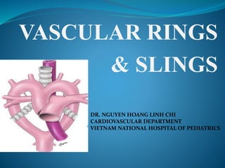
  • 1. VASCULAR RINGS & SLINGS DR. NGUYEN HOANG LINH CHI CARDIOVASCULAR DEPARTMENT VIETNAM NATIONAL HOSPITAL OF PEDIATRICS
  • 2. EMBRYOLOGY  Develops during 3rd -8th week of the fetal life  Ventral aorta partly fuse  Aortic sac  Unfused parts remain as Rt & Lt horns of the sac  The first arteries to appear in the embryo  Right & left primitive aorta  Continuous with endocardial tube  Each one – dorsal & ventral portion
  • 3. EMBRYOLOGY  Six pairs of arterial arches appear, connecting the dorsal & ventral aorta  All are not present at the same time  Selective regression & persistance of these arch vessels forms the major arteries of head, neck & thorax
  • 4. EMBRYOLOGY  The 1st, 2nd, 5th arches disappear - 1st arch : dissappeared, small portion  maxillary artery - 2nd arch : dissappeared, remain portion  hyoid + stapedial arteries - 5th arch : never forms/form incompletetly  regresses
  • 5. EMBRYOLOGY  3rd arch  Common carotid artery  4th arch : - Right : proximal part of RSA - Left: part of aortic arch between LCA & LSA  6th arch  Pulmonary arteries & Ductus arteriosus - Right side: + Proximal part  proximal segment of RPA + Distal portion  dissapears - Left side: + Proximal part  proximal segment of LPA + Distal part : forms ductus arterious
  • 6. EMBRYOLOGY  Aortic arch + Proximal segment from aortic sac + Middle segment from the Lt 4th arch arch + Distal segment from the Lt dorsal aortic
  • 7. EMBRYOLOGY  Brachiocephalic A  Rt horn of the aortic sac  Rt subclavian : + Rt 4th arch artery + Rt dorsal aorta + 7th intersegmental A  Lt subclavian : 7th intersegmental A
  • 9. VASCULAR RING DEFINITION Vascular ring is a congenital anomaly in which the aortic arch and its branches completely or incompletely encircle and compress the trachea or esophagus or both INCIDENCE:  Based on surgical case series  True prevalence : ascertain ( ~ 1- 3% CHD) - Double aortic arch : most common - Rt Aortic arch (RAA) & aberrant LtSA
  • 10. VASCULAR RING CLASSIFICATION  Complete Vascular Ring - Double aortic ring - Rt aortic arch (RAA) & retroesophageal component + Retroesophageal LSA & ligamentum/ductus arteriosum + Mirror-image branching & retroesophageal ligamentum arteriosum + Retroesophageal Lt brachiocephalic artery - Lt aortic arch (LAA) & Rt DAo & Rt PDA/ligamenttum arteriosus - Cervical aortic arch complex  Incomplete Vascular Ring - Lt aortic arch & Retroesophageal RSA - Tracheal compression brachiocephalic or Lt common carotid artery - Ductus arteriosus sling - Malrotation of heart & PDA  Pulmonary artery Sling
  • 11. VASCULAR RING KEY FEATURES  Arch location  Branching of aorta  Compression of airway  Diverticulum of Kommerel  dEscending aorta
  • 13. VASCULAR RING CLASSIFICATION  Complete Vascular Ring - Double aortic ring - Rt aortic arch (RAA) & retroesophageal component + Retroesophageal LSA & ligamentum arteriosum + Mirror-image branching & retroesophageal ligamentum arteriosum + Retroesophageal Lt brachiocephalic artery - Lt aortic arch (LAA) & Rt DAo & Rt PDA/ligamenttum arteriosus - Cervical aortic arch complex  Incomplete Vascular Ring - Lt aortic arch & Retroesophageal RSA - Tracheal compression b brachiocephalic or Lt common carotid artery - Ductus arteriosus sling - Malrotation of heart & PDA  Pulmonary artery Sling
  • 14. DOUBLE AORTIC ARCH •Most common vascular ring (40%) • Both of embryonic right and left arches persist •Ascending aorta arises normally  leaves the pericardium  divides 2 branches (Lt & Rt arch)  join posteriorly to Dao • Lt arch : -pass anterior, left of trachea  join by ductus /ligamentum arteriosus - gives 2 vessels: LCA , LSA • Rt arch: -pass posterior, right of esophageus  join -left-side Dao -Gives 2 vessels : RCA, RSA • Right arch dominant is most common (75%) , •Left arch dominant (25%), 2 arch is equal (5%) •Associated anomalies : uncommon (TOF,
  • 15. DOUBLE AORTIC ARCH CLINICAL PRESENTATIONS RESPIRATORY SYMPTOMPSs FEEDING SYMPTOMS - Inspiratory stridor ( onset <3 moth) - “ Noisy breathing ” or wheezing -Chronic cough -Recurrent respiratory infections -Hoarse cry -ALTE/apnea -Gagging or choking -Recurrent emesis -Dysphagia ( solid foods, in older children ) -Failure to thrive PHYSICAL EXAM -Often normal -Poor weight gain ( if severe compression) -Pulmonary exam : wheezing, stridor, dyspnea, retraction
  • 16. DOUBLE AORTIC ARCH DIAGNOSIS  Chest X-ray  Barium constrat esophagrams  Bronchography  Echocardiography  Magnetic resonance imaging (MRI)  Computed tomography  Angiography
  • 18. Barium constrat esophagrams Figure 51-7 , p1843,Kirklin, Barratt-Boyes-Cardiac Surgery 2013
  • 23. Bronchography STAGING -Percentage is evaluated by using ETT of different sizes the largest ETT that can be place with 20cm pressure is evaluated against a scale developed by Myers and Cotton: + Grade 1: Obstruction of 0-50% of the lumen + Grade 2: Obstruction of 51-70% of the lumen + Grade 3: Obstruction of 71-99% of the lumen + Grade 4: Obstruction of 100% ( no visible lumen)
  • 24. DOUBLE AORTIC ARCH Figure 51-9 , p1846,Kirklin, Barratt-Boyes-Cardiac Surgery 2013 SURGICAL TREATMENT
  • 25. VASCULAR RING CLASSIFICATION  Complete Vascular Ring - Double aortic ring - Rt aortic arch (RAA) & retroesophageal component : + Retroesophageal LSA & ligamentum/ductus arteriosum + Mirror-image branching & retroesophageal ligamentum arteriosum + Retroesophageal Lt brachiocephalic artery + RAA & Retroesophageal LSA - Lt aortic arch (LAA) & Rt DAo & Rt PDA/ligamenttum arteriosus - Cervical aortic arch complex  Incomplete Vascular Ring - Lt aortic arch & Retroesophageal RSA - Tracheal compression brachiocephalic or Lt common carotid artery - Ductus arteriosus sling - Malrotation of heart & PDA  Pulmonary artery Sling
  • 26. RAA + ABERRANT LEFT SUBCLAVIAN ARTERY  Second most common vascular ring (30%)  Regression of left aortic arch segment between LCA & LSCA (4th arch)  LSCA originates as last branch from the aortic arch  LSA pass posterior, left of esophagus.  Left ductus ligament originates from bulbous dilation at the base of LSA (diverticulum of Kommerell) & attaches to proximal LPA the ring compresses esophagus + trachea  DAo can be left/right side  Usually an isolated anomaly
  • 27. RAA+ ABERRANT LEFT SUBCLAVIAN ARTERY
  • 28. RAA+ ABERRANT LEFT SUBCLAVIAN ARTERY BARIUM ESOPHAGRAM Figure 51-7 , p1843,Kirklin, Barratt-Boyes-Cardiac Surgery 2013
  • 29. RAA+ ABERRANT LEFT SUBCLAVIAN ARTERY  AORTOGRAM: Figure 51-8 , p1845,Kirklin, Barratt-Boyes-Cardiac Surgery 2013
  • 31.  SURGICAL TREATMENT RAA+ ABERRANT LEFT SUBCLAVIAN ARTERY Figure 51-10 , p1847,Kirklin, Barratt-Boyes-Cardiac Surgery 2013
  • 32. VASCULAR RING CLASSIFICATION  Complete Vascular Ring - Double aortic ring - Rt aortic arch (RAA) & retroesophageal component + Retroesophageal LSA & ligamentum arteriosum + Mirror-image branching & retroesophageal ligamentum arteriosum + Retroesophageal Lt brachiocephalic artery + RAA & Retroesophageal LSA - Lt aortic arch (LAA) & Rt DAo & Rt PDA/ligamenttum arteriosus - Cervical aortic arch complex  Incomplete Vascular Ring - Lt aortic arch & Retroesophageal RSA - Tracheal compression brachiocephalic or Lt common carotid artery - Ductus arteriosus sling - Malrotation of heart & PDA  Pulmonary artery Sling
  • 33. RAA with Mirror-image Branching & Retroesophageal Ligamentum Arteriosus  Regression of left dorsal aorta between the left 7th intersegmental artery (LSA) & left 6th arch (ductus)
  • 34. VASCULAR RING CLASSIFICATION  Complete Vascular Ring - Double aortic ring - Rt aortic arch (RAA) & retroesophageal component + Retroesophageal LSA & ligamentum arteriosum + Mirror-image branching & retroesophageal ligamentum arteriosum + Retroesophageal Lt brachiocephalic artery - Lt aortic arch (LAA) & Rt DAo & Rt PDA/ligamenttum arteriosus - Cervical aortic arch complex  Incomplete Vascular Ring - Lt aortic arch & Retroesophageal RSA - Tracheal compression brachiocephalic or Lt common carotid artery - Ductus arteriosus sling - Malrotation of heart & PDA  Pulmonary Vascular Sling
  • 35. RAA with RETROESOPHAGEAL LEFT INNOMINATE ARTERY Left arch regresses between right arch and LCA
  • 36. ANOMALOUS LEFT PULMONARY ARTERY “PULMONARY VASCULAR SLING” • Left PA arises extrapericardially from posterosuperior wall of RPA & courses behind the trachea , in front of the esophagus to enter the hilum of the left lung + compresses the right main bronchus • RPA direct continuation of the pulmonary trunk . • Left lung hilum is lower than normal in relation to the pulmonary trunk •Ductus/ligamentum arteriosus follows a normal course from pulmonary trunk RPA to anomalous LPA  join the DAo  Ring-Sling complex
  • 37. PULMONARY ARTERY SLING  DIAGNOSIS :  CXR (AP and lateral) - Tracheal narowing and/or displacement( lateral) - Aortic arch location - Tracheal position : abnormal deviation to the left (due to the aorta coursin over the right main bronchus OR Mildline position with double aortic arch - Atelectasis, hyperinflation, or pneumonia may be present
  • 39. PULMONARY ARTERY SLING BARIUM ESOPHAGRAM -Most important and reliabe diagnostic tool -Posterior (+ anterior) compression of sophagus on lateral
  • 40. PULMONARY ARTERY SLING  ECHOCARDIOGRAPHY
  • 41. PULMONARY ARTERY SLING  CT/MRI : identify vascular structures and anatomy of tracheobronchial
  • 42. PULMONARY ARTERY SLING ANGIOGRAPHY : considered “gold standard”, but rarely needed for diagnosis
  • 43. PULMONARY ARTERY SLING  BRONCHOSCOPY: - Use as a diagnostic tool is controversial - Recommended for diagnosis of a vascular sling rule out concomitant tracheal rings ( and useful for abberant innominate artery )
  • 44. PULMONARY ARTERY SLING  TREATMENT - Asymptomatic patient need no surgical treatment, even when anomalies are found incidentally - Medical management is recommended for infants with mild symptoms - Respiratory distress, history of recurrent pulmonary infections, apneic spells, FTT are indications for surgical intervention
  • 45. PULMONARY ARTERY SLING p1853,Kirklin, Barratt-Boyes-Cardiac Surgery 2013
  • 46. PULMONARY ARTERY SLING p1855,Kirklin, Barratt-Boyes-Cardiac Surgery 2013
  • 48. TRACHEAL COMPRESSION BY INNOMINATE ARTERY
  • 49. TRACHEAL COMPRESSION BY INNOMINATE ARTERY Figure 51-12 , p1849,Kirklin, Barratt-Boyes-Cardiac Surgery 2013