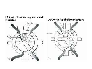
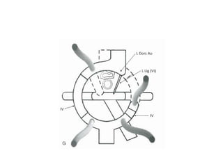
This document discusses various anomalies of the aortic arch including abnormal branching patterns, abnormal positions of the arch, and the presence of extra arches. It defines key types of anomalies such as double aortic arch, right aortic arch, and interrupted aortic arch. For each type, the document describes associated embryology, diagnostic methods including echocardiography and angiography, and potential treatments.

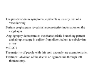


![CTEV [ clubfoot] DR ARUN LAL ,DR MOHAMED ASHRAF travancore medical college k...](https://cdn.slidesharecdn.com/ss_thumbnails/ctevclubfootdrarunlaldrmohamedashraftravancoremedicalcollegekollamkeralaindia-260208063247-18fc466c-thumbnail.jpg?width=640&height=640&fit=bounds)


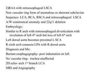
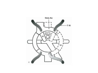
![PERI-PROSTHETIC FRACTURE NAIL-PLATE CONSTRUCT [NPC].pptx](https://cdn.slidesharecdn.com/ss_thumbnails/drarunkumardrmohamedashrafperiprostheticfrasturenail-plateconstructnpc-260209164459-7e9d15a1-thumbnail.jpg?width=640&height=640&fit=bounds)


