Download to read offline
![IOSR Journal of Computer Engineering (IOSR-JCE)
e-ISSN: 2278-0661,p-ISSN: 2278-8727, Volume 17, Issue 6, Ver. I (Nov – Dec. 2015), PP 152-156
www.iosrjournals.org
DOI: 10.9790/0661-1761152156 www.iosrjournals.org 152 | Page
Automated Detection of Microaneurysm, Hard Exudates, and
Cotton Wool Spots in Retinal fundus Images
Raju Maher1
, Sangramsing Kayte1
, Suvarnsing Bhable2
, Jaypalsing Kayte2
1,2
Department of Computer Science and Information Technology
Dr. Babasaheb Ambedkar Marathwada University, Aurangabad
Abstract: The The automatic identification of Image processing techniques for abnormalities in retinal images.
Its very importance in diabetic retinopathy screening. Manual annotations of retinal images are rare and
exclusive to obtain. The ophthalmoscope used direct analysis is a small and portable apparatus contained of a
light source and a set of lenses view the retina. The existence of diabetic retinopathy detected can be examining
the retina for its individual features. The first presence of diabetic retinopathy is the form of Microaneurysms.
This paper describes different works needed to the automatic identification of hard exudates and cotton wool
spots in retinal images for diabetic retinopathy detection and support vector machine (SVM) for classifying
images. This system is evaluated on a large dataset containing 130 retinal images. The proposed method Results
show that exudates were detected from a database with 96.9% sensitivity, specificity 96.1% and
97.38%accuracy.
Keywords: Diabetic retinopathy Retinal images, Biomedical image Processing, exudate, CAD.
I. Introduction
The Diabetic Retinopathy (DR) is one of the most important causes of blindness and vision defects in
developed countries[1]. Early detection is crucial for the prevention of visual loss and blindness in diabetes
patients. In this disease, several visual retinal abnormalities appear in the retinal fundus, representing a visual
indicator of changes in the vision[2]. The automatic detection of visual signs provides an actual way to obtain an
early detection of diabetic retinopathy and to avoid future complications. In retinal images, early diabetic
retinopathy lesions may be also classified into “red lesion” and ”bright lesions”, such as hard exudates and
cotton wool spots.3 The identification of these bright abnormalities acquiring manual remarks of these
abnormalities in retinal images is a boring and time consuming task and its especially for large amounts of
training data that are needed. The number of people afflicted with the disease continues to grow it. It occurs
when the pancreas does not secrete enough insulin or the body is unable to process it properly. This results in an
abnormal increase in the glucose level in the blood. Over time this high level of glucose causes damage to blood
vessels. This damage affects both eyes and nervous system, as well as heart, kidneys and other organs [4].The
diabetes have two types. Diabetic retinopathy is the most normal cause of new cases of blindness is an effect of
diabetic retinopathy and its prevalence is set to continue rising. Estimated 50–65 new cases of blindness per
100,000 people happened every year [3]. Microaneurysms and other areas of abnormal retinal blood vessels may
leak fluid, causing the retina to bleed or swell. They are collapse of retinal blood vessels may result in fluid
leaking into the center of the retina. Abnormal blood vessels that grow on the surface of the retina
(neovascularization) which can bleed and scar.
Diabetic retinopathy is divided into some stages mild, moderate, severe and proliferative DR. A brief
description of the different stages of DR.
a) Mild non-proliferative retinopathy: Microaneurysms, i.e., small swellings in the tiny blood vessels of the
retina will be formed in this stage.
b) Moderate non-proliferative retinopathy: As the disease progresses, some blood vessels that nourish the retina
are blocked.
c) Severe non-proliferative retinopathy: Many more blood vessels are blocked, depriving several areas of the
retina of their blood supply. The affected areas of the retina begin to show sign of ischemia (lack of oxygen)
such as blot hemorrhages, bleeding of the veins and intraretinal microvascular abnormalities.
d) Proliferative retinopathy: At this advanced stage, the non-proliferative factors produced by the retina begin to
trigger the growth of new blood vessels. These new blood vessels are abnormal and fragile.
Progresses from mild non-proliferative abnormalities, characterized by increased vascular permeability, to
moderate and severe non-proliferative diabetic retinopathy (NPDR), characterized by vascular closure, to
proliferative diabetic retinopathy (PDR) [7]. An active learning framework has been used in different
applications, such as detection of mine-like objects in sonar imagery, labeling video data or content-based
information retrieval. In this paper, we propose a CAD system for the detection and differentiation of hard
exudates, cotton wool spots in retinal images incorporating an active learner in the training process [8].](https://image.slidesharecdn.com/v01761152156-160702093522/85/V01761152156-1-320.jpg)
![IOSR Journal of Computer Engineering (IOSR-JCE)
e-ISSN: 2278-0661,p-ISSN: 2278-8727, Volume 17, Issue 6, Ver. I (Nov – Dec. 2015), PP 152-156
www.iosrjournals.org
DOI: 10.9790/0661-1761152156 www.iosrjournals.org 152 | Page
Automated Detection of Microaneurysm, Hard Exudates, and
Cotton Wool Spots in Retinal fundus Images
Raju Maher1
, Sangramsing Kayte1
, Suvarnsing Bhable2
, Jaypalsing Kayte2
1,2
Department of Computer Science and Information Technology
Dr. Babasaheb Ambedkar Marathwada University, Aurangabad
Abstract: The The automatic identification of Image processing techniques for abnormalities in retinal images.
Its very importance in diabetic retinopathy screening. Manual annotations of retinal images are rare and
exclusive to obtain. The ophthalmoscope used direct analysis is a small and portable apparatus contained of a
light source and a set of lenses view the retina. The existence of diabetic retinopathy detected can be examining
the retina for its individual features. The first presence of diabetic retinopathy is the form of Microaneurysms.
This paper describes different works needed to the automatic identification of hard exudates and cotton wool
spots in retinal images for diabetic retinopathy detection and support vector machine (SVM) for classifying
images. This system is evaluated on a large dataset containing 130 retinal images. The proposed method Results
show that exudates were detected from a database with 96.9% sensitivity, specificity 96.1% and
97.38%accuracy.
Keywords: Diabetic retinopathy Retinal images, Biomedical image Processing, exudate, CAD.
I. Introduction
The Diabetic Retinopathy (DR) is one of the most important causes of blindness and vision defects in
developed countries[1]. Early detection is crucial for the prevention of visual loss and blindness in diabetes
patients. In this disease, several visual retinal abnormalities appear in the retinal fundus, representing a visual
indicator of changes in the vision[2]. The automatic detection of visual signs provides an actual way to obtain an
early detection of diabetic retinopathy and to avoid future complications. In retinal images, early diabetic
retinopathy lesions may be also classified into “red lesion” and ”bright lesions”, such as hard exudates and
cotton wool spots.3 The identification of these bright abnormalities acquiring manual remarks of these
abnormalities in retinal images is a boring and time consuming task and its especially for large amounts of
training data that are needed. The number of people afflicted with the disease continues to grow it. It occurs
when the pancreas does not secrete enough insulin or the body is unable to process it properly. This results in an
abnormal increase in the glucose level in the blood. Over time this high level of glucose causes damage to blood
vessels. This damage affects both eyes and nervous system, as well as heart, kidneys and other organs [4].The
diabetes have two types. Diabetic retinopathy is the most normal cause of new cases of blindness is an effect of
diabetic retinopathy and its prevalence is set to continue rising. Estimated 50–65 new cases of blindness per
100,000 people happened every year [3]. Microaneurysms and other areas of abnormal retinal blood vessels may
leak fluid, causing the retina to bleed or swell. They are collapse of retinal blood vessels may result in fluid
leaking into the center of the retina. Abnormal blood vessels that grow on the surface of the retina
(neovascularization) which can bleed and scar.
Diabetic retinopathy is divided into some stages mild, moderate, severe and proliferative DR. A brief
description of the different stages of DR.
a) Mild non-proliferative retinopathy: Microaneurysms, i.e., small swellings in the tiny blood vessels of the
retina will be formed in this stage.
b) Moderate non-proliferative retinopathy: As the disease progresses, some blood vessels that nourish the retina
are blocked.
c) Severe non-proliferative retinopathy: Many more blood vessels are blocked, depriving several areas of the
retina of their blood supply. The affected areas of the retina begin to show sign of ischemia (lack of oxygen)
such as blot hemorrhages, bleeding of the veins and intraretinal microvascular abnormalities.
d) Proliferative retinopathy: At this advanced stage, the non-proliferative factors produced by the retina begin to
trigger the growth of new blood vessels. These new blood vessels are abnormal and fragile.
Progresses from mild non-proliferative abnormalities, characterized by increased vascular permeability, to
moderate and severe non-proliferative diabetic retinopathy (NPDR), characterized by vascular closure, to
proliferative diabetic retinopathy (PDR) [7]. An active learning framework has been used in different
applications, such as detection of mine-like objects in sonar imagery, labeling video data or content-based
information retrieval. In this paper, we propose a CAD system for the detection and differentiation of hard
exudates, cotton wool spots in retinal images incorporating an active learner in the training process [8].](https://image.slidesharecdn.com/v01761152156-160702093522/75/V01761152156-1-2048.jpg)
![Automated Detection of Microaneurysm, Hard Exudates, and Cotton Wool Spots in Retinal…
DOI: 10.9790/0661-1761152156 www.iosrjournals.org 153 | Page
II. Method
The In this work, 130 retinal photographs are used from DIARETDB0 a database including a selected
of high-quality medical images which are representatives of the diabetic retinopathy and have been verified by
experts for detection of moderate non-proliferative diabetic retinopathy (NPDR), severe NPDR, proliferative
diabetic retinopathy and normal cases, have been studied. Feature extraction is the most important part of the
proposed system. The inputs are used as extracted features to the classifiers. The first pre-processed images after
a contrast enhancement process is carried out Feature extraction. Our pre-processing step primarily consists of
image contrast improvement based on histogram equalization, morphological operators and followed by
binarization[11]. The image resolution varied from 768x576 to 2048x1536 pixels while the field of view
coverage varied 50 degree. From this data set, three images containing hard exudates, cotton wool spots the
initial training set.
A human observer, performed manual annotations of hard exudates, cotton wool spots.10 unseen
images were selected as the test set to evaluate the final algorithm performance. These images contained 98 hard
exudates, 22 cotton wool spots. In the candidate extraction step, the objects that are possible bright lesions are
extracted from the images using a earlier described technique.3 The green channel of the RGB image is
convolved with 14 digital filters based on Gaussian derivatives filter[6]. These filters are variation and
translation of the image and were selected from a larger set of second order irreducible invariants using a feature
selection algorithm [3]. This system is an iterative procedure where at each iteration the active learner is called
to select an unlabeled sample from a pool of unlabeled data and an expert is asked for its label The next step is
to classify each candidate as hard exudate, cotton wool spots incorporating an morphological techniques in the
training process. The idea is to select efficiently a set of training samples from the unlabeled data in an active
way to boost the performance of the classifier and reduce the number of samples that need to be labeled.
(a) (b) (c)
Figure 1(a) Original fundus image (b) Green Channel Image(c) lesson detection.
These features represent visual concepts that the ophthalmologists use to differentiate among the retinal
lesions. The features provides information about the size, shape, color and contrast of the candidates, as well as
background information such as the nearness to the vessels and the proximity to the closest red lesion. To
examine these values, algorithms that vessel segmentation [8-9] and perform red lesion classification were
previously applied to the retinal images [8-9]. No feature selection algorithms were applied in the process.
Table 1. Each object features extracted of candidate.
Features of images Description
1 Area (size in pixel).
2 Length of the perimeter of the candidate.
3 Distance to the closest red lesion.
For the classifier c, we choose a four-class linear discriminant classifier. This classifier sets linear
decision boundaries between the different classes in order to separate hard exudates, cotton wool spots, drusen
and non-lesions. As the dimensionality of the problem is high, the selection of a linear classifier permits
obtaining adequate classification performance as well as reducing time complexity.The final algorithm for the
training process in the classification of candidates as hard exudates, cotton wool spots After applying the KNN
algorithm, a classifier typically trained for the identification of(bright lesson) hard exudates, cotton wool spots is
obtained.](https://image.slidesharecdn.com/v01761152156-160702093522/85/V01761152156-2-320.jpg)
![Automated Detection of Microaneurysm, Hard Exudates, and Cotton Wool Spots in Retinal…
DOI: 10.9790/0661-1761152156 www.iosrjournals.org 154 | Page
III. Experiments And Results
A. Experiments
The proposed detection methods are tested and evaluated on DIARETDB1 [8-9], a publicly available
database of colored fundus images and corresponding ground truth images. Lesion based evaluation and image
based evaluation are employed to measure the accuracy of the proposed detection method at the pixel level.
Figure shows the results of microaneurysms and exudates detection for normal DR. In example the green
component, of the RGB fundus image, was chosen to obtain the microaneurysms. Similar to the exudates
detection algorithm, first the prominent structures within retina images, Such as blood vessel tree and optic disc
are to be removed.
Table 2. Lesion based result.
Classifier KNN
Total Images 130
True positives 87
False positives 2
False negatives 4
True negatives 37
Sensitivity 95.6%
Specificity 94.87%
Accuracy 95.38%
Several parameters such as True Positive (TP), True Negative (TN), False Positive (FP) and False
Negative (FN) are calculated. These parameters are calculated by comparing the classifier outcome with the
number of normal and abnormal images from the database. For an abnormal image, the result is true positive if
the outcome of classification is abnormal and the result is False Negative (FN) if the classifier output is normal.
For normal image, the result is True Negative (TN), if the classifier output is normal and False Positive (FP) if
the classification outcome is abnormal. In a given image dataset, these parameters, TP,TN, FP, FN are used in
the calculation of the accuracy, Sensitivity (SN) and specificity (SP)[2]. Performance of the classifier can be
measured in terms of sensitivity, specificity and accuracy. The results were compared with the performance
obtained using random sampling. This method randomly selects the next sample from the pool of unlabeled
data. It relates to a passive learning model where the training set is a casual sampling of the data.
B. Results
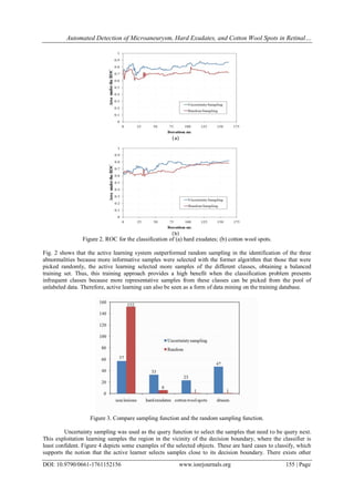
The results for each performance of the sampling bright lesion detection method is good even for lesion
based evaluation, as the proposed hybrid microaneurysm detection method resulted in a very high sensitivity
with reasonable specificity, an ophthalmologist can take its assistance in detecting Microaneurysms, exudates
and cotton wool spot in the mass screening of diabetic retinopathy. It achieves a sensitivity of 95.6% and a
specificity of 94.87% and accuracy of 95.38%, the performance of the microaneurysm detection method can be
enhanced further by augmenting the amount of training data for the microaneurysm candidate object
classification.
IV. Discussion
This paper presents an automated method to detect microaneurysm and exudates and cotton wool spots
in digital fundus images. A new candidate microaneurysm detection scheme based on matched filtering and
local relative entropy is proposed algorithm selected representative samples from database. The performance of
this microaneurysm detection method is compared with mathematical morphology based microaneurysm
detection method. A detection scheme that combines both detection methods is tested as well as detect. The
results of the method on a per image basis show that the hybrid detection scheme achieved an accuracy of
95.38%, sensitivity of 95.6% combined with 94.87% specificity. It indicates a large decrease in retinal images
that have to be shown to an ophthalmologist.](https://image.slidesharecdn.com/v01761152156-160702093522/85/V01761152156-3-320.jpg)
![Automated Detection of Microaneurysm, Hard Exudates, and Cotton Wool Spots in Retinal…
DOI: 10.9790/0661-1761152156 www.iosrjournals.org 156 | Page
query functions, called explorative learning, which sample from regions where no labeled samples are
presented.4 It could be interesting to apply an approach using alternatively exploitation and explorative learning
to sample in different regions of the sample space.
V. Conclusions
This paper presents an automated method to detect microaneurysm and exudates in digital fundus
images. A new candidate microaneurysm detection scheme based on matched filtering and local relative entropy
is proposed. The performance of this microaneurysm detection method is compared with mathematical
morphology based microaneurysm detection method this permits the design of a classifier with automatically
selected useful samples. Results show that the proposed method increases the classification accuracy compared
to random sampling. Local relative entropy the number of labeled examples required labeled databases that are
now available in many application areas. An ophthalmologist can take its help in detecting microaneurysms,
exudates and cotton wool spot in the mass screening of diabetic retinopathy.
References
[1] Kinyoun, J., Barton, F., Fisher, M., Hubbard, L., Aiello, L., and Ferris, F., “Detection of diabetic macular edema. Ophthalmoscopy
versus photography–Early Treatment Diabetic Retinopathy Study Report Number 5. The ETDRS Research Group.” Ophthalmology
96, 746–750 (1989).
[2] Early Treatment Diabetic Retinopathy Study Research Group, “Early Photocoagulation for Diabetic Retinopathy: ETDRS report 9,”
Ophthalmology 98, 766–785 (1991).
[3] Raju Sahebrao Maher, Sangramsing N. Kayte, Sandip T. Meldhe, Mukta Dhopeshwarkar, “Automated Diagnosis Non-proliferative
Diabetic Retinopathy in Fundus Images using Support Vector Machine” International Journal of Computer Applications (0975 –
8887)Volume 125 – No.15, September 2015.
[4] Dura, E., Zhang, Y., Liao, X., Dobeck, G. J., and Carin, L., “Active learning for detection of mine-like objects in side-scan sonar
imagery,” IEEE Journal of Oceanic Engineering 30(2), 360–371 (2005).
[5] Yan, R., Yang, J., and Hauptmann, A., “Automatically labeling video data using multi-class active learning,” in [Proc. Ninth IEEE
International Conference on Computer Vision], 516–523 (2003).
[6] Zhang, C. and Chen, T., “An active learning framework for content-based information retrieval,” IEEE Transactions on Multimedia
4(2), 260–268 (2002).
[7] Raju Sahebrao Maher, Dnyaneshwar S. Panchal, Sangramsing Kayte, Dr. Mukta Dhopeshwarkar” Automatic Identification of
Various Stages of Diabetic Retinopathy Using Retinal Fundus Images” International Journal of Advanced Research in Computer
Science and Software Engineering, Volume 5, Issue 9, September 2015.
[8] Abr`amoff, M. D. and Niemeijer, M., “The automatic detection of the optic disc location in retinal images using optic disc location
regression,” in [Engineering in Medicine and Biology Society, 2006. EMBS ’06.] , 4432–4435 (2006).
[5] Dima, C. and Hebert, M., “Active learning for outdoor obstacle detection,” in [Proceedings of Robotics: Science and Systems],
(2005).
[6] Niemeijer, M., van Ginneken, B., Staal, J., Suttorp-Schulten, M., and Abr`amoff, M. D., “Automatic detection of red lesions in
digital color fundus photographs,” IEEE Transactions on Medical Imaging 24(5) , 584–592 (2005).
[7] Raju Maher, Sangramsing Kayte, Dnyaneshwar Panchal, Pankaj Sathe, Sandip Meldhe, “A Decision Support System for Automatic
Screening of Non-proliferative Diabetic Retinopathy” International Journal of Emerging Research in Management and Technology,
Volume-4,Issue-10, October-2015
[8] Raju Maher, Dr.Mukta Dhopeshwarkar “Automated Detection of Non-proliferative Diabetes Retinopathy Using Fundus Images”
International Journal of Advanced Research in Computer Science and Software Engineering Volume 5, Issue 3, March 2015.
[9] Sangramsing N. Kayte, Siddharth B. Dabhade and Bharatratna P. Gaikwad, “Design and Development for Detection of Blood
Vessels, Microneurysms and Exudates from the Retina”, Proceedings of the National Conference on Advancements in the Era of
Multi-Disciplinary Systems (AEMDS-2013), Elsevier Publications 2013,ISBN: 978-93-5107-057-3,PP.394](https://image.slidesharecdn.com/v01761152156-160702093522/85/V01761152156-5-320.jpg)

This document summarizes an automated method for detecting microaneurysms, hard exudates, and cotton wool spots in retinal fundus images. 130 retinal images were used from a public database to train and evaluate classifiers. Features were extracted from pre-processed images and candidate lesions were classified using support vector machines and k-nearest neighbors algorithms. The proposed method achieved 95.6% sensitivity, 94.87% specificity, and 95.38% accuracy in detecting lesions, outperforming random sampling. Active learning was shown to select more informative training samples than random sampling, improving classifier performance.


![Getting Started with Apache Spark: Big Data Made Simple [Free Meetup]](https://cdn.slidesharecdn.com/ss_thumbnails/apachesparkgettingstarted-260203175547-8361bcc3-thumbnail.jpg?width=640&height=640&fit=bounds)

