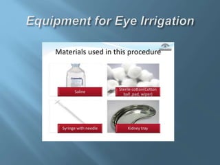
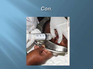
Eye irrigation is used to flush
particles and harmful chemicals
from the conjunctiva and cornea
 Chemical injury to the eye
 Removal of small particulate matter from the eye
 Treatment of foreign body sensation when no particulates
are visible (sometimes successful)
 The cornea or conjunctiva may be
mechanically abraded by the tip of the IV
tubing, by the scleral lens, or by an
irrigating stream pointed directly at the
cornea.
 Irrigating solution, eg, normal (0.9%) saline,
 IV tubing and IV pole
 Drainage basin and towels to collect irrigation
fluid runoff
 Face/eye protection, gloves, and gown for
operator(s)
 Local anesthetic (eg, 0.5% proparacaine eye-drops)
 Expanded pH paper or pH test strips
 Gauze pads, eyelid retractors
 Cotton-tipped applicators (swabs)
 Scleral (irrigating) lens
 Place the patient supine on the bed or stretcher.
 Hang bags of saline irrigation fluid several feet
above the patient’s head (proper fluid flow
depends on this height).
 Place a plastic drainage basin under the
patient’s eye to collect the irrigation fluid and
towels on the stretcher.
 An assistant may be used to retract the eyelids
during irrigation and should stand on the
opposite side of the stretcher.
 Patient history
 Record
 Patient comfort
 Checking pH of the eye
 Glove use
 Equipment
 Clean and dry work surface or
 Dressing
 Towel or waterproof cape;
 Sterile plastic undine (Fig 2) or intravenous
infusion set;
 Sterile kidney dish;
 Universal indicator strips;
 Local anesthetic drops, as prescribed according
to local policy
 Instil local anaesthetic eye drops.
 With the patient lying down, protect the neck and shoulders
with a towel or sheet.
 Place the bowl or kidney dish against the cheek, on the
affected side, with the head tilted sideways towards it.
 Fill the feeding cup or syringe with the irrigating fluid and
test the temperature on your hand.
 Ask the patient to fix his/her gaze ahead.
 Open the eyelids. If necessary, gently use eyelid retractors.
 Pour or syringe the fluid slowly and steadily, from no more
than 5 centimeters away, onto the front surface of the eye,
inside the lower eyelid and under the upper eyelid.
 If possible, Evert the upper eyelid to access all of the upper
conjunctiva fornix.
 Ask the patient to move the eye in all directions while the
irrigation is maintained.
 Check and record the visual acuity when the procedure is
finished.
 In alkali and acid burns, refer the patient to an
ophthalmologist for assessment.
eye irrigation.pptx

eye irrigation.pptx

  • 3.
    Eye irrigation isused to flush particles and harmful chemicals from the conjunctiva and cornea
  • 4.
     Chemical injuryto the eye  Removal of small particulate matter from the eye  Treatment of foreign body sensation when no particulates are visible (sometimes successful)
  • 5.
     The corneaor conjunctiva may be mechanically abraded by the tip of the IV tubing, by the scleral lens, or by an irrigating stream pointed directly at the cornea.
  • 7.
     Irrigating solution,eg, normal (0.9%) saline,  IV tubing and IV pole  Drainage basin and towels to collect irrigation fluid runoff  Face/eye protection, gloves, and gown for operator(s)  Local anesthetic (eg, 0.5% proparacaine eye-drops)  Expanded pH paper or pH test strips  Gauze pads, eyelid retractors  Cotton-tipped applicators (swabs)  Scleral (irrigating) lens
  • 8.
     Place thepatient supine on the bed or stretcher.  Hang bags of saline irrigation fluid several feet above the patient’s head (proper fluid flow depends on this height).  Place a plastic drainage basin under the patient’s eye to collect the irrigation fluid and towels on the stretcher.  An assistant may be used to retract the eyelids during irrigation and should stand on the opposite side of the stretcher.
  • 10.
     Patient history Record  Patient comfort  Checking pH of the eye  Glove use
  • 11.
     Equipment  Cleanand dry work surface or  Dressing  Towel or waterproof cape;  Sterile plastic undine (Fig 2) or intravenous infusion set;  Sterile kidney dish;  Universal indicator strips;  Local anesthetic drops, as prescribed according to local policy
  • 12.
     Instil localanaesthetic eye drops.  With the patient lying down, protect the neck and shoulders with a towel or sheet.  Place the bowl or kidney dish against the cheek, on the affected side, with the head tilted sideways towards it.  Fill the feeding cup or syringe with the irrigating fluid and test the temperature on your hand.  Ask the patient to fix his/her gaze ahead.  Open the eyelids. If necessary, gently use eyelid retractors.
  • 13.
     Pour orsyringe the fluid slowly and steadily, from no more than 5 centimeters away, onto the front surface of the eye, inside the lower eyelid and under the upper eyelid.  If possible, Evert the upper eyelid to access all of the upper conjunctiva fornix.  Ask the patient to move the eye in all directions while the irrigation is maintained.  Check and record the visual acuity when the procedure is finished.  In alkali and acid burns, refer the patient to an ophthalmologist for assessment.