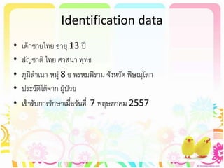
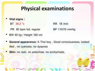
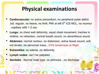
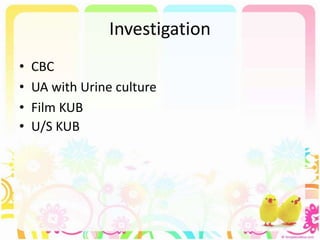
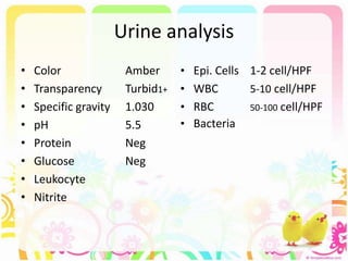
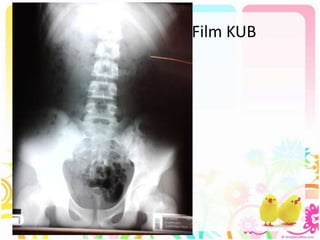
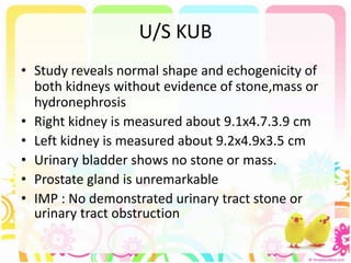
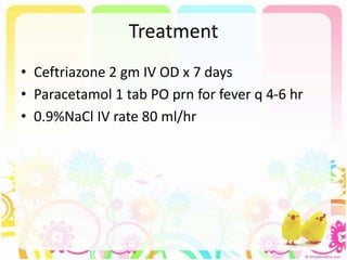
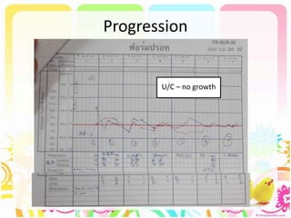
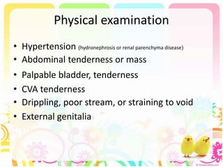
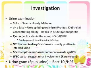
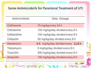
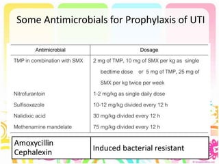
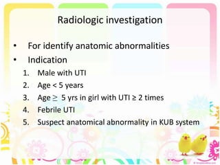
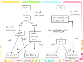
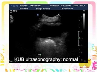
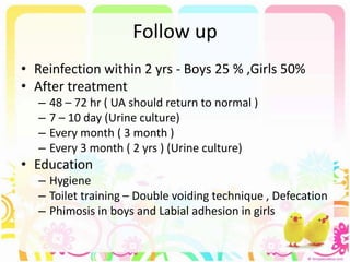
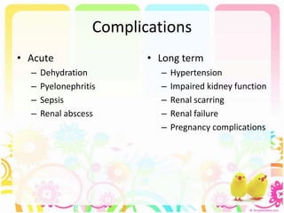
Case conference
Urinary tract infection
Int Patcharapon Udomluck
Identification data
• เด็กชายไทย อายุ 13 ปี
• สัญชาติ ไทย ศาสนา พุทธ
• ภูมิลาเนา หมู่ 8 อ พรหมพิราม จังหวัด พิษณุโลก
• ประวัติได้จาก ผู้ป่วย
• เข้ารับการรักษาเมื่อวันที่ 7 พฤษภาคม 2557
Chief complaint & Present illness
• ปวดเอวด้านขวา 1 วันก่อนมา
• 1 วันก่อน ปวดท้องด้านขวา และปวดเอว อาการค่อยๆปวด ปวด
ลักษณะบีบๆ ปวดนานเป็นชั่วโมง ร้าวไปหลัง มีไข้ มีปัสสาวะแสบขัด
ปัสสาวะเข้มขึ้นสีแดงคล้า ปัสสาวะบ่อย ขยับแล้วปวดมากขึ้น ทานยา
แก้ปวดไม่เบา จึงมาโรงพยาบาล
Past history & family history
• Past history
– ปฏิเสธโรคประจาตัว, ปฏิเสธประวัติผ่าตัด
– เคยมีประวัติ ต่อยมวย โดนเพื่อนเตะที่สีข้างด้านขวา เมื่อ 1 เดือนก่อน
หลังจากนั้นมีปวดบ้าง ไม่ช้า ไม่ได้มาโรงพยาบาล
• Personal history
– ปฏิเสธ ดื่มสุรา หรือสูบบุหรี่
– ปฏิเสธแพ้ยาแพ้อาหาร
• Family history
– ปฏิเสธ โรคประจาตัวในครอบครัว ปฏิเสธโรคนิ่วในครอบครัว
Physical examinations
• Vital signs :
BT 38.2 oc RR 18 /min
PR 80 bpm full, regular BP 110/70 mmHg
• BW 45 kg / Height 160 cm
• General appearance: A Thai boy , Good consciousness, looked
illed , no cyanosis, no dyspnea
• Skin: no rash, no petechiae, no ecchymosis,
Physical examinations
• Head: normal shape, no evidence of head trauma
• Eyes: not pale conjunctiva, no conjunctivitis , no icteric sclera ,
no sunken eyeballs, no dry lips and mucosa
• Ears: normal pinna, normal external auditory canal, no
discharge
• Nose: no rhinorrhea
• Throat & mouth: no oral ulcer, no injected pharynx , no tonsil
enlargement
Physical examinations
• Cardiovascular: no active precordium, no peripheral pulse deficit
,full, regular, no heave, no thrill, PMI at left 5th lCS MCL, no murmur
, capillary refill < 2 sec
• Lungs: no chest wall deformity, equal chest movement, trachea in
midline, no retraction, normal breath sound, no adventitious sound
• Abdomen: normal contour, no distension, active bowel sound, soft,
not tender, no abnormal mass , CVA tenderness at Right
• Extremities: no edema, no deformity
• Lymph nodes: can’t be palpated
• Genitalia : Normal male type, no phimosis , no discharge
Physical examinations
Neurological systems
• Mental status : good consciousness , E4V5M6
• Sensory : no decrease sensation
• Motor : normal tone , motor power gr. V all
Problem list
• Acute febrile illness with symptom of urinary
tract infection
– Frequently urination
– Dysuria
– Red color urine
– Right flank pain + CVA tenderness at Right
• History of trauma
Investigation
• CBC
• UA with Urine culture
• Film KUB
• U/S KUB
CBC
• WBC 6,340 cell/ul
• Neu 54.9 %
• Lymph 36 %
• Mono 7.3 %
• Eos 1.6 %
• Baso 0.2 %
• Anisocytosis few
• Microcytosis few
• RBC 5,790,000 /ul
• Hb 12.8 g/dL
• Hct 40.1 %
• MCV 69.3 fL
• RDW 14.2 %
• Platelet 267,000 /ul
Urine analysis
• Color Amber
• Transparency Turbid1+
• Specific gravity 1.030
• pH 5.5
• Protein Neg
• Glucose Neg
• Leukocyte
• Nitrite
• Epi. Cells 1-2 cell/HPF
• WBC 5-10 cell/HPF
• RBC 50-100 cell/HPF
• Bacteria
Film KUB
U/S KUB
• Study reveals normal shape and echogenicity of
both kidneys without evidence of stone,mass or
hydronephrosis
• Right kidney is measured about 9.1x4.7.3.9 cm
• Left kidney is measured about 9.2x4.9x3.5 cm
• Urinary bladder shows no stone or mass.
• Prostate gland is unremarkable
• IMP : No demonstrated urinary tract stone or
urinary tract obstruction
Treatment
• Ceftriazone 2 gm IV OD x 7 days
• Paracetamol 1 tab PO prn for fever q 4-6 hr
• 0.9%NaCl IV rate 80 ml/hr
Progression
U/C – no growth
Follow up U/A
• Color Yellow
• Transparency Clear
• Specific gravity 1.010
• pH 6.0
• Protein Neg
• Glucose Neg
• Leukocyte
• Nitrite
• Epi. Cells 0-1 cell/HPF
• WBC 3-5 cell/HPF
• RBC 5-10 cell/HPF
• Bacteria
Urinary tract infection
in Pediatrics
Prevalence
• During the 1st yr Male : female = 2.8-5.4:1
• Beyond 1 yr Male : female = 1:10
• Girls
– First UTI usually occurs by the age of 5 yr
with peaks during infancy and toilet training
– After the first UTI, 60-80% of girls will develop a second
UTI develop within 18 mo.
Etiology
• Females
– Escherichia coli (75-90 %) , Klebsiella and Proteus
• Males
– Proteus (30 % ) risk to Triple phosphate stone
• Other pathogen
– Pseudomonas
– Staphylococcus saprophyticus
– S. epidermidis
– Coag-neg staphy
• Viral infections, particularly adenovirus, may also occur,
especially as a cause of cystitis
Pathogenesis and Pathology
• Ascending infections
– Arise from the fecal flora  colonize the perineum 
urethra  enter the bladder
– Uncircumcised boys, the bacteria beneath the prepuce
– Normally the simple and compound papillae in the kidney
have an antireflux mechanism BUT Some compound
papillae, typically located in the upper and lower poles of
the kidney, allow intrarenal reflux
– Voiding dysfunction, Infrequent  Urinary stasis
• Hematogenous spread (Rare) – renal infection
Risk Factors for Urinary Tract Infection
• Female
• Uncircumcised male
• Vesicoureteral reflux
• Toilet training
• Voiding dysfunction
• Obstructive uropathy
• Urethral instrumentation
• Wiping from back to front
• Bubble bath
• Tight clothing (underwear)
• Pinworm infestation
• Constipation
• P fimbriated bacteria
• Anatomic abnormality
• Neuropathic bladder
• Sexual activity
• Pregnancy
Classification
• Symptomatic bacteriuria
– Upper UTI - Pyelonephritis
– Lower UTI - Cystitis
• Asymptomatic bacteriuria
Symptomatic bacteriuria
Pyelonephritis Cystitis
- Flank pain
- Fever
- Malaise
- N/V
- Occasionally diarrhea
- Infants = nonspecific
symptoms such as
jaundice, poor feeding,
irritability, and weight
loss
- Dysuria
- Urgency
- Frequency
- Suprapubic pain
- Incontinence
- Malodorous urine
- * No fever
Asymptomatic bacteriuria
• Positive urine culture without any
manifestations of infection and occurs almost
exclusively in girls
• If left untreated in Pregnant women , can
result in a symptomatic UTI
Physical examination
• Hypertension (hydronephrosis or renal parenchyma disease)
• Abdominal tenderness or mass
• Palpable bladder, tenderness
• CVA tenderness
• Drippling, poor stream, or straining to void
• External genitalia
Investigation
• Urine examination
– Color : Clear or cloudy, Malodor
– pH : Base – Urea splitting organism (Proteus, Klebsiella)
– Concentrating ability – Impair in acute pyelonephritis
– Pyuria (leukocytes in the urine) > 5 cell/HPF
• * Can be present or not in urine infection
– Nitrites and leukocyte esterase - usually positive in
infected urine
– Microscopic hematuria is common in acute cystitis
– WBC casts - suggest renal involvement (Rarely seen)
• Urine gram (Spun urine) – Bact 10 /HPF
Diagnosis
• Urine culture (Necessary for confirmation)
• Sample collection
– Bag collection
– Clean voided (Midstream urine)
– Suprapubic puncture
– Catheterization
• Placing the sample in a refrigerator 4 c within 2 hr
Investigation
• CBC - Leukocytosis, neutrophilia
• Elevated ESR and CRP are common ( nonspecific
markers of bacterial infection )
• With a renal abscess, WBC > 20,000 to 25,000/mm3
• Blood cultures should be considered
– Because sepsis is common in pyelonephritis
Principle of management
1. Treatment of acute infection
2. Prevention of further infection
3. Adequate investigation
4. Arrangement of further treatment
5. Follow up - Prevention of recurrence and
long-term complications
Treatment
Indication for hospitalize
• Age <2 months
• Sepsis or potential bacteremia
• Immunocompromised patient
• Vomiting or inability to tolerate oral
medication
• Lack of adequate outpatient follow-up
• Failure to respond to outpatient therapy
Treatment
• Mild symptom or doubtful diagnosis
 Oral antibiotic before the results of culture are
known (Repeat culture - if the results are
uncertain)
 OPD case
• Severe symptom
 Urine culture with treatment immediately (IV)
 Hospitalization
Some Antimicrobrils for Oral Treatment of UTI
Nitrofurantoin 5-7 mg/kg/day hr in 3 to 4 divided doses
Some Antimicrobrils for Parenteral Treatment of UTI
3-5 12,8 h
Treatment
• Acute pyelonephritis
 Hospitalization
 14-day course of broad-spectrum antibiotics capable
of reaching significant tissue levels is preferable
• Cystitis
 7 – 10 day of antibiotic
• Not recommend Short course or Single dose
• The safety and efficacy of oral ciprofloxacin in
children is under study
Prophylaxis antibiotic
• Indication
– Vesicoureteral reflux
– Age < 2 yr with Acute pyelonephritis  Prophylaxis
antibiotic 6 month
– Neonates and infants with febrile UTI and abnormal renal
scan
– Recurrence > 3 times/year esp.with bladder instability
– Neurogenic bladder
– Obstructive uropathy
Prophylaxis antibiotic
• Duration
–VUR case and Case risk to Urine stasis
(Neurogenic bladder, Calculi)
• Prophylaxis - Until Age 6 yr with Normal renal growth ,
no new scar and no recurrence acute pyelonephritis
–No VUR
• Prophylaxis antibiotic 3 – 6 month
Some Antimicrobials for Prophylaxis of UTI
Amoxycillin
Cephalexin Induced bacterial resistant
Radiologic investigation
• For identify anatomic abnormalities
• Indication
1. Male with UTI
2. Age < 5 years
3. Age ≥ 5 yrs in girl with UTI ≥ 2 times
4. Febrile UTI
5. Suspect anatomical abnormality in KUB system
Imaging studies
1. Ultrasonography (U/S)
2. Voiding cystourethrography (VCUG)
3. Intravenous pyelogram (IVP)
4. DMSA (2,3 dimercaptosuccinic a) scan
Recurrent UTI, pyelonephritis ,
BUN Cr rising, HT
ปกติ
KUB ultrasonography: normal
IVP
• Nephrogram  Pelvicalyceal systems 
Ureter  Bladder
• Acute obstruct
 Prolong and dense nephrogram
 Dilatation over obstruction point
 Delayed excretion in Pelvicalyceal system
 Filling defect
IVP
Normal Pelvicalyceal
system (Cup shaped)
Hydronephrosis
UPJ Obstruct
VCUG: normal
VCUG: VUR
Posterior urethral valves
Follow up
• Reinfection within 2 yrs - Boys 25 % ,Girls 50%
• After treatment
– 48 – 72 hr ( UA should return to normal )
– 7 – 10 day (Urine culture)
– Every month ( 3 month )
– Every 3 month ( 2 yrs ) (Urine culture)
• Education
– Hygiene
– Toilet training – Double voiding technique , Defecation
– Phimosis in boys and Labial adhesion in girls
Complications
• Acute
– Dehydration
– Pyelonephritis
– Sepsis
– Renal abscess
• Long term
– Hypertension
– Impaired kidney function
– Renal scarring
– Renal failure
– Pregnancy complications
Other type of cystitis
• Acute hemorrhagic cystitis
– Caused by E. coli / attributed also to Adenovirus types 11 and 21
– More frequent in males
– Self-limiting, with hematuria lasting 4 days
• Eosinophilic cystitis
– Exposure to an allergen
– Filling defects in the bladder caused by masses that consist
histologically of inflammatory infiltrates with eosinophils
– Treatment – antihistamines , NSAIDs
• Interstitial cystitis
– Irritative voiding symptoms + negative U/C
– Diagnosis by cystoscopic  mucosal ulcers with bladder distention
– Treatments  included bladder hydrodistention and laser ablation of
ulcerated areas, but no treatment yields sustained relief
Obstructive uropathy
• Calyceal – Pelvis – Ureter – Bladder – Urethra
• Intrinsic causes
– Vesicoureteral reflux
– Congenital anomalies
– Tumor (Wilm, Papilloma)
– Stone
– Fibrosis or stricture
• Extrinsic causes
– Aberrant vessel
– Tumor
Congenital anomalies of urinary system
• Kidney
– Number
– Size
– Location (Ectopic)
– Fusion (Horseshoe)
• Ureter
– Duplication
– UPJ obstruct
– Ureterocele
• Bladder
– Duplication
– Diverticulum
• Urethral
– Urethral vale
– Duplication
– Diverticulum
– Epispadias
– Hypospadias
Vesicoureteral reflux
• 2 type
– Primary VUR - Congenital familial disorder
• Abnormalities of ureterovesical junction
 lateral and cephalad displacement of ureteric orifice
+ Short intramural ureter “ Golf hole “
Vesicoureteral reflux
– Secondary VUR - Outflow obstruction
• Anatomical obstruction - posterior urethral valve
• Functional obstruction - neurogenic bladder , bladder
trabeculation , diverticula
• Severe VUR  Intrarenal reflux
Simple type
collecting duct
obligue & slitlike opening
Compound type
collecting duct
Perpendicula &round opening
Vesicoureteral reflux
• Grading
• Intrarenal Reflux  Renal Scarring
 Hypertension , Chronic renal failure
Treatment Recommendations for Vesicoureteral Reflux
Diagnosed Following a Urinary Tract Infection
American Urological Association Pediatric Vesicoureteral Reflux Guidelines Panel Report

Uti case ped

  • 1.
    Case conference Urinary tractinfection Int Patcharapon Udomluck
  • 2.
    Identification data • เด็กชายไทยอายุ 13 ปี • สัญชาติ ไทย ศาสนา พุทธ • ภูมิลาเนา หมู่ 8 อ พรหมพิราม จังหวัด พิษณุโลก • ประวัติได้จาก ผู้ป่วย • เข้ารับการรักษาเมื่อวันที่ 7 พฤษภาคม 2557
  • 3.
    Chief complaint &Present illness • ปวดเอวด้านขวา 1 วันก่อนมา • 1 วันก่อน ปวดท้องด้านขวา และปวดเอว อาการค่อยๆปวด ปวด ลักษณะบีบๆ ปวดนานเป็นชั่วโมง ร้าวไปหลัง มีไข้ มีปัสสาวะแสบขัด ปัสสาวะเข้มขึ้นสีแดงคล้า ปัสสาวะบ่อย ขยับแล้วปวดมากขึ้น ทานยา แก้ปวดไม่เบา จึงมาโรงพยาบาล
  • 4.
    Past history &family history • Past history – ปฏิเสธโรคประจาตัว, ปฏิเสธประวัติผ่าตัด – เคยมีประวัติ ต่อยมวย โดนเพื่อนเตะที่สีข้างด้านขวา เมื่อ 1 เดือนก่อน หลังจากนั้นมีปวดบ้าง ไม่ช้า ไม่ได้มาโรงพยาบาล • Personal history – ปฏิเสธ ดื่มสุรา หรือสูบบุหรี่ – ปฏิเสธแพ้ยาแพ้อาหาร • Family history – ปฏิเสธ โรคประจาตัวในครอบครัว ปฏิเสธโรคนิ่วในครอบครัว
  • 5.
    Physical examinations • Vitalsigns : BT 38.2 oc RR 18 /min PR 80 bpm full, regular BP 110/70 mmHg • BW 45 kg / Height 160 cm • General appearance: A Thai boy , Good consciousness, looked illed , no cyanosis, no dyspnea • Skin: no rash, no petechiae, no ecchymosis,
  • 6.
    Physical examinations • Head:normal shape, no evidence of head trauma • Eyes: not pale conjunctiva, no conjunctivitis , no icteric sclera , no sunken eyeballs, no dry lips and mucosa • Ears: normal pinna, normal external auditory canal, no discharge • Nose: no rhinorrhea • Throat & mouth: no oral ulcer, no injected pharynx , no tonsil enlargement
  • 7.
    Physical examinations • Cardiovascular:no active precordium, no peripheral pulse deficit ,full, regular, no heave, no thrill, PMI at left 5th lCS MCL, no murmur , capillary refill < 2 sec • Lungs: no chest wall deformity, equal chest movement, trachea in midline, no retraction, normal breath sound, no adventitious sound • Abdomen: normal contour, no distension, active bowel sound, soft, not tender, no abnormal mass , CVA tenderness at Right • Extremities: no edema, no deformity • Lymph nodes: can’t be palpated • Genitalia : Normal male type, no phimosis , no discharge
  • 8.
    Physical examinations Neurological systems •Mental status : good consciousness , E4V5M6 • Sensory : no decrease sensation • Motor : normal tone , motor power gr. V all
  • 9.
    Problem list • Acutefebrile illness with symptom of urinary tract infection – Frequently urination – Dysuria – Red color urine – Right flank pain + CVA tenderness at Right • History of trauma
  • 10.
    Investigation • CBC • UAwith Urine culture • Film KUB • U/S KUB
  • 11.
    CBC • WBC 6,340cell/ul • Neu 54.9 % • Lymph 36 % • Mono 7.3 % • Eos 1.6 % • Baso 0.2 % • Anisocytosis few • Microcytosis few • RBC 5,790,000 /ul • Hb 12.8 g/dL • Hct 40.1 % • MCV 69.3 fL • RDW 14.2 % • Platelet 267,000 /ul
  • 12.
    Urine analysis • ColorAmber • Transparency Turbid1+ • Specific gravity 1.030 • pH 5.5 • Protein Neg • Glucose Neg • Leukocyte • Nitrite • Epi. Cells 1-2 cell/HPF • WBC 5-10 cell/HPF • RBC 50-100 cell/HPF • Bacteria
  • 13.
  • 15.
    U/S KUB • Studyreveals normal shape and echogenicity of both kidneys without evidence of stone,mass or hydronephrosis • Right kidney is measured about 9.1x4.7.3.9 cm • Left kidney is measured about 9.2x4.9x3.5 cm • Urinary bladder shows no stone or mass. • Prostate gland is unremarkable • IMP : No demonstrated urinary tract stone or urinary tract obstruction
  • 16.
    Treatment • Ceftriazone 2gm IV OD x 7 days • Paracetamol 1 tab PO prn for fever q 4-6 hr • 0.9%NaCl IV rate 80 ml/hr
  • 17.
  • 18.
    Follow up U/A •Color Yellow • Transparency Clear • Specific gravity 1.010 • pH 6.0 • Protein Neg • Glucose Neg • Leukocyte • Nitrite • Epi. Cells 0-1 cell/HPF • WBC 3-5 cell/HPF • RBC 5-10 cell/HPF • Bacteria
  • 19.
  • 20.
    Prevalence • During the1st yr Male : female = 2.8-5.4:1 • Beyond 1 yr Male : female = 1:10 • Girls – First UTI usually occurs by the age of 5 yr with peaks during infancy and toilet training – After the first UTI, 60-80% of girls will develop a second UTI develop within 18 mo.
  • 21.
    Etiology • Females – Escherichiacoli (75-90 %) , Klebsiella and Proteus • Males – Proteus (30 % ) risk to Triple phosphate stone • Other pathogen – Pseudomonas – Staphylococcus saprophyticus – S. epidermidis – Coag-neg staphy • Viral infections, particularly adenovirus, may also occur, especially as a cause of cystitis
  • 22.
    Pathogenesis and Pathology •Ascending infections – Arise from the fecal flora  colonize the perineum  urethra  enter the bladder – Uncircumcised boys, the bacteria beneath the prepuce – Normally the simple and compound papillae in the kidney have an antireflux mechanism BUT Some compound papillae, typically located in the upper and lower poles of the kidney, allow intrarenal reflux – Voiding dysfunction, Infrequent  Urinary stasis • Hematogenous spread (Rare) – renal infection
  • 24.
    Risk Factors forUrinary Tract Infection • Female • Uncircumcised male • Vesicoureteral reflux • Toilet training • Voiding dysfunction • Obstructive uropathy • Urethral instrumentation • Wiping from back to front • Bubble bath • Tight clothing (underwear) • Pinworm infestation • Constipation • P fimbriated bacteria • Anatomic abnormality • Neuropathic bladder • Sexual activity • Pregnancy
  • 26.
    Classification • Symptomatic bacteriuria –Upper UTI - Pyelonephritis – Lower UTI - Cystitis • Asymptomatic bacteriuria
  • 27.
    Symptomatic bacteriuria Pyelonephritis Cystitis -Flank pain - Fever - Malaise - N/V - Occasionally diarrhea - Infants = nonspecific symptoms such as jaundice, poor feeding, irritability, and weight loss - Dysuria - Urgency - Frequency - Suprapubic pain - Incontinence - Malodorous urine - * No fever
  • 28.
    Asymptomatic bacteriuria • Positiveurine culture without any manifestations of infection and occurs almost exclusively in girls • If left untreated in Pregnant women , can result in a symptomatic UTI
  • 29.
    Physical examination • Hypertension(hydronephrosis or renal parenchyma disease) • Abdominal tenderness or mass • Palpable bladder, tenderness • CVA tenderness • Drippling, poor stream, or straining to void • External genitalia
  • 30.
    Investigation • Urine examination –Color : Clear or cloudy, Malodor – pH : Base – Urea splitting organism (Proteus, Klebsiella) – Concentrating ability – Impair in acute pyelonephritis – Pyuria (leukocytes in the urine) > 5 cell/HPF • * Can be present or not in urine infection – Nitrites and leukocyte esterase - usually positive in infected urine – Microscopic hematuria is common in acute cystitis – WBC casts - suggest renal involvement (Rarely seen) • Urine gram (Spun urine) – Bact 10 /HPF
  • 31.
    Diagnosis • Urine culture(Necessary for confirmation) • Sample collection – Bag collection – Clean voided (Midstream urine) – Suprapubic puncture – Catheterization • Placing the sample in a refrigerator 4 c within 2 hr
  • 33.
    Investigation • CBC -Leukocytosis, neutrophilia • Elevated ESR and CRP are common ( nonspecific markers of bacterial infection ) • With a renal abscess, WBC > 20,000 to 25,000/mm3 • Blood cultures should be considered – Because sepsis is common in pyelonephritis
  • 34.
    Principle of management 1.Treatment of acute infection 2. Prevention of further infection 3. Adequate investigation 4. Arrangement of further treatment 5. Follow up - Prevention of recurrence and long-term complications
  • 35.
    Treatment Indication for hospitalize •Age <2 months • Sepsis or potential bacteremia • Immunocompromised patient • Vomiting or inability to tolerate oral medication • Lack of adequate outpatient follow-up • Failure to respond to outpatient therapy
  • 36.
    Treatment • Mild symptomor doubtful diagnosis  Oral antibiotic before the results of culture are known (Repeat culture - if the results are uncertain)  OPD case • Severe symptom  Urine culture with treatment immediately (IV)  Hospitalization
  • 37.
    Some Antimicrobrils forOral Treatment of UTI Nitrofurantoin 5-7 mg/kg/day hr in 3 to 4 divided doses
  • 38.
    Some Antimicrobrils forParenteral Treatment of UTI 3-5 12,8 h
  • 39.
    Treatment • Acute pyelonephritis Hospitalization  14-day course of broad-spectrum antibiotics capable of reaching significant tissue levels is preferable • Cystitis  7 – 10 day of antibiotic • Not recommend Short course or Single dose • The safety and efficacy of oral ciprofloxacin in children is under study
  • 40.
    Prophylaxis antibiotic • Indication –Vesicoureteral reflux – Age < 2 yr with Acute pyelonephritis  Prophylaxis antibiotic 6 month – Neonates and infants with febrile UTI and abnormal renal scan – Recurrence > 3 times/year esp.with bladder instability – Neurogenic bladder – Obstructive uropathy
  • 41.
    Prophylaxis antibiotic • Duration –VURcase and Case risk to Urine stasis (Neurogenic bladder, Calculi) • Prophylaxis - Until Age 6 yr with Normal renal growth , no new scar and no recurrence acute pyelonephritis –No VUR • Prophylaxis antibiotic 3 – 6 month
  • 42.
    Some Antimicrobials forProphylaxis of UTI Amoxycillin Cephalexin Induced bacterial resistant
  • 43.
    Radiologic investigation • Foridentify anatomic abnormalities • Indication 1. Male with UTI 2. Age < 5 years 3. Age ≥ 5 yrs in girl with UTI ≥ 2 times 4. Febrile UTI 5. Suspect anatomical abnormality in KUB system
  • 44.
    Imaging studies 1. Ultrasonography(U/S) 2. Voiding cystourethrography (VCUG) 3. Intravenous pyelogram (IVP) 4. DMSA (2,3 dimercaptosuccinic a) scan
  • 45.
    Recurrent UTI, pyelonephritis, BUN Cr rising, HT ปกติ
  • 46.
  • 47.
    IVP • Nephrogram Pelvicalyceal systems  Ureter  Bladder • Acute obstruct  Prolong and dense nephrogram  Dilatation over obstruction point  Delayed excretion in Pelvicalyceal system  Filling defect
  • 48.
    IVP Normal Pelvicalyceal system (Cupshaped) Hydronephrosis UPJ Obstruct
  • 50.
  • 51.
  • 52.
  • 55.
    Follow up • Reinfectionwithin 2 yrs - Boys 25 % ,Girls 50% • After treatment – 48 – 72 hr ( UA should return to normal ) – 7 – 10 day (Urine culture) – Every month ( 3 month ) – Every 3 month ( 2 yrs ) (Urine culture) • Education – Hygiene – Toilet training – Double voiding technique , Defecation – Phimosis in boys and Labial adhesion in girls
  • 56.
    Complications • Acute – Dehydration –Pyelonephritis – Sepsis – Renal abscess • Long term – Hypertension – Impaired kidney function – Renal scarring – Renal failure – Pregnancy complications
  • 57.
    Other type ofcystitis • Acute hemorrhagic cystitis – Caused by E. coli / attributed also to Adenovirus types 11 and 21 – More frequent in males – Self-limiting, with hematuria lasting 4 days • Eosinophilic cystitis – Exposure to an allergen – Filling defects in the bladder caused by masses that consist histologically of inflammatory infiltrates with eosinophils – Treatment – antihistamines , NSAIDs • Interstitial cystitis – Irritative voiding symptoms + negative U/C – Diagnosis by cystoscopic  mucosal ulcers with bladder distention – Treatments  included bladder hydrodistention and laser ablation of ulcerated areas, but no treatment yields sustained relief
  • 58.
    Obstructive uropathy • Calyceal– Pelvis – Ureter – Bladder – Urethra • Intrinsic causes – Vesicoureteral reflux – Congenital anomalies – Tumor (Wilm, Papilloma) – Stone – Fibrosis or stricture • Extrinsic causes – Aberrant vessel – Tumor
  • 59.
    Congenital anomalies ofurinary system • Kidney – Number – Size – Location (Ectopic) – Fusion (Horseshoe) • Ureter – Duplication – UPJ obstruct – Ureterocele • Bladder – Duplication – Diverticulum • Urethral – Urethral vale – Duplication – Diverticulum – Epispadias – Hypospadias
  • 60.
    Vesicoureteral reflux • 2type – Primary VUR - Congenital familial disorder • Abnormalities of ureterovesical junction  lateral and cephalad displacement of ureteric orifice + Short intramural ureter “ Golf hole “
  • 61.
    Vesicoureteral reflux – SecondaryVUR - Outflow obstruction • Anatomical obstruction - posterior urethral valve • Functional obstruction - neurogenic bladder , bladder trabeculation , diverticula • Severe VUR  Intrarenal reflux Simple type collecting duct obligue & slitlike opening Compound type collecting duct Perpendicula &round opening
  • 62.
    Vesicoureteral reflux • Grading •Intrarenal Reflux  Renal Scarring  Hypertension , Chronic renal failure
  • 63.
    Treatment Recommendations forVesicoureteral Reflux Diagnosed Following a Urinary Tract Infection American Urological Association Pediatric Vesicoureteral Reflux Guidelines Panel Report

Editor's Notes

  • #21 3-5% of girls and 1% of boys. BOYS = more common in uncircumcised boys
  • #22 UTIs are caused mainly by colonic bacteria UTIs have been considered an important risk factor for the development of renal insufficiency or end-stage renal disease Major anomalies = urinary diversion , neurogenic bladder, Catheter related ความสำคัญของการวินิจฉัย และให้การรักษา UTI ในเด็ก เนื่องจาก UTI ในเด็กมีความเสี่ยงต่อ renal failure และ ESRD ในอนาคตได้
  • #23 antireflux mechanism that prevents urine from flowing in a retrograde manner into the collecting tubules
  • #25 Anatomic abnormality (e.g., labial adhesion) The bladder  uninhibited contractions forcing urine out (Overactive bladder)  high-pressure, turbulent urine flow or incomplete bladder emptying  UTI Voiding dysfunction Toilet-trained child Constipation Obstructive uropathy  Hydronephrosis  Urinary stasis  UTI
  • #27 Upper UTI Renal parenchyma involvement = Acute pyelonephritis No parenchymal involvement = Pyelitis Pyelonephritis(may result in renal injury, which is termed pyelonephritic scarring) Cystitis does not cause fever and does not result in renal injury.
  • #29 This condition is benign and does not cause renal injury Some girls are mistakenly identified as having asymptomatic bacteriuria, whereas they actually are symptomatic, experiencing day or night incontinence or perineal discomfort
  • #31 Xanthogranulomatous pyelonephritis is a rare type of renal infection characterized by granulomatous inflammation with giant cells and foamy histiocytes. It may present clinically as a renal mass or an acute or chronic infection. Renal calculi, obstruction, and infection with Proteus or E. coli contribute to the development of this lesion, which usually requires total or partial nephrectomy.
  • #32 A urinalysis should be obtained from the same specimen as that cultured Infection can occur in absence of pyuria If the child is asymptomatic and the urinalysis result is normal  unlikely urine infected if the child is symptomatic, even if the urinalysis result is negative  a UTI is possible, Urine gram ถ้านำปัสสาวะไม่ปั่น พบเชื้อ 1 ตัว/ 1 HPF  colonies > 10^5
  • #33 Urine culture is necessary for confirmation and appropriate therapy. Thus, the diagnosis of UTI depends on having the proper sample of urine. - In uncircumcised males, the prepuce must be retracted; if the prepuce is not retractable, this method of urine collection is unreliable. Catheterization - The use of a No. 5 French polyethylene feeding tube in infants or a No. 8 French tube with proper lubrication in older children minimizes the chance of urethral trauma and contamination If the urine sits at room temperature for more than 60 min, overgrowth of a minor contaminant may suggest a UTI, when in fact the urine may not be infected. Catheterization shortly after spontaneous voiding produces a measure of the residual urine in the bladder and helps assess problems related to bladder emptying.
  • #38 if a midstream culture grew between 104 and 105 colonies of a gram-negative organism, a second culture may be obtained by catheterization before treatment is initiated Cystitis possible progression to pyelonephritis Before the results of a culture and sensitivities are available  Oral antibiotic treat  Bactrim / Nitrofurantoin / Amoxy Severe symptom  Children who are dehydrated, are unable to drink fluids, or in whom sepsis is a possibility should be admitted to the hospital for intravenous rehydration and intravenous antibiotic therapy.
  • #39 TMP/SMX bactrim  against most strains of E. coli. Nitrofurantoin  being active against Klebsiella-Enterobacter organisms contraindication in infant age < 1 mth  Hemolysis in G6PD , Liver, Renal The oral fluoroquinolone ciprofloxacin is an alternative agent for resistant microorganisms, particularly Pseudomonas, in patients older than 17 yr Short course with Pseudomonas UTI in child However, the clinical use of fluoroquinolones in children should be restricted because of potential cartilage damage that occurred in research with immature animals. The safety and efficacy of oral ciprofloxacin in children is under study.
  • #40 Ceftri MAX 2 gm Aminoglycosides --> potential ototoxicity and nephrotoxicity Gentamicin --> serum creatinine must be obtained before initiating treatment < 1 mth 2.5 mg /kg q 12 h > 1 mth 2.5 mg/kg q 8 h
  • #41 Neonates IV antibiotic 3-5 days  Continue Oral antibiotic until 10 – 14 days Child IV
  • #45 เนื่องจาก พบ abnormalities ในผู้ป่วย UTI ถึง 30 -50 % เพื่อตรวจ renal scar ในเด็กที่มี่ obstructive หรือ severe reflux เพื่อ early detection และให้การรักษาได้เร็ว ทำให้มี renal normal growth ได้ จักรชัย จึงธีรพานิช, urinary tract infection.ประไพพิมพ์ ธีระคุปต์และคณะ: ปัญหาสารน้ำอิเลกโทรไลต์และโรคไตในเด็ก, 2004, หน้า 323-337
  • #46 U/S  R/O Hydronephrosis , Renal or Perirenal abscess (Contrast)VCUG  Male  Radiographic VCUG with Fluoroscopic control  Can be classified Grade of VUR + Dx Urethral obstruction from Posterior urethral valve  Female  Radioisotopic VCUG  Prevent over expose of radiation at Ovaries (50-100x) Renal scan (DMSA)  scan parenchymal filling defect = Acute pyelonephritis / Most sens and accurate for RENAL SCARRing When the diagnosis of acute pyelonephritis is uncertain, renal scanning with technetium-labeled DMSA or glucoheptonate is useful
  • #47 หลังการรักษาติดเชื้อแล้วต้องให้ prophylaxis antibiotic จนกว่าจะได้ผลตรวจ Radionuclide scintigraphy with Tc-99-DTPA  Evaluate function and urodynamics Radionuclide scintigraphy with Tc 99 glucoheptonate  Evaluate pyelonephritis
  • #57 TAKE HOME Febrile infant without any localizing sign should take urinalysis. UTI in children associated with GU anomaly Obstructive anomaly 0-4% VUR 8-40% Further investigations and follow up should be concerned Recurrent UTI should always look for risk factor  Control voiding and defecation  ถ่ายให้ตรงเวลา ถ่ายนาน
  • #58 Renal scar risk to HT – 20% within 10-20yr Renal scar  Progressive renal damage  ESRD 10%, common in Age < 1 yr with anomalies (Post urethral valve)
  • #64 Grade I : reflux into a nondilated distal ureter Grade II : reflux into the upper collecting system without dilatation Grade III : reflux into dilated ureter and/or blunting of calyceal fornices Grade IV : reflux into a grossly dilated ureter Grade V : massive reflux with ureteral dilatation and tortuosity and effacement of the calyceal details
  • #65 Grade I – Self within 5 yr Grade II – Self 80% Grade III – Self 46% Unilate – Spon resove > Bilate ระยะหลังพบวาการผาตัดแกไข VUR ในผูปวยที่มี severe reflux เมื่อเทียบกับการให antibiotic prophylaxis ไมพบความแตกตางกันในแงของ renal growth และ UTI recurrence rate