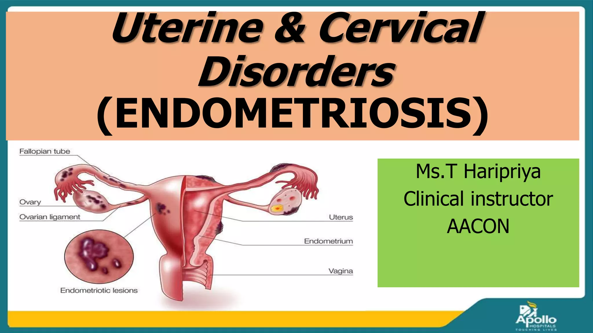
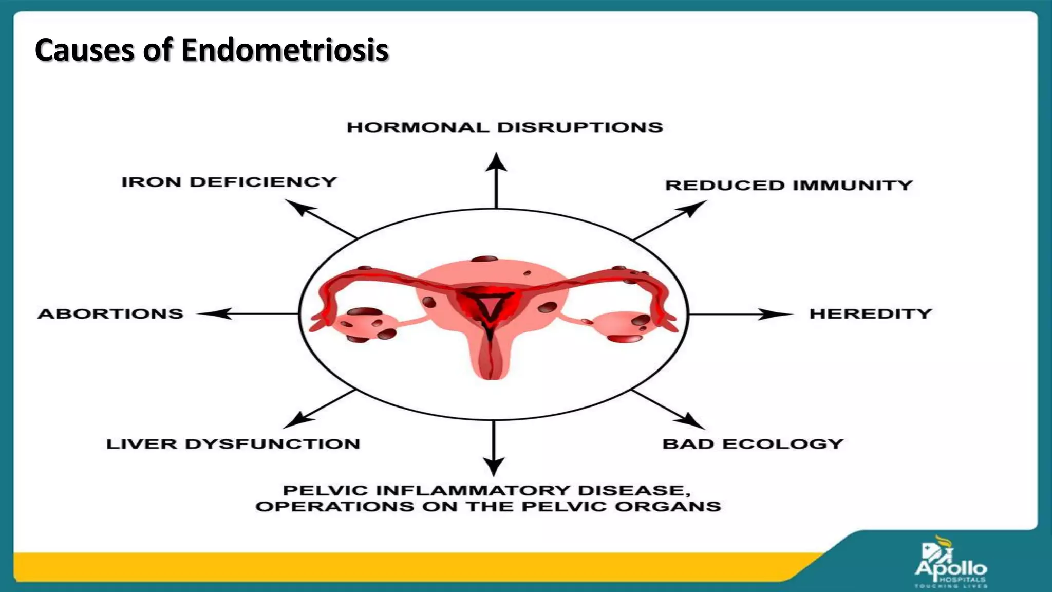
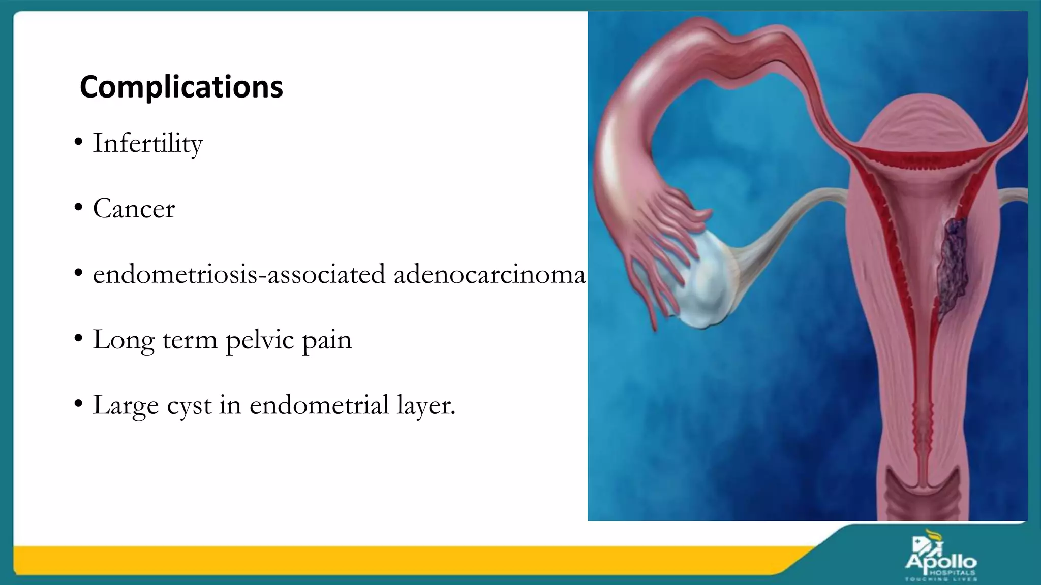
Uterine & Cervical
Disorders
(ENDOMETRIOSIS)
Ms.T Haripriya
Clinical instructor
AACON
Introduction
•
Endometriosis is a condition in which cells similar to those in
the endometrium, the layer of tissue that normally covers
the inside of the uterus, grows outside of it. Most often this
is on the ovaries, fallopian tubes, and tissue around the
uterus and ovaries; however, in rare cases it may also occur
in other parts of the body.
Definition
1. Presence of functioning endometrium (glands & stroma) in sites other
than uterine mucosa is called endometriosis. - D.C. Dutta
2. A benign but progressive condition characterized by endometrial
glands and stroma found in locations other than the endometrium.
- Roger P. Smith, M.D
Sites of endometriosis
Types of Endometriosis
1. Superficial peritoneal lesion: This is the most common kind. It is lesions
on peritoneum, a thin film that lines your pelvic cavity.
2. Endometrioma (ovarian lesion): These dark, fluid-filled cysts, also called
chocolate cysts, form deep in your ovaries. They don’t respond well to
treatment and can damage healthy tissue.
3. Deeply infiltrating endometriosis: This type grows under your
peritoneum and can involve organs near your uterus, such as your bowels or
bladder. About 1% to 5% of women with endometriosis have it.
Causes of Endometriosis
Diagnostic findings
 Complete history collection
 Physical examination
 Laparoscopy: It is a surgical process. This process of diagnosis also allows for
surgical treatment through laparoscopy. 6 to 13 percent of women have shown
the invisible lesions of endometriosis in the biopsy.
 Ultrasound: a pelvic ultrasound detects the larger endometriotic cysts as in
ovaries called endometriomas.
 Vaginal ultrasound is used in detecting deeper endometriomas and before
operating on them. This is one of the most easily accessible, inexpensive and
required no preparation.
 Magnetic resonance imaging: it is a noninvasive technique. But due to its
limited availability and cost, it is not widely recommended. But it clearly and
accurately diagnoses smaller lesions.
Treatment Management
Cont.
1. Hormonal Therapy: pills for contraception reduces pain. Estrogen-
progesterone birth control pill is the first line of treatment for most women with
endometriosis.
2. Progesterone: does not allow the growth of the endometrium. Progestin
drugs are given through hormonal therapy. Dienogest is one such. When estrogen
can’t be given along side progesterone, only progesterone will be prescribed.
3. Steroids: steroids are given to inhibit the growth of the endometrium.
4.NSAIDs such as ibuprofen and naproxen relieves the pain. Morphine sulphate
tablet an opioid is suggested as a painkiller. This mimics the action of endorphins.
5. Surgery is largely effective in treating endometriosis than medications. The
removal of endometrial tissue is done without any damage to the underlying
organs.
Complications
• Infertility
• Cancer
• endometriosis-associated adenocarcinoma
• Long term pelvic pain
• Large cyst in endometrial layer.
Preventive Measures
 Anti-inflammatory healing diet
 Correct hormonal imbalances
 Balance blood sugar levels
 Magnesium
 Vitamin D
 Essential oil packs
 Zinc
 turmeric
Uterine & cervical
Polyps
Introduction
Uterine polyps are growths attached to the inner wall of the uterus that
expand into the uterus. Uterine polyps, also known as endometrial polyps, form
as a result of cells in the lining of the uterus (endometrium) overgrowing. These
polyps are usually noncancerous (benign), although some can be cancerous or
can turn into cancer (precancerous polyps).
Cervical polyps are growths on the cervical canal, the passage that
connects the uterus to the vagina. They’re often reddish, purplish, or grayish in
color. They may be shaped like a finger, bulb, or thin stem. They can range in
size from a few millimeters to several centimeters long.
Cervical Polyps
Uterine Polyps
Clinical Manifestations
The symptoms of uterine polyps include:
 Irregular menstrual periods (unpredictable timing and flow).
 Unusually heavy flow during menstrual periods (heavy menstrual
bleeding).
 Bleeding or spotting between periods ( intermenstrual bleeding).
 Infertility (being unable to become pregnant or carry a pregnancy to
term).
 Vaginal spotting or bleeding after menopause (red, pink or brown blood).
 Bleeding after intercourse.
Diagnostic Findings
 complete history collection
 Physical examination
 Transvaginal ultrasound: imaginary study that provide an image of the
inside of the uterus, including fallopian tubes.
 Sono hysterography: After initial transvaginal ultrasound, provider may
send a sterile fluid into the uterus through a thin tube called a catheter. The
fluid causes the uterus to expand, providing a clearer image of any growths
within your uterine cavity during the ultrasound procedure.
Cont.
 Hysteroscopy: It is a insertion a long, thin tube with a lighted telescope
(hysteroscope) through your vagina and cervix into your uterus. The
hysteroscope allows your provider to examine the inside of your uterus.
Hysteroscopy is sometimes used in combination with surgery to remove
uterine polyps.
 Endometrial biopsy: uses a soft plastic instrument to collect tissue
from the inner walls of the uterus. The sample is tested in a laboratory to
detect any abnormal cells.
 Curettage: uses a long metal instrument called a curette to collect tissue
from the inner walls of your uterus. The curette has a small loop on the
end that's used to scrape tissue or polyps. The tissue or polyps that are
removed may be sent to the laboratory for testing to determine if cancer
cells are present.
MANAGEMENT AND TREATMENT
 Medications: Progestins and gonadotropin-releasing hormone agonists
help control / balanced hormone levels, like progestins or gonadotropin-
releasing hormone agonists, may be used to relieve symptoms. The
symptoms usually return after you stop taking the medication.
 Uterine Polypectomy: It is remove polyp during hysteroscopy. A
hysteroscope allows to insert tools that can be used to excise (cut) and
remove polyps. The advantage of hysteroscopy is that it’s precise. It can
then send the tissue to a lab for testing to check for signs of cancer.
 Hysterectomy (removal of the uterus).
uterine and cervical disorders.pptx

uterine and cervical disorders.pptx

  • 1.
    Uterine & Cervical Disorders (ENDOMETRIOSIS) Ms.THaripriya Clinical instructor AACON
  • 2.
    Introduction • Endometriosis is acondition in which cells similar to those in the endometrium, the layer of tissue that normally covers the inside of the uterus, grows outside of it. Most often this is on the ovaries, fallopian tubes, and tissue around the uterus and ovaries; however, in rare cases it may also occur in other parts of the body.
  • 3.
    Definition 1. Presence offunctioning endometrium (glands & stroma) in sites other than uterine mucosa is called endometriosis. - D.C. Dutta 2. A benign but progressive condition characterized by endometrial glands and stroma found in locations other than the endometrium. - Roger P. Smith, M.D
  • 4.
  • 5.
    Types of Endometriosis 1.Superficial peritoneal lesion: This is the most common kind. It is lesions on peritoneum, a thin film that lines your pelvic cavity. 2. Endometrioma (ovarian lesion): These dark, fluid-filled cysts, also called chocolate cysts, form deep in your ovaries. They don’t respond well to treatment and can damage healthy tissue. 3. Deeply infiltrating endometriosis: This type grows under your peritoneum and can involve organs near your uterus, such as your bowels or bladder. About 1% to 5% of women with endometriosis have it.
  • 6.
  • 8.
    Diagnostic findings  Completehistory collection  Physical examination  Laparoscopy: It is a surgical process. This process of diagnosis also allows for surgical treatment through laparoscopy. 6 to 13 percent of women have shown the invisible lesions of endometriosis in the biopsy.  Ultrasound: a pelvic ultrasound detects the larger endometriotic cysts as in ovaries called endometriomas.  Vaginal ultrasound is used in detecting deeper endometriomas and before operating on them. This is one of the most easily accessible, inexpensive and required no preparation.  Magnetic resonance imaging: it is a noninvasive technique. But due to its limited availability and cost, it is not widely recommended. But it clearly and accurately diagnoses smaller lesions.
  • 9.
  • 10.
    Cont. 1. Hormonal Therapy:pills for contraception reduces pain. Estrogen- progesterone birth control pill is the first line of treatment for most women with endometriosis. 2. Progesterone: does not allow the growth of the endometrium. Progestin drugs are given through hormonal therapy. Dienogest is one such. When estrogen can’t be given along side progesterone, only progesterone will be prescribed. 3. Steroids: steroids are given to inhibit the growth of the endometrium. 4.NSAIDs such as ibuprofen and naproxen relieves the pain. Morphine sulphate tablet an opioid is suggested as a painkiller. This mimics the action of endorphins. 5. Surgery is largely effective in treating endometriosis than medications. The removal of endometrial tissue is done without any damage to the underlying organs.
  • 11.
    Complications • Infertility • Cancer •endometriosis-associated adenocarcinoma • Long term pelvic pain • Large cyst in endometrial layer.
  • 12.
    Preventive Measures  Anti-inflammatoryhealing diet  Correct hormonal imbalances  Balance blood sugar levels  Magnesium  Vitamin D  Essential oil packs  Zinc  turmeric
  • 13.
  • 14.
    Introduction Uterine polyps aregrowths attached to the inner wall of the uterus that expand into the uterus. Uterine polyps, also known as endometrial polyps, form as a result of cells in the lining of the uterus (endometrium) overgrowing. These polyps are usually noncancerous (benign), although some can be cancerous or can turn into cancer (precancerous polyps). Cervical polyps are growths on the cervical canal, the passage that connects the uterus to the vagina. They’re often reddish, purplish, or grayish in color. They may be shaped like a finger, bulb, or thin stem. They can range in size from a few millimeters to several centimeters long.
  • 15.
  • 17.
    Clinical Manifestations The symptomsof uterine polyps include:  Irregular menstrual periods (unpredictable timing and flow).  Unusually heavy flow during menstrual periods (heavy menstrual bleeding).  Bleeding or spotting between periods ( intermenstrual bleeding).  Infertility (being unable to become pregnant or carry a pregnancy to term).  Vaginal spotting or bleeding after menopause (red, pink or brown blood).  Bleeding after intercourse.
  • 18.
    Diagnostic Findings  completehistory collection  Physical examination  Transvaginal ultrasound: imaginary study that provide an image of the inside of the uterus, including fallopian tubes.  Sono hysterography: After initial transvaginal ultrasound, provider may send a sterile fluid into the uterus through a thin tube called a catheter. The fluid causes the uterus to expand, providing a clearer image of any growths within your uterine cavity during the ultrasound procedure.
  • 20.
    Cont.  Hysteroscopy: Itis a insertion a long, thin tube with a lighted telescope (hysteroscope) through your vagina and cervix into your uterus. The hysteroscope allows your provider to examine the inside of your uterus. Hysteroscopy is sometimes used in combination with surgery to remove uterine polyps.  Endometrial biopsy: uses a soft plastic instrument to collect tissue from the inner walls of the uterus. The sample is tested in a laboratory to detect any abnormal cells.  Curettage: uses a long metal instrument called a curette to collect tissue from the inner walls of your uterus. The curette has a small loop on the end that's used to scrape tissue or polyps. The tissue or polyps that are removed may be sent to the laboratory for testing to determine if cancer cells are present.
  • 21.
    MANAGEMENT AND TREATMENT Medications: Progestins and gonadotropin-releasing hormone agonists help control / balanced hormone levels, like progestins or gonadotropin- releasing hormone agonists, may be used to relieve symptoms. The symptoms usually return after you stop taking the medication.  Uterine Polypectomy: It is remove polyp during hysteroscopy. A hysteroscope allows to insert tools that can be used to excise (cut) and remove polyps. The advantage of hysteroscopy is that it’s precise. It can then send the tissue to a lab for testing to check for signs of cancer.  Hysterectomy (removal of the uterus).