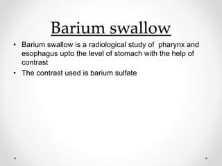
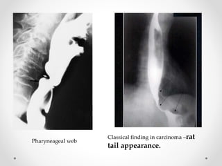
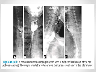
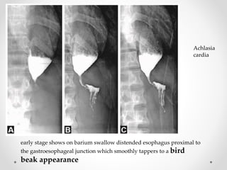
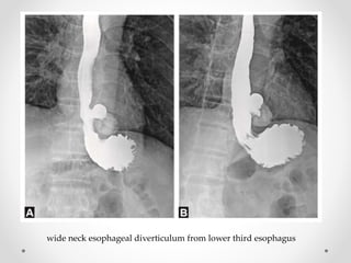
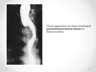
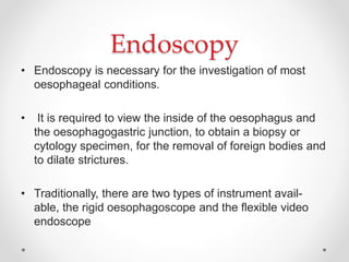
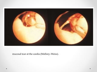
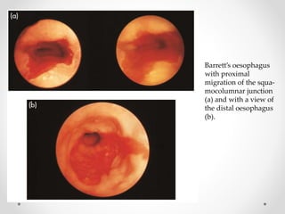
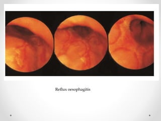
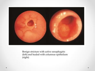
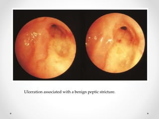
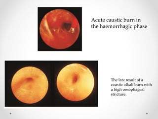
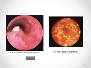
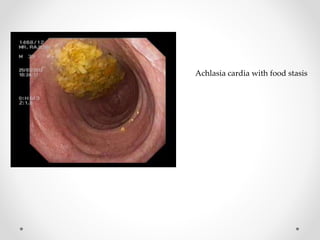
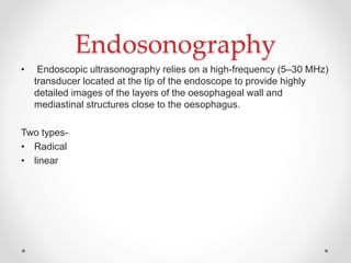
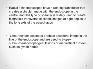
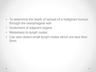
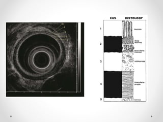
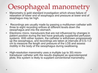
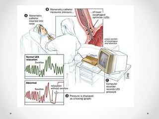
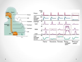
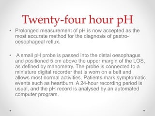
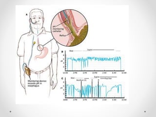
Oesophagus
Investigations
PRIYA ANUSHA DSOUZA
Radiography
• CXR
• BARIUM SWALLOW
• CT
Chest X-Ray
False teeth impacted in the
oesophagus.
(Note: modern dentures
are usually radiolucent.)
Mediastinal shadow due to
a large, fluid-filled
oesophagus.
Barium swallow
• Barium swallow is a radiological study of pharynx and
esophagus upto the level of stomach with the help of
contrast
• The contrast used is barium sulfate
Indications for barium swallow
• Dysphagia
• Heart burn, retrosternal pain, regurgitation &
odynophagia.
• Hiatus hernia
• Reflux oesophagitis
• Stricture formation.
• Esophageal carcinoma.
• Motility disorder like
i. Achalasia
ii. diffuse esophageal spasms.
• Pressure or invasion from extrinsic lesions.
• Assessment of abnormality of
i. pharyngo esophageal junction including zenkers diverticulum
ii. cricoid webs
iii. cricopharyngeal Achalasia.
Contraindiactions
• Suspected leakage from esophagus into the
mediastinum or pleura and peritoneal cavities.
• Tracheo-esophageal fistula
Pharyneageal web
Classical finding in carcinoma –rat
tail appearance.
Achlasia
cardia
early stage shows on barium swallow distended esophagus proximal to
the gastroesophageal junction which smoothly tappers to a bird
beak appearance
wide neck esophageal diverticulum from lower third esophagus
Barium swallow shows
dilation of the
esophagus (white
arrow) proximal to the
smooth narrowing at
lower esophageal
sphincter (black arrow)
irregular areas of narrowing
and dilatation -----
“corkscrew”
“rosary bead"
esophagus
The esophageal muscle is
hypertrophied, but
histologically normal
Diffuse esophageal
spasm
Oesophageal varices
Supine right side up
position, high density
thin barium should be
used.
Varices are best
demonstrated in
mucosal relief study
after using Buscopan/
valsalva maneuver.
Barium meal examination of four different cases of esophageal carcinoma show
middle third involvement in A and B and involvement of lower esophagus in C
and D. The lesions show irregular narrowing of lumen with shouldering effect and
rolled margins.
Barium in the esophagus
exhibits the 'reverse 3 sign'
outlining the medial site of the
aortic indentation in the
descending aorta
Coarctation of Aorta
Classic appearance of a large oesophageal
gastrointestinal stromal tumour on
barium swallow
CT
CT scan demonstrate the structural esophageal
abnormalities
Computed tomography scan showing a
primary tumour of the lower oesophagus.
Contrast-enhanced CT (CECT) chest shows (A) Circumferential wall thickening
of mid esophagus with loss of fat planes with bronchus, pulmonary artery and
aorta. The esophageal lumen is narrowed. Contrast-enhanced CT in another
case shows; (B) Well-defined asymmetric circumferential wall thickening of the
esophagus in midthoracic region causing marked luminal narrowing
Positron emission tomography
• Positron emission tomography (PET) in the context of
cancer staging relies on the generally high metabolic
activity (particularly in the glycolytic pathway) of tumours
compared with normal tissues
• The patient is given a small dose of the radio
pharmaceutical agent 18F-fluorodeoxyglucose (FDG).
This enters cells and is phosphorylated. FDG-6-
phosphate cannot be metabolised further and, because it
is a highly polar molecule, it cannot easily diffuse back
out of the cell. After intravenous injection of FDG, it
continuously accumulates in metabolically active cells.
Endoscopy
• Endoscopy is necessary for the investigation of most
oesophageal conditions.
• It is required to view the inside of the oesophagus and
the oesophagogastric junction, to obtain a biopsy or
cytology specimen, for the removal of foreign bodies and
to dilate strictures.
• Traditionally, there are two types of instrument avail-
able, the rigid oesophagoscope and the flexible video
endoscope
The normal lower oesophageal sphincter:
(a) open; (b) closed.
mucosal tear at the cardia (Mallory–Weiss).
Barrett’s oesophagus
with proximal
migration of the squa-
mocolumnar junction
(a) and with a view of
the distal oesophagus
(b).
Reflux oesophagitis
Benign stricture with active oesophagitis
(left) and healed with columnar epithelium
(right)
Ulceration associated with a benign peptic stricture.
Acute caustic burn in
the haemorrhagic phase
The late result of a
caustic alkali burn with
a high oesophageal
stricture.
oesophageal candidiasis
Achlasia cardia with food stasis
Ulcero-proliferative in the oesophaus
Hiatus hernia Esophageal perforation
Chromoendoscopy
• Chromoendoscopy involves the topical application of
stains or pigments to improve tissue localisation,
charectorisation or diagnosis during endoscopy
• Absorptive stains- Lugol’s Iodine, methylene blue
• Contrast stain- Indigocarmine
• Reactive stains- Congo red
Carcinoma in situ
showing the varied
presentations:
(a) occult form;
(b) erythroplakia;
(c) leukoplakia.
The right-hand
pictures in (a) and (b)
demonstrate the use
of vital staining with
methylene blue
Endosonography
• Endoscopic ultrasonography relies on a high-frequency (5–30 MHz)
transducer located at the tip of the endoscope to provide highly
detailed images of the layers of the oesophageal wall and
mediastinal structures close to the oesophagus.
Two types-
• Radical
• linear
• Radial echoendoscopes have a rotating transducer that
creates a circular image with the endoscope in the
centre, and this type of scanner is widely used to create
diagnostic transverse sectional images at right angles to
the long axis of the oesophagus
• Linear echoendoscopes produce a sectoral image in the
line of the endoscope and are used to biopsy
submucosal oesophageal lesions or mediastinal masses
such as lymph nodes
• To determine the depth of spread of a malignant tumour
through the oesophageal wall
• Involvment of adjacent organs
• Metastasis to lymph nodes
• Can also detect small lymph nodes which are less than
5mm
Endosonography demonstrating an
‘early’ tumour.
Note the preservation of the outer
dark wall layer that represents the
muscle coat.
Endosonography demonstrating
an ‘advanced’ local tumour.
Note the breach of the outer
white line that represents the
inter- face between the
oesophageal wall and the
mediastinum.
Endosonography
demonstrating
malignant nodes.
These are usually
large, hypoechoic
and round
compared with
normal nodes.
Oesophageal manometry
• Manometry is gold standard investigation which shows failure of
relaxation of lower end of esophagus and pressure at lower end of
esophagus may be high.
• Recordings are usually made by passing a multilumen catheter with
three to eight recording orifices at different levels down the
oesophagus and into the stomach.
• Electronic micro- transducers that are not influenced by changes in
patient position during the test have gradually supplanted perfusion
systems. With either system, the catheter is withdrawn progressively
up the oesophagus, and recordings are taken at intervals of 0.5–1.0
cm to measure the length and pressure of the LOS and assess
motility in the body of the oesophagus during swallowing.
• High-resolution manometry uses a multiple (up to 30) micro-
transducer catheter with the results displayed as spaciotemporal
plots; this system is likely to supplant conventional manometry.
Twenty-four hour pH
• Prolonged measurement of pH is now accepted as the
most accurate method for the diagnosis of gastro-
oesophageal reflux.
• A small pH probe is passed into the distal oesophagus
and positioned 5 cm above the upper margin of the LOS,
as defined by manometry. The probe is connected to a
miniature digital recorder that is worn on a belt and
allows most normal activities. Patients mark symptomatic
events such as heartburn. A 24-hour recording period is
usual, and the pH record is analysed by an automated
computer program.
Investigations - esophagus

Investigations - esophagus

  • 1.
  • 2.
  • 3.
    Chest X-Ray False teethimpacted in the oesophagus. (Note: modern dentures are usually radiolucent.)
  • 4.
    Mediastinal shadow dueto a large, fluid-filled oesophagus.
  • 5.
    Barium swallow • Bariumswallow is a radiological study of pharynx and esophagus upto the level of stomach with the help of contrast • The contrast used is barium sulfate
  • 6.
    Indications for bariumswallow • Dysphagia • Heart burn, retrosternal pain, regurgitation & odynophagia. • Hiatus hernia • Reflux oesophagitis • Stricture formation. • Esophageal carcinoma. • Motility disorder like i. Achalasia ii. diffuse esophageal spasms. • Pressure or invasion from extrinsic lesions. • Assessment of abnormality of i. pharyngo esophageal junction including zenkers diverticulum ii. cricoid webs iii. cricopharyngeal Achalasia.
  • 7.
    Contraindiactions • Suspected leakagefrom esophagus into the mediastinum or pleura and peritoneal cavities. • Tracheo-esophageal fistula
  • 8.
    Pharyneageal web Classical findingin carcinoma –rat tail appearance.
  • 10.
    Achlasia cardia early stage showson barium swallow distended esophagus proximal to the gastroesophageal junction which smoothly tappers to a bird beak appearance
  • 11.
    wide neck esophagealdiverticulum from lower third esophagus
  • 12.
    Barium swallow shows dilationof the esophagus (white arrow) proximal to the smooth narrowing at lower esophageal sphincter (black arrow)
  • 13.
    irregular areas ofnarrowing and dilatation ----- “corkscrew” “rosary bead" esophagus The esophageal muscle is hypertrophied, but histologically normal Diffuse esophageal spasm
  • 14.
    Oesophageal varices Supine rightside up position, high density thin barium should be used. Varices are best demonstrated in mucosal relief study after using Buscopan/ valsalva maneuver.
  • 15.
    Barium meal examinationof four different cases of esophageal carcinoma show middle third involvement in A and B and involvement of lower esophagus in C and D. The lesions show irregular narrowing of lumen with shouldering effect and rolled margins.
  • 16.
    Barium in theesophagus exhibits the 'reverse 3 sign' outlining the medial site of the aortic indentation in the descending aorta Coarctation of Aorta
  • 17.
    Classic appearance ofa large oesophageal gastrointestinal stromal tumour on barium swallow
  • 18.
    CT CT scan demonstratethe structural esophageal abnormalities
  • 19.
    Computed tomography scanshowing a primary tumour of the lower oesophagus.
  • 20.
    Contrast-enhanced CT (CECT)chest shows (A) Circumferential wall thickening of mid esophagus with loss of fat planes with bronchus, pulmonary artery and aorta. The esophageal lumen is narrowed. Contrast-enhanced CT in another case shows; (B) Well-defined asymmetric circumferential wall thickening of the esophagus in midthoracic region causing marked luminal narrowing
  • 21.
    Positron emission tomography •Positron emission tomography (PET) in the context of cancer staging relies on the generally high metabolic activity (particularly in the glycolytic pathway) of tumours compared with normal tissues • The patient is given a small dose of the radio pharmaceutical agent 18F-fluorodeoxyglucose (FDG). This enters cells and is phosphorylated. FDG-6- phosphate cannot be metabolised further and, because it is a highly polar molecule, it cannot easily diffuse back out of the cell. After intravenous injection of FDG, it continuously accumulates in metabolically active cells.
  • 23.
    Endoscopy • Endoscopy isnecessary for the investigation of most oesophageal conditions. • It is required to view the inside of the oesophagus and the oesophagogastric junction, to obtain a biopsy or cytology specimen, for the removal of foreign bodies and to dilate strictures. • Traditionally, there are two types of instrument avail- able, the rigid oesophagoscope and the flexible video endoscope
  • 24.
    The normal loweroesophageal sphincter: (a) open; (b) closed.
  • 26.
    mucosal tear atthe cardia (Mallory–Weiss).
  • 27.
    Barrett’s oesophagus with proximal migrationof the squa- mocolumnar junction (a) and with a view of the distal oesophagus (b).
  • 28.
  • 29.
    Benign stricture withactive oesophagitis (left) and healed with columnar epithelium (right)
  • 30.
    Ulceration associated witha benign peptic stricture.
  • 31.
    Acute caustic burnin the haemorrhagic phase The late result of a caustic alkali burn with a high oesophageal stricture.
  • 32.
  • 33.
  • 34.
  • 35.
  • 36.
    Chromoendoscopy • Chromoendoscopy involvesthe topical application of stains or pigments to improve tissue localisation, charectorisation or diagnosis during endoscopy • Absorptive stains- Lugol’s Iodine, methylene blue • Contrast stain- Indigocarmine • Reactive stains- Congo red
  • 37.
    Carcinoma in situ showingthe varied presentations: (a) occult form; (b) erythroplakia; (c) leukoplakia. The right-hand pictures in (a) and (b) demonstrate the use of vital staining with methylene blue
  • 39.
    Endosonography • Endoscopic ultrasonographyrelies on a high-frequency (5–30 MHz) transducer located at the tip of the endoscope to provide highly detailed images of the layers of the oesophageal wall and mediastinal structures close to the oesophagus. Two types- • Radical • linear
  • 40.
    • Radial echoendoscopeshave a rotating transducer that creates a circular image with the endoscope in the centre, and this type of scanner is widely used to create diagnostic transverse sectional images at right angles to the long axis of the oesophagus • Linear echoendoscopes produce a sectoral image in the line of the endoscope and are used to biopsy submucosal oesophageal lesions or mediastinal masses such as lymph nodes
  • 41.
    • To determinethe depth of spread of a malignant tumour through the oesophageal wall • Involvment of adjacent organs • Metastasis to lymph nodes • Can also detect small lymph nodes which are less than 5mm
  • 43.
    Endosonography demonstrating an ‘early’tumour. Note the preservation of the outer dark wall layer that represents the muscle coat. Endosonography demonstrating an ‘advanced’ local tumour. Note the breach of the outer white line that represents the inter- face between the oesophageal wall and the mediastinum.
  • 44.
    Endosonography demonstrating malignant nodes. These areusually large, hypoechoic and round compared with normal nodes.
  • 45.
    Oesophageal manometry • Manometryis gold standard investigation which shows failure of relaxation of lower end of esophagus and pressure at lower end of esophagus may be high. • Recordings are usually made by passing a multilumen catheter with three to eight recording orifices at different levels down the oesophagus and into the stomach. • Electronic micro- transducers that are not influenced by changes in patient position during the test have gradually supplanted perfusion systems. With either system, the catheter is withdrawn progressively up the oesophagus, and recordings are taken at intervals of 0.5–1.0 cm to measure the length and pressure of the LOS and assess motility in the body of the oesophagus during swallowing. • High-resolution manometry uses a multiple (up to 30) micro- transducer catheter with the results displayed as spaciotemporal plots; this system is likely to supplant conventional manometry.
  • 48.
    Twenty-four hour pH •Prolonged measurement of pH is now accepted as the most accurate method for the diagnosis of gastro- oesophageal reflux. • A small pH probe is passed into the distal oesophagus and positioned 5 cm above the upper margin of the LOS, as defined by manometry. The probe is connected to a miniature digital recorder that is worn on a belt and allows most normal activities. Patients mark symptomatic events such as heartburn. A 24-hour recording period is usual, and the pH record is analysed by an automated computer program.