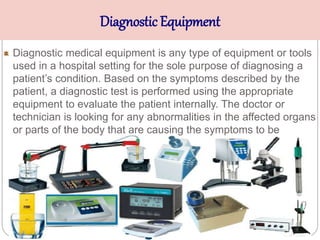
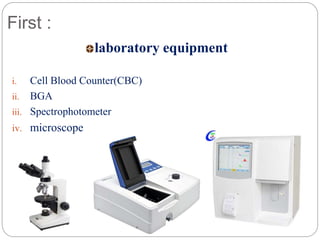
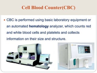
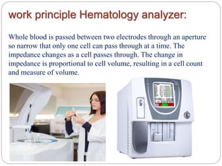
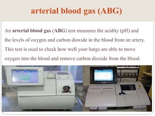
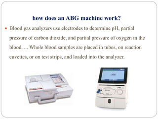
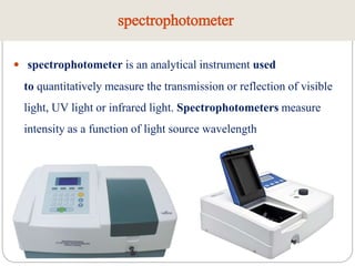
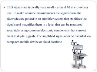
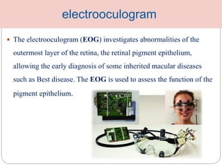
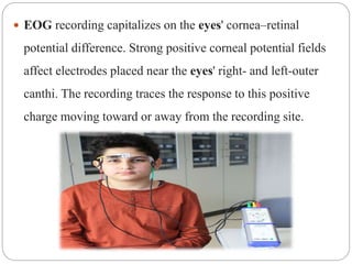
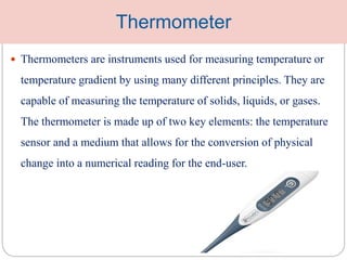
STUDENT NAME : Ibtihaj AbuSnyma
DIAGNOSTIC DEVICES
Medical Diagnostic
Systems
Diagnostic Equipment
Diagnostic medical equipment is any type of equipment or tools
used in a hospital setting for the sole purpose of diagnosing a
patient’s condition. Based on the symptoms described by the
patient, a diagnostic test is performed using the appropriate
equipment to evaluate the patient internally. The doctor or
technician is looking for any abnormalities in the affected organs
or parts of the body that are causing the symptoms to be
exhibited.
Diagnostic Equipment
For example
 laboratory equipment
 Diagnostic equipment
Diagnostic medical equipment is found in
outpatient care centers for adult and pediatrics,
in emergency rooms, as well as inpatient
hospital rooms and intensive care units.
 The following list is not exhaustive, but it provides an overview
of some of the most commonly used diagnostic devices.
First :
laboratory equipment
i. Cell Blood Counter(CBC)
ii. BGA
iii. Spectrophotometer
iv. microscope
Cell Blood Counter(CBC)
 CBC is performed using basic laboratory equipment or
an automated hematology analyzer, which counts red
and white blood cells and platelets and collects
information on their size and structure.
work principle Hematology analyzer:
Whole blood is passed between two electrodes through an aperture
so narrow that only one cell can pass through at a time. The
impedance changes as a cell passes through. The change in
impedance is proportional to cell volume, resulting in a cell count
and measure of volume.
arterial blood gas (ABG)
An arterial blood gas (ABG) test measures the acidity (pH) and
the levels of oxygen and carbon dioxide in the blood from an artery.
This test is used to check how well your lungs are able to move
oxygen into the blood and remove carbon dioxide from the blood.
how does an ABG machine work?
 Blood gas analyzers use electrodes to determine pH, partial
pressure of carbon dioxide, and partial pressure of oxygen in the
blood. ... Whole blood samples are placed in tubes, on reaction
cuvettes, or on test strips, and loaded into the analyzer.
spectrophotometer
 spectrophotometer is an analytical instrument used
to quantitatively measure the transmission or reflection of visible
light, UV light or infrared light. Spectrophotometers measure
intensity as a function of light source wavelength
what is the basic principle of
spectrophotometer?
 Spectrophotometry is a method to measure how much a
chemical substance absorbs light by measuring the intensity of
light as a beam of light passes through sample solution. The
basic principle is that each compound absorbs or transmits light
over a certain range of wavelength
Microscope
A microscope is an instrument that is used to magnify small objects.
Some microscopes can even be used to observe an object at the
cellular level, allowing scientists to see the shape of a cell, its nucleus,
mitochondria, and other organelles.
SECOND:
Diagnostic equipment
i. ECG
ii. EEG
iii. EMG
iv. Patient monitors
v. pulse oximeter
vi. pH meter
vii. Thermometer
In Emergency Department &Operations
Room.
In Emergency Department & Maternity
Department
.
In Emergency Department.
Intensive Care
Unit.
In Emergency Department.
Patient monitors
 Patient monitors are standard pieces of medical equipment
that keep accurate track of a patient’s condition and state of
health during and after surgery. They are necessary for adult,
pediatric & neonatal patients.
pulse oximeter
 A pulse oximeter is a small, lightweight device used to monitor the
amount of oxygen carried in the body. This noninvasive tool
attaches painlessly to your fingertip, sending two wavelengths of
light through the finger to measure your pulse rate and how much
oxygen is in your system. Once the oximeter finishes its
assessment, its screen will display the percent of oxygen in your
blood coming from your heart.
How does a pulse oximeter work?
When you insert your finger into a pulse oximeter, it beams different
wavelengths of light through your finger (you won’t feel a thing). It’s
targeting hemoglobin, a protein molecule in your blood that carries
oxygen. Hemoglobin absorbs different amounts and wavelengths of
light depending on the level of oxygen it’s carrying. Your pulse
oximeter will give you a numerical reading ,a percentage that indicates
the level of oxygen saturation in your blood.
Electrocardiographs
 Electrocardiographs measure the electrical activity of the heart.
During this examination, heart rate can be recorded, as well as the
regularity of the beats. These are two key indicators of any issues in
the heart. Physicians can even read an electrocardiograph to
determine the size and position of each heart chamber. And finally, a
major use for the electrocardiograph is to diagnose damage to the
heart and the impact and efficacy of drug treatment or device
implant.
 The ECG is a very valuable piece of diagnostic equipment, since
it makes it possible for doctors to precisely analyze heart
functioning. Electrodes are connected to the machine via small
wires and attached to the chest, arms, or legs of the patient. The
electrodes record and measure electrical activity in the body to
represent heart activity in real-time. A computer printout, known
as an electrocardiogram, can indicate irregular cardiac rhythms,
muscle weakness, and tissue damage.
EEG
 An EEG machine is a device that records the electrical activity
of the brain. It contain electrodes that can detect brain activity
when placed on a subject’s scalp. The electrodes record the
brain wave patterns and the EEG machine sends the data to a
computer or cloud server.
How Does an EEG Machine Work?
 The EEG electrodes pick up on electrical activity produced by neurons. EEG
machines use an array of electrodes because the brain produces different
signals from different brain regions. The number of electrodes corresponds
with the number of channels an EEG machine has. The more channels, the
higher the resolution of EEG data captured. A 32 channel EEG machine
captures a more detailed picture of brainwave activity than an 8 channel EEG
machine, in other words.
 EEG signals are typically very small – around 10 microvolts or
less. To make accurate measurements the signals from the
electrodes are passed to an amplifier system that stabilizes the
signals and magnifies them to a level that can be measured
accurately using common electronic components that convert
them to digital signals. The amplified signals can be recorded via
computer, mobile device or cloud database
Electromyography
 Electromyography (EMG) is a diagnostic procedure to assess
the health of muscles and the nerve cells that control them
(motor neurons). EMG results can reveal nerve dysfunction,
muscle dysfunction or problems with nerve-to-muscle signal
transmission.
 Motor neurons transmit electrical signals that cause muscles to
contract. An EMG uses tiny devices called electrodes to
translate these signals into graphs, sounds or numerical values
that are then interpreted by a specialist.
 During a needle EMG, a needle electrode inserted directly into a
muscle records the electrical activity in that muscle.
 A nerve conduction study, another part of an EMG, uses electrode
stickers applied to the skin (surface electrodes) to measure the
speed and strength of signals traveling between two or more
points.
electrooculogram
 The electrooculogram (EOG) investigates abnormalities of the
outermost layer of the retina, the retinal pigment epithelium,
allowing the early diagnosis of some inherited macular diseases
such as Best disease. The EOG is used to assess the function of the
pigment epithelium.
 EOG recording capitalizes on the eyes' cornea–retinal
potential difference. Strong positive corneal potential fields
affect electrodes placed near the eyes' right- and left-outer
canthi. The recording traces the response to this positive
charge moving toward or away from the recording site.
pH meter
 pH meter is an instrument used to measure acidity or alkalinity of a
solution - also know as Principle of operation
 Potentiometric pH meters measure the voltage between two
electrodes and display the result converted into the corresponding
pH value. They comprise a simple electronic amplifier and a pair of
electrodes, or alternatively a combination electrode, and some form
of display calibrated in pH units
Thermometer
 Thermometers are instruments used for measuring temperature or
temperature gradient by using many different principles. They are
capable of measuring the temperature of solids, liquids, or gases.
The thermometer is made up of two key elements: the temperature
sensor and a medium that allows for the conversion of physical
change into a numerical reading for the end-user.

DIAGNOSTIC DEVICES.pptx

  • 1.
    STUDENT NAME :Ibtihaj AbuSnyma DIAGNOSTIC DEVICES Medical Diagnostic Systems
  • 2.
    Diagnostic Equipment Diagnostic medicalequipment is any type of equipment or tools used in a hospital setting for the sole purpose of diagnosing a patient’s condition. Based on the symptoms described by the patient, a diagnostic test is performed using the appropriate equipment to evaluate the patient internally. The doctor or technician is looking for any abnormalities in the affected organs or parts of the body that are causing the symptoms to be exhibited.
  • 3.
    Diagnostic Equipment For example laboratory equipment  Diagnostic equipment
  • 4.
    Diagnostic medical equipmentis found in outpatient care centers for adult and pediatrics, in emergency rooms, as well as inpatient hospital rooms and intensive care units.
  • 5.
     The followinglist is not exhaustive, but it provides an overview of some of the most commonly used diagnostic devices.
  • 6.
    First : laboratory equipment i.Cell Blood Counter(CBC) ii. BGA iii. Spectrophotometer iv. microscope
  • 7.
    Cell Blood Counter(CBC) CBC is performed using basic laboratory equipment or an automated hematology analyzer, which counts red and white blood cells and platelets and collects information on their size and structure.
  • 8.
    work principle Hematologyanalyzer: Whole blood is passed between two electrodes through an aperture so narrow that only one cell can pass through at a time. The impedance changes as a cell passes through. The change in impedance is proportional to cell volume, resulting in a cell count and measure of volume.
  • 9.
    arterial blood gas(ABG) An arterial blood gas (ABG) test measures the acidity (pH) and the levels of oxygen and carbon dioxide in the blood from an artery. This test is used to check how well your lungs are able to move oxygen into the blood and remove carbon dioxide from the blood.
  • 10.
    how does anABG machine work?  Blood gas analyzers use electrodes to determine pH, partial pressure of carbon dioxide, and partial pressure of oxygen in the blood. ... Whole blood samples are placed in tubes, on reaction cuvettes, or on test strips, and loaded into the analyzer.
  • 11.
    spectrophotometer  spectrophotometer isan analytical instrument used to quantitatively measure the transmission or reflection of visible light, UV light or infrared light. Spectrophotometers measure intensity as a function of light source wavelength
  • 12.
    what is thebasic principle of spectrophotometer?  Spectrophotometry is a method to measure how much a chemical substance absorbs light by measuring the intensity of light as a beam of light passes through sample solution. The basic principle is that each compound absorbs or transmits light over a certain range of wavelength
  • 13.
    Microscope A microscope isan instrument that is used to magnify small objects. Some microscopes can even be used to observe an object at the cellular level, allowing scientists to see the shape of a cell, its nucleus, mitochondria, and other organelles.
  • 14.
    SECOND: Diagnostic equipment i. ECG ii.EEG iii. EMG iv. Patient monitors v. pulse oximeter vi. pH meter vii. Thermometer In Emergency Department &Operations Room. In Emergency Department & Maternity Department . In Emergency Department. In Emergency Department. Intensive Care Unit. In Emergency Department. In Emergency Department.
  • 15.
    Patient monitors  Patientmonitors are standard pieces of medical equipment that keep accurate track of a patient’s condition and state of health during and after surgery. They are necessary for adult, pediatric & neonatal patients.
  • 16.
    pulse oximeter  Apulse oximeter is a small, lightweight device used to monitor the amount of oxygen carried in the body. This noninvasive tool attaches painlessly to your fingertip, sending two wavelengths of light through the finger to measure your pulse rate and how much oxygen is in your system. Once the oximeter finishes its assessment, its screen will display the percent of oxygen in your blood coming from your heart.
  • 17.
    How does apulse oximeter work? When you insert your finger into a pulse oximeter, it beams different wavelengths of light through your finger (you won’t feel a thing). It’s targeting hemoglobin, a protein molecule in your blood that carries oxygen. Hemoglobin absorbs different amounts and wavelengths of light depending on the level of oxygen it’s carrying. Your pulse oximeter will give you a numerical reading ,a percentage that indicates the level of oxygen saturation in your blood.
  • 18.
    Electrocardiographs  Electrocardiographs measurethe electrical activity of the heart. During this examination, heart rate can be recorded, as well as the regularity of the beats. These are two key indicators of any issues in the heart. Physicians can even read an electrocardiograph to determine the size and position of each heart chamber. And finally, a major use for the electrocardiograph is to diagnose damage to the heart and the impact and efficacy of drug treatment or device implant.
  • 19.
     The ECGis a very valuable piece of diagnostic equipment, since it makes it possible for doctors to precisely analyze heart functioning. Electrodes are connected to the machine via small wires and attached to the chest, arms, or legs of the patient. The electrodes record and measure electrical activity in the body to represent heart activity in real-time. A computer printout, known as an electrocardiogram, can indicate irregular cardiac rhythms, muscle weakness, and tissue damage.
  • 20.
    EEG  An EEGmachine is a device that records the electrical activity of the brain. It contain electrodes that can detect brain activity when placed on a subject’s scalp. The electrodes record the brain wave patterns and the EEG machine sends the data to a computer or cloud server.
  • 21.
    How Does anEEG Machine Work?  The EEG electrodes pick up on electrical activity produced by neurons. EEG machines use an array of electrodes because the brain produces different signals from different brain regions. The number of electrodes corresponds with the number of channels an EEG machine has. The more channels, the higher the resolution of EEG data captured. A 32 channel EEG machine captures a more detailed picture of brainwave activity than an 8 channel EEG machine, in other words.
  • 22.
     EEG signalsare typically very small – around 10 microvolts or less. To make accurate measurements the signals from the electrodes are passed to an amplifier system that stabilizes the signals and magnifies them to a level that can be measured accurately using common electronic components that convert them to digital signals. The amplified signals can be recorded via computer, mobile device or cloud database
  • 23.
    Electromyography  Electromyography (EMG)is a diagnostic procedure to assess the health of muscles and the nerve cells that control them (motor neurons). EMG results can reveal nerve dysfunction, muscle dysfunction or problems with nerve-to-muscle signal transmission.  Motor neurons transmit electrical signals that cause muscles to contract. An EMG uses tiny devices called electrodes to translate these signals into graphs, sounds or numerical values that are then interpreted by a specialist.
  • 24.
     During aneedle EMG, a needle electrode inserted directly into a muscle records the electrical activity in that muscle.  A nerve conduction study, another part of an EMG, uses electrode stickers applied to the skin (surface electrodes) to measure the speed and strength of signals traveling between two or more points.
  • 25.
    electrooculogram  The electrooculogram(EOG) investigates abnormalities of the outermost layer of the retina, the retinal pigment epithelium, allowing the early diagnosis of some inherited macular diseases such as Best disease. The EOG is used to assess the function of the pigment epithelium.
  • 26.
     EOG recordingcapitalizes on the eyes' cornea–retinal potential difference. Strong positive corneal potential fields affect electrodes placed near the eyes' right- and left-outer canthi. The recording traces the response to this positive charge moving toward or away from the recording site.
  • 27.
    pH meter  pHmeter is an instrument used to measure acidity or alkalinity of a solution - also know as Principle of operation  Potentiometric pH meters measure the voltage between two electrodes and display the result converted into the corresponding pH value. They comprise a simple electronic amplifier and a pair of electrodes, or alternatively a combination electrode, and some form of display calibrated in pH units
  • 28.
    Thermometer  Thermometers areinstruments used for measuring temperature or temperature gradient by using many different principles. They are capable of measuring the temperature of solids, liquids, or gases. The thermometer is made up of two key elements: the temperature sensor and a medium that allows for the conversion of physical change into a numerical reading for the end-user.