1. Excitable tissues include nerve cells, nerve fibers, muscle fibers, and some plant cells that generate action potentials along their cell membranes in response to stimulation.
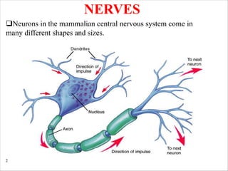
2. Nerve cells have a cell body containing organelles, dendrites that receive synaptic inputs, and a single axon that conducts electrical impulses to synaptic terminals.
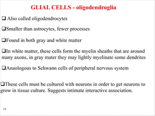
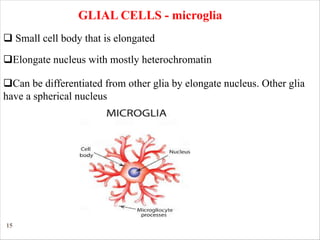
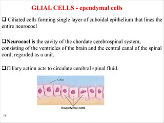
3. Glial cells such as astrocytes, oligodendrocytes, microglia, and ependymal cells provide support and insulation to neurons in the nervous system.
This document summarizes muscle physiology, including:
1. The functions of muscle tissue such as movement, stability, and respiration.
2. The properties of muscle tissue including excitability, conductivity, contractility, extensibility, and elasticity.
3. The types and classifications of muscles as well as the roles of agonist and antagonist muscles.
4. Key aspects of muscle anatomy and the sliding filament mechanism of muscle contraction.
1) Skeletal muscle contraction occurs via the sliding filament theory where the binding of myosin heads to actin causes sarcomeres to shorten.
2) Motor neurons innervate muscle fibers to produce either contraction or relaxation in response to neural signals.
3) Calcium release from the sarcoplasmic reticulum in response to action potentials is key to initiating muscle contraction by exposing binding sites on the actin thin filaments.
Degeneration & regeneration of nerve fiber.ppt by Dr. PANDIAN M.Pandian M
Â
This document discusses the degeneration and regeneration of nerve fibers following injury. It describes the various types of nerve injuries classified based on severity from first to fifth degree. When an axon is injured, degenerative changes occur in the distal segment, proximal segment, and nerve cell body. The distal segment undergoes Wallerian degeneration where the axon breaks down. Regeneration is possible if the nerve cell body and nucleus remain intact and the cut ends are within 3mm and aligned. Peripheral nerves can regenerate guided by Schwann cells, while regeneration is more limited in the central nervous system.
1. Neurons communicate via graded potentials over short distances and action potentials over long distances. Action potentials are generated when voltage-gated sodium channels open, causing rapid depolarization, followed by voltage-gated potassium channels opening to cause repolarization.
2. At chemical synapses, neurotransmitters are released from presynaptic terminals and bind to receptors on the postsynaptic cell, eliciting an excitatory or inhibitory response.
3. Faster conducting myelinated fibers like A fibers transmit touch and position sense while smaller unmyelinated C fibers transmit pain and temperature sensations. Fiber diameter, myelination and temperature influence conduction velocity.
The document discusses the history and discoveries of nerve physiology. It describes how Joseph Erlanger and Herbert Gasser developed tools to measure nerve impulses using oscilloscopes. Their work led to their shared Nobel Prize in 1944. Later, Hodgkin, Huxley, and Eccles advanced understanding of ionic mechanisms in nerves through experiments on squid nerves. Neher and Sakmann also received a Nobel Prize for developing a technique to measure currents through single ion channels. The document then provides detailed explanations and diagrams about the resting membrane potential, action potentials, graded potentials, and the mechanisms of nerve signal propagation.
Skeletal muscle makes up 40-50% of total body weight and is attached by tendons to bones. Skeletal muscle cells are multinucleated and striated, have visible banding patterns, and are voluntary muscles under conscious control. Skeletal muscles produce force for locomotion and postural support. Microscopically, skeletal muscle contains myofibrils with thick and thin filaments that slide during muscle contraction and relaxation. Skeletal muscle contraction occurs through summation of motor unit contractions and tetanization at higher stimulation frequencies.
This document provides information on nerve muscle physiology. It discusses the structure and function of nerves, neurons, and muscles. It explains how nerve signals trigger action potentials in muscles, causing contraction. It describes the sliding filament theory of muscle contraction and different types of muscle fibers. Stimulation methods like strength duration curves are discussed to assess denervated and healthy muscles. Electrical stimulation can aid tissue repair by mimicking the body's natural current of injury.
This document summarizes the action potential in neurons. It describes how an action potential is initiated by voltage-gated sodium channels opening during depolarization. This allows sodium ions to rush into the neuron, further depolarizing the membrane. Then, the sodium channels quickly inactivate while voltage-gated potassium channels open, allowing potassium ions to leave the neuron and repolarize the membrane back to its resting potential. The precise opening and closing of sodium and potassium channels underlies the generation and propagation of action potentials along neuronal membranes.
Properties of nerve fiber by Pandian M, Dept Physiology DYPMCKOP, this ppt fo...Pandian M
Â
Describe the types, functions & properties of nerve fibres
3.2.1 Classify nerve fibres
3.2.2 Classify nerve fibres based on the diameter & conduction velocity
3.2.3 Describe the salient features of Erlanger & Gasser
classification of nerve fibres
3.2.4 State the functions of type A, B & C nerve fibres
3.2.5 Compare & contrast the numerical classification with the
Erlanger & Gasser classification in the sensory nerve fibres
This document discusses the three types of muscle tissue: skeletal, cardiac, and smooth muscle. It provides details on their characteristics, such as whether they are striated or not, voluntary or involuntary, and their locations and functions in the body. Skeletal muscle is voluntary, attached to bones, and enables movement. Cardiac muscle is involuntary and makes up the heart wall. Smooth muscle is involuntary and located in organs like the stomach.
In this powerpoint, i have mentioned all the information with diagrams and functions in a very easy way. I am always there to solve any of the queries. Thank you.
The document discusses the motor unit, which consists of a motor neuron and the muscle fibers it innervates. It explains that motor units are recruited in order from smaller to larger units as the strength of the neural signal increases. Precise movements involve fewer muscle fibers per neuron, around 2-3, while imprecise movements involve more fibers, around 100 per neuron. Examples are requested of precise and imprecise movements.
The document describes various aspects of muscle contraction including:
1) Excitation-contraction coupling which involves depolarization of the muscle membrane leading to calcium release and muscle contraction.
2) The roles of the sarcoplasmic reticulum, t-tubules, and troponin-tropomyosin complex in regulating calcium levels and exposing actin binding sites during contraction.
3) The sliding filament theory of how myosin heads binding to actin causes muscle shortening through an ATP-driven cycling of cross-bridge formation and breaking.
This document provides a summary of the action potential in excitable tissues like nerves and muscles. It explains that excitable cells maintain a resting membrane potential with a negative charge inside and positive outside due to potassium ion leakage and intracellular proteins. An action potential is triggered by stimuli and causes rapid depolarization as sodium ions enter the cell, repolarization as potassium ions exit, returning the membrane to its resting potential. Precise ion channel openings and closings enable impulse transmission and functions like muscle contraction. Sodium-potassium pumps then restore ion gradients using ATP.
During a muscle contraction, thick myosin filaments and thin actin filaments slide across one another via cross-bridge cycling where myosin heads bind to and pull on actin filaments. This causes the sarcomere, the basic unit of muscle contraction, to shorten as the Z-bands move closer together without a change in filament lengths. The sliding filament model explains muscle contraction through cyclical cross-bridge binding powered by ATP hydrolysis allowing the power stroke and detachment of myosin from actin to enable repeated contractions.
This document discusses the physiology of muscle contraction. It describes the three types of muscle tissue - skeletal, cardiac, and smooth muscle. It explains that muscle contraction is initiated by calcium ion release, which allows the sliding of actin and myosin filaments. This sliding filament theory involves myosin binding to actin and using ATP hydrolysis to generate a power stroke, shortening the muscle. Repeated cycling of this cross-bridge formation causes muscle contraction.
Smooth muscle lacks visible cross-striations and contains actin and myosin arranged irregularly. It contracts through calcium binding to calmodulin rather than troponin. Smooth muscle is either single or multi-unit. Single unit smooth muscle contracts as a syncytium through gap junctions and shows spontaneous rhythmic contractions. Multi-unit smooth muscle contracts through discrete localized contractions in response to nerve stimulation. Smooth muscle action potentials are driven by calcium influx and can include plateaus, producing prolonged contractions. Contraction results from calcium binding to calmodulin and phosphorylating myosin light chains, with relaxation through dephosphorylation.
The document discusses the anatomy and histology of the central nervous system. It describes the different types of neurons, their classification based on structure and function. It also discusses the supporting glial cells like astrocytes, oligodendrocytes, microglia and ependymal cells. It explains the structure and function of synapses and myelin sheath formation in the CNS.
This document summarizes the key properties and functions of the three main types of muscle tissue: skeletal, cardiac, and smooth muscle. It describes their locations, structures, contraction mechanisms, and functions. Skeletal muscle is striated and voluntary, attaching to bones via tendons to enable movement. Cardiac muscle is also striated and pumps blood throughout the body. Smooth muscle is non-striated and involuntary, found in organs to enable processes like digestion. The document provides detailed descriptions of muscle fibers, sarcomeres, calcium handling, and more.
The resting membrane potential (RMP) refers to the stable voltage difference between the inside and outside of a cell membrane when the cell is not actively transmitting signals. The RMP results from selective permeability of ions like potassium and sodium across the membrane. At rest, the neuron's RMP is approximately -70mV due to higher intracellular potassium concentration creating a diffusion potential of -94mV, and lower intracellular sodium contributing +61mV. Additional contribution from the sodium-potassium pump, which actively transports ions against their gradients, results in the overall RMP of -90mV in neurons.
The document discusses the structure and function of neurons. It describes how neurons transmit signals through dendrites, the cell body and axon. An action potential is generated when the membrane potential reaches threshold. This involves the opening of voltage-gated sodium and potassium channels. Action potentials propagate along axons through continuous conduction in unmyelinated axons or saltatory conduction in myelinated axons between nodes of Ranvier. Factors like myelination, axon diameter and temperature can influence conduction velocity.
The document discusses membrane potential and action potentials in neurons. It provides details on:
- The resting membrane potential is established by ion gradients maintained by the sodium-potassium pump. There is a net negative charge inside and positive outside the membrane.
- An action potential is initiated when the membrane reaches its threshold potential due to sodium influx. It involves stages of depolarization, repolarization and refractory periods.
- The all-or-none principle states that an action potential will only be generated if the threshold is reached. Speed and propagation depends on myelination.
- Different cell types like cardiac and smooth muscles exhibit variations in their action potential waveforms.
Physiological properties of nerve fibersmariaidrees3
Â
Nerve fibers have low thresholds for excitation compared to other cells. When stimulated above the threshold, nerve fibers conduct all-or-none action potentials that propagate along the fiber. Below threshold, only local electrotonic potentials occur. Myelinated fibers conduct action potentials rapidly through saltatory conduction, where the impulse jumps between nodes of Ranvier. Nerve fibers are refractory immediately after an action potential and cannot conduct another for a period.
Dr. Nilesh Kate's document provides an overview of smooth muscle physiology. It discusses the functional anatomy and organization of smooth muscle, including that it is non-striated, involuntary muscle that exists in bundles. It describes the two types of smooth muscle - single unit and multi unit - and their characteristics. The document outlines the structure of smooth muscle fibers and covers the processes of excitability, contraction, and relaxation. It explains excitation and inhibition of smooth muscle can occur through nerves, hormones, pacemakers, stretching or temperature changes. In summary, the document provides a comprehensive review of smooth muscle types, organization, function and physiology.
This document discusses nerve and muscle physiology, including:
1. It describes the different types of ion channels in the plasma membrane, including leak channels, chemically-gated channels, mechanically-gated channels, and voltage-gated channels.
2. It explains the concepts of resting membrane potential, graded potentials, and action potentials in excitable cells like nerves and muscles. Key factors that determine resting membrane potential are the concentration gradients and permeability of ions like sodium and potassium.
3. It provides details on the initiation and propagation of action potentials, which involve the opening and closing of voltage-gated sodium and potassium channels in response to changes in membrane potential.
The document summarizes the mechanism of skeletal muscle contraction. It describes how an action potential leads to a rise in intracellular calcium levels through excitation-contraction coupling. This triggers the sliding filament theory where actin and myosin filaments slide past each other through cross-bridge cycling powered by ATP hydrolysis. Calcium binds to troponin C, allowing the power stroke to occur as myosin heads pull the actin filaments towards the center of the sarcomere. Relaxation occurs as calcium is re-sequestered in the sarcoplasmic reticulum, breaking the cross-bridges.
This document provides an introduction to skeletal muscle structure and function. It discusses the following key points in 3 sentences or less:
Skeletal muscle forms about 50% of body weight, is voluntary and striated, and its main functions are tension development and shortening under nervous system control. Skeletal muscle is composed of bundles of muscle fibers which contain myofibrils made up of repeating structural units called sarcomeres, the basic contractile units of muscle. Each sarcomere contains thin actin filaments and thick myosin filaments arranged in a repeating pattern that gives skeletal muscle its striated appearance visible under electron microscopy.
1. The document describes the structure and properties of excitable tissues like nerves and muscles. It discusses the anatomy of neurons including the cell body, dendrites, axon, and synaptic terminals.
2. Key properties of nerves are described, including excitability, conductivity, the all-or-none principle, accommodation, and infatiguability. The mechanisms of the resting membrane potential and action potential are summarized.
3. The stages of an action potential are outlined as depolarization, repolarization, after-depolarization, and after-hyperpolarization. Factors influencing stimulus effectiveness are also noted.
The nervous system includes the brain, spinal cord, and a complex network of nerves. This system sends messages back and forth between the brain and the body.
The brain is what controls all the body's functions. The spinal cord runs from the brain down through the back. It contains threadlike nerves that branch out to every organ and body part. This network of nerves relays messages back and forth from the brain to different parts of the body.What Are the Parts of the Nervous System?
The nervous system is made up of the central nervous system and the peripheral nervous system:
The central nervous system includes the brain and spinal cord.
The peripheral nervous system includes the nerves that run throughout the whole body.How Does the Nervous System Work?
The nervous system uses tiny cells called neurons (NEW-ronz) to send messages back and forth from the brain, through the spinal cord, to the nerves throughout the body.
Billions of neurons work together to create a communication network. Different neurons have different jobs. For example, sensory neurons send information from the eyes, ears, nose, tongue, and skin to the brain. Motor neurons carry messages away from the brain to the rest of the body to allow muscles to move. These connections make up the way we think, learn, move, and feel. They control how our bodies work — regulating breathing, digestion, and the beating of our hearts.
This document provides an overview of the central nervous system. It discusses the main components and functions.
The central nervous system consists of the brain and spinal cord. The brain is made up of the cerebrum, diencephalon, brainstem and cerebellum. The spinal cord contains ascending and descending tracts that transmit sensory and motor signals between the brain and body.
The brain and spinal cord contain grey matter with neuron cell bodies and white matter with myelinated axons. Neuroglia provide support to neurons. The brain and spinal cord are protected by meninges and cerebrospinal fluid.
Neurons are the basic functional units and come in different types. They transmit signals through electrical
The document summarizes key aspects of the nervous system seminar. It describes the basic structure and function of neurons, glial cells, and nerve fibers. It discusses the organization of the nervous system including the central nervous system structures like the brain, spinal cord, and meninges. Key topics covered include the resting membrane potential, action potentials, synapses, and reflexes. Classification and properties of different nerve fibers are also summarized.
The nervous system is composed of neurons and glial cells. Neurons communicate via electrical and chemical signals to control all body functions. The nervous system is divided into the central nervous system (brain and spinal cord) and peripheral nervous system (nerves). The peripheral system connects the central system to the rest of the body. Within the central system, sensory neurons carry stimuli from receptors to the brain and spinal cord, motor neurons carry signals from the central system to effectors like muscles and glands, and interneurons connect sensory and motor neurons.
The document provides an overview of the nervous system:
1. It describes the nervous system as a network of billions of nerve cells that functions as the control center of the body, integrating homeostasis, movement, and other functions.
2. The peripheral nervous system communicates between the central nervous system and the rest of the body, and can be divided into sensory and motor divisions.
3. Within neurons, the cell body contains organelles and receives inputs, while the axon conducts electrical signals to transmit outputs to other neurons.
The document provides an overview of the nervous system, including its basic functions, organization, and components. Key points:
1) The nervous system is a network of nerve cells that functions as the control center of the body, integrating homeostasis, movement, and other functions.
2) It has two main divisions - the central nervous system (CNS) comprising the brain and spinal cord, and the peripheral nervous system outside of the CNS.
3) Neurons are the basic functional units that conduct electrical signals to transmit information via chemical neurotransmitters at synapses.
Neurons are the basic building blocks of the nervous system and communicate via electrical and chemical signals. There are three main types of neurons - sensory neurons that receive information, interneurons that process information within the central nervous system, and motor neurons that send signals to muscles and glands. Glial cells provide support and insulation for neurons and are involved in nutrient supply, debris removal, and synaptic function. Neurons and glial cells work cooperatively to carry out the vital functions of the nervous system.
The document summarizes the structure and organization of the nervous system. It describes how the nervous system is composed of neurons and glial cells. Neurons have a cell body, dendrites, an axon, and axon terminals. The axon of most neurons is coated by a myelin sheath formed by Schwann cells in the peripheral nervous system and oligodendrocytes in the central nervous system. The nervous system is organized anatomically into the central nervous system (brain and spinal cord) and peripheral nervous system. Functionally, neurons are classified as afferent (sensory), efferent (motor), or interneurons.
The following power point presentation talks about neural control and coordination in humans. In this, we study about neurons, the conduction of nerve impulse, about Central Nervous System and also about sense organs
This document summarizes nervous tissue, including its cells and classification. It describes the two main cell types: neurons, which transmit electrical signals, and neuroglia cells, which support and protect neurons. Neurons are composed of a cell body, dendrites that receive signals, and an axon that transmits signals. There are different types of neurons classified by their structure. Neuroglia include oligodendrocytes, astrocytes, ependymal cells, microglia, Schwann cells, and satellite cells that each have distinct functions in supporting the nervous system. Together, neurons and glia allow the nervous system to coordinate voluntary and involuntary body functions through electrical signaling.
1. The nervous system is divided into the central nervous system and peripheral nervous system. The central nervous system is the brain and spinal cord, and the peripheral nervous system includes cranial and spinal nerves.
2. Neurons conduct electrical and chemical signals to transmit information, while glial cells provide support to neurons. Myelination affects how fast impulses are conducted along neurons.
3. Neurotransmitters are released at synapses to chemically transmit signals between neurons. The signal can be excitatory and increase the chance of firing an action potential, or inhibitory and decrease excitability.
Anatomy-Nervous-System Anatomy and Physiology updated.pptxJRRolfNeuqelet
Â
The nervous system is made up of neurons and neuroglia. Neurons transmit signals as electrical impulses between parts of the body, while neuroglia support and protect neurons. There are two main cell types - neurons, which generate and transmit nerve impulses, and neuroglia, which provide nutrients and insulation. The nervous system coordinates activities through neuronal communication via electrical and chemical signals at synapses to allow for reflexes and voluntary control of the body.
The nervous system consists of neurons and neuroglial cells. Neurons transmit nerve impulses through electrical and chemical signals. The neuron has a cell body, dendrites which receive signals, and an axon which transmits signals. Schwann cells wrap around axons and form myelin sheaths to aid impulse conduction. The nervous system regulates sensation, movement, and organ function through sensory, motor and interneurons. Nerve impulses rely on ion exchange and are transmitted across synapses using neurotransmitters. The central and peripheral nervous systems work together to control all bodily functions.
The nervous system consists of neurons and neuroglial cells. Neurons transmit nerve impulses through electrical and chemical signals. The neuron has a cell body, dendrites which receive signals, and an axon which transmits signals. Schwann cells wrap around axons and form myelin sheaths to insulate axons. Myelin allows faster impulse transmission. The nervous system regulates sensation, movement, and organ function through sensory, motor and interneurons. Nerve impulses rely on ion exchange and travel through the nervous system via pathways and reflex arcs.
The nervous system consists of neurons and neuroglial cells. Neurons transmit nerve impulses through electrical and chemical signals. The neuron has a cell body, dendrites which receive signals, and an axon which transmits signals. Schwann cells wrap around axons and produce myelin sheaths for insulation and faster signal transmission. The nervous system has sensory, inter, and motor neurons and performs functions like receiving information and transmitting instructions. Diseases can disrupt myelin sheaths and impair signaling.
What is different about activities on the two sides of the synapse?Salman Ul Islam
Â
The document discusses the differences between the presynaptic and postsynaptic sides of the synapse. Specifically, it notes that the presynaptic side contains calcium channels and synaptic vesicles filled with neurotransmitters, while the postsynaptic side contains ion channels with receptors for neurotransmitters. It then provides an overview of how neurotransmitters are released from the presynaptic terminal and bind to receptors on the postsynaptic membrane, allowing for transmission of signals between neurons.
The document discusses the structure and function of the nervous system. It describes how the nervous system is divided into the central nervous system (CNS), which includes the brain and spinal cord, and the peripheral nervous system (PNS). Neurons are the basic functional units that transmit signals in the nervous system. The document outlines the main parts of neurons including the cell body, dendrites, axon, and myelin sheath. It also describes the different types of neurons and specialized cells that support neurons called neuroglial cells.
basic nervous system-CNS-PNS -cell bodie- axon-dendron-grye matter- white mat...shailesh sangle
Â
The nervous system is a complex network of cells, tissues, and organs that coordinates and regulates the body's responses to internal and external stimuli. It is responsible for the control and coordination of all the body's functions, including movement, sensation, thought, and behavior.
The nervous system can be divided into two main parts: the central nervous system (CNS) and the peripheral nervous system (PNS). The CNS consists of the brain and spinal cord, while the PNS consists of all the nerves that extend from the CNS to the rest of the body.
The nervous system is made up of different types of cells, including neurons and glial cells. Neurons are specialized cells that transmit signals through the body in the form of electrical impulses. Glial cells, on the other hand, support and protect the neurons and help maintain the proper functioning of the nervous system.
The nervous system is responsible for many vital functions, including:
Sensory processing: The nervous system receives sensory information from the environment and the body's internal organs, and processes and interprets this information to generate appropriate responses.
Motor control: The nervous system controls the muscles and other organs of the body to produce movement and other responses.
Cognitive functions: The nervous system is responsible for the processes of learning, memory, language, and other complex mental activities.
Autonomic functions: The nervous system regulates the body's automatic functions, such as breathing, heart rate, digestion, and other bodily processes that are not under conscious control.
Overall, the nervous system is a complex and intricate system that plays a critical role in maintaining the body's homeostasis and overall well-being.
IF YOU ARE INTRESTED TO SHOP MAMA EARTH'S PRODUCTES ....
Mamaearth is an Indian personal care brand that offers natural, toxin-free, and sustainable products for all age groups. Their product range includes skincare, haircare, baby care, and men's grooming products. The brand is focused on promoting a healthy and sustainable lifestyle, and all their products are cruelty-free and environmentally friendly.
COPY THAT LINK AND PASTE ON SEARCH BAR OF GOOGLE
https://ekaro.in/enkr20230328s23089700
CHOOSE YOUR AND ORDER NOW.......
thank you....
This document summarizes the composition, properties, functions and volume of blood. It discusses that blood consists of cells suspended in plasma. The main cells are red blood cells, white blood cells and platelets. Plasma is mostly water with dissolved proteins and electrolytes. Bone marrow produces blood cells. Key functions of blood include transport, defense, regulation and hemostasis. The normal blood volume in adults is 5-6 liters. The hematocrit level indicates the proportion of red blood cells and can be used to screen for anemia. The blood and plasma volumes are regulated to maintain homeostasis.
The document discusses skeletal muscle and its structure and function. It contains 3 types of muscle: skeletal, smooth, and cardiac. Skeletal muscle is striated, involuntary muscle that moves the body and maintains posture. It develops tension and produces heat. Skeletal muscle fibers are cylindrical, multinucleated cells containing myofibrils with overlapping actin and myosin filaments that slide past each other to cause contraction. Contraction is initiated by calcium release from the sarcoplasmic reticulum in response to action potentials.
This document discusses heart murmurs, which are abnormal sounds caused by turbulent blood flow through the heart. It describes the different types of murmurs:
1) Systolic murmurs occur between the first and second heart sounds and can be caused by valve stenosis or insufficiency.
2) Diastolic murmurs occur between the second heart sound and next first sound, caused by stenosis or insufficiency of different heart valves.
3) Continuous murmurs occur during both systole and diastole, associated with conditions like a patent ductus arteriosus.
It provides examples of specific murmurs caused by abnormalities of the aortic, mitral, and tricuspid valves.
1. Erythrocytes, also known as red blood cells, are biconcave discs that contain hemoglobin and have an average lifespan of 120 days. Their primary function is to carry oxygen from the lungs to tissues and carbon dioxide from tissues back to the lungs.
2. Polycythemia is a condition of increased red blood cell count or hemoglobin concentration in the blood. Primary polycythemia is caused by excessive red blood cell production while secondary polycythemia occurs in response to tissue hypoxia such as at high altitudes.
3. Hemoglobin is the iron-containing protein in red blood cells that carries oxygen. It is composed of globin proteins and heme groups. Abnormalities
The document discusses heart sounds, including the four main types: S1, S2, S3, and S4. S1 and S2 are normally heard with each heartbeat while S3 and S4 may sometimes be heard. S1 occurs with the closure of the atrioventricular valves at the beginning of systole. S2 occurs with the closure of the semilunar valves at the end of systole. S3 occurs during early ventricular filling and S4 occurs during atrial contraction just before S1 of the next cycle. The sounds are associated with different parts of the cardiac cycle and changes in sounds can provide information about cardiac function and health issues.
Hemostasis occurs via four mechanisms: vasoconstriction, formation of a platelet plug, formation of a blood clot, and repair of the damaged blood vessel. Blood coagulation involves a cascade of reactions through the extrinsic and intrinsic pathways. The extrinsic pathway is initiated by tissue damage and the intrinsic pathway is initiated by contact with foreign surfaces. Both pathways lead to the conversion of prothrombin to thrombin which converts fibrinogen to fibrin to form a clot. Calcium ions, platelets, vitamin K, and the coagulation factors are essential for effective hemostasis and blood coagulation.
The cardiac cycle describes the repeating pattern of heart contraction and relaxation that pumps blood throughout the body. It consists of systole, when the heart contracts to pump blood, and diastole, when the heart relaxes and refills with blood. Each cycle normally occurs at a rate of about 75 times per minute. The cycle includes phases of isometric contraction and relaxation when pressure changes but volume does not, and ejection and filling phases when blood is pumped out or flows into the heart. Precise events in pressure, volume, valve opening and closing, and sounds occur throughout each phase of the cardiac cycle.
1. Carbon dioxide is transported in the blood from tissues to the lungs in both dissolved form and chemically combined as bicarbonate and carbamino compounds.
2. In the tissues, carbon dioxide diffuses into red blood cells where the carbonic anhydrase enzyme converts it mainly to bicarbonate, which then diffuses out of the cells in exchange for chloride ions in a process called the chloride shift.
3. This transport of carbon dioxide and shift of bicarbonate and chloride regulates blood pH and allows for exchange of carbon dioxide in the lungs, where it is released for exhalation.
This document summarizes the regulation of breathing. It discusses both chemical and non-chemical regulation. The chemical regulation is primarily via central and peripheral chemoreceptors that monitor changes in blood gases like CO2, O2 and pH. The central chemoreceptors are located in the brain and respond mainly to CO2, while peripheral chemoreceptors in the carotid and aortic bodies respond more to changes in O2, CO2 and pH. Non-chemical regulation involves signals from various receptors and parts of the body that influence breathing rate and depth.
The document discusses compliance of the lungs and chest wall, measurement of respiratory compliance (static vs dynamic), factors that affect lung compliance such as lung size and surface tension, diseases that decrease compliance, work of breathing including elastic and resistive components, factors that increase work of breathing, effects of gravity on ventilation and pulmonary blood flow including differences in regional lung volumes and flows, pneumothorax types including closed, open, and tension pneumothorax and their effects, and artificial pneumothorax as a therapeutic procedure.
This document discusses various types of hypoxia including hypoxic, anaemic, stagnant, and histotoxic hypoxia. It also covers topics like acclimatization to high altitudes, cyanosis, oxygen therapy, and carbon monoxide poisoning. The key points are:
1. Hypoxia is defined as oxygen deficiency at the tissue level and can be caused by problems with ventilation, gas exchange, blood flow, or cellular respiration.
2. Acclimatization to high altitudes involves mechanisms like increased ventilation, blood oxygen carrying capacity, circulation, and tissue oxygen delivery over weeks.
3. Cyanosis is a blue skin discoloration caused by abnormally high amounts of deoxygen
Oxygen transport (carriage)_by_the_bl000zulujunior
Â
1. Oxygen transport by the blood involves oxygen flowing from the alveoli to the tissues via hemoglobin in the blood. The oxygen carrying capacity of hemoglobin is increased about 70 times compared to dissolved oxygen alone.
2. The oxygen-hemoglobin dissociation curve shows the relationship between oxygen pressure and hemoglobin saturation. It is sigmoid shaped due to cooperative binding of oxygen.
3. Factors like acidosis, increased temperature, and 2,3-DPG can shift the curve rightward, increasing oxygen unloading to tissues. Fetal hemoglobin and alkalosis shift the curve leftward, decreasing oxygen unloading.
This document discusses gas exchange in the lungs. It covers the laws of gases, the partial pressure of gases, standardization of gas volumes, gas dissolution in fluids, gas diffusion through membranes, Fick's law of gas diffusion, the alveolar-capillary membrane, gas exchange across this membrane, equilibration of respiratory gases in the lungs, and the diffusion capacity of the lung. The key points are that oxygen diffuses from the alveoli into the blood while carbon dioxide diffuses out, and the solubility of carbon dioxide is much higher than oxygen allowing for faster diffusion.
The document discusses pulmonary surfactant, which reduces surface tension in the lungs. It describes the composition of surfactant, which contains phospholipids like phosphatidylcholine and proteins. Phosphatidylcholine is the most abundant lipid and reduces surface tension. Surfactant is synthesized in alveolar type II cells and stored in lamellar bodies. Hormones like glucocorticoids and estrogen increase surfactant production through various enzymatic pathways. Surfactant is essential for lung function as it prevents alveolar collapse and facilitates gas exchange.
Breathing is controlled by respiratory centers located in the brain stem and spinal cord. The medullary respiratory centers include the dorsal respiratory group (DRG), ventral respiratory group (VRG), and Botzinger complex. The DRG contains inspiratory neurons that promote inspiration, while the VRG contains expiratory neurons that promote expiration. The pontine respiratory group in the pons modulates the medullary centers. Rhythmic breathing occurs through reciprocal inhibition between inspiratory and expiratory neurons. Inspiration is an active process involving contraction of the diaphragm and rib muscles, while expiration is usually passive due to lung elasticity.
This document discusses factors that contribute to alveolar stability in the lungs. Alveolar stability is maintained by two main factors: surfactant and alveolar interdependence. Surfactant acts to reduce surface tension in small alveoli, preventing their collapse. Alveolar interdependence describes how neighboring alveoli support each other's volume. Together, surfactant and interdependence help maintain equal pressure across alveoli of different sizes. The document also discusses the elastic properties of the respiratory system, including how the recoiling forces of the lungs and chest wall interact to determine intrapulmonary pressure at different lung volumes.
share - Lions, tigers, AI and health misinformation, oh my!.pptxTina Purnat
Â
• Pitfalls and pivots needed to use AI effectively in public health
• Evidence-based strategies to address health misinformation effectively
• Building trust with communities online and offline
• Equipping health professionals to address questions, concerns and health misinformation
• Assessing risk and mitigating harm from adverse health narratives in communities, health workforce and health system
- Video recording of this lecture in English language: https://youtu.be/kqbnxVAZs-0
- Video recording of this lecture in Arabic language: https://youtu.be/SINlygW1Mpc
- Link to download the book free: https://nephrotube.blogspot.com/p/nephrotube-nephrology-books.html
- Link to NephroTube website: www.NephroTube.com
- Link to NephroTube social media accounts: https://nephrotube.blogspot.com/p/join-nephrotube-on-social-media.html
micro teaching on communication m.sc nursing.pdfAnurag Sharma
Â
Microteaching is a unique model of practice teaching. It is a viable instrument for the. desired change in the teaching behavior or the behavior potential which, in specified types of real. classroom situations, tends to facilitate the achievement of specified types of objectives.
These lecture slides, by Dr Sidra Arshad, offer a quick overview of the physiological basis of a normal electrocardiogram.
Learning objectives:
1. Define an electrocardiogram (ECG) and electrocardiography
2. Describe how dipoles generated by the heart produce the waveforms of the ECG
3. Describe the components of a normal electrocardiogram of a typical bipolar lead (limb II)
4. Differentiate between intervals and segments
5. Enlist some common indications for obtaining an ECG
6. Describe the flow of current around the heart during the cardiac cycle
7. Discuss the placement and polarity of the leads of electrocardiograph
8. Describe the normal electrocardiograms recorded from the limb leads and explain the physiological basis of the different records that are obtained
9. Define mean electrical vector (axis) of the heart and give the normal range
10. Define the mean QRS vector
11. Describe the axes of leads (hexagonal reference system)
12. Comprehend the vectorial analysis of the normal ECG
13. Determine the mean electrical axis of the ventricular QRS and appreciate the mean axis deviation
14. Explain the concepts of current of injury, J point, and their significance
Study Resources:
1. Chapter 11, Guyton and Hall Textbook of Medical Physiology, 14th edition
2. Chapter 9, Human Physiology - From Cells to Systems, Lauralee Sherwood, 9th edition
3. Chapter 29, Ganong’s Review of Medical Physiology, 26th edition
4. Electrocardiogram, StatPearls - https://www.ncbi.nlm.nih.gov/books/NBK549803/
5. ECG in Medical Practice by ABM Abdullah, 4th edition
6. Chapter 3, Cardiology Explained, https://www.ncbi.nlm.nih.gov/books/NBK2214/
7. ECG Basics, http://www.nataliescasebook.com/tag/e-c-g-basics
Local Advanced Lung Cancer: Artificial Intelligence, Synergetics, Complex Sys...Oleg Kshivets
Â
Overall life span (LS) was 1671.7±1721.6 days and cumulative 5YS reached 62.4%, 10 years – 50.4%, 20 years – 44.6%. 94 LCP lived more than 5 years without cancer (LS=2958.6±1723.6 days), 22 – more than 10 years (LS=5571±1841.8 days). 67 LCP died because of LC (LS=471.9±344 days). AT significantly improved 5YS (68% vs. 53.7%) (P=0.028 by log-rank test). Cox modeling displayed that 5YS of LCP significantly depended on: N0-N12, T3-4, blood cell circuit, cell ratio factors (ratio between cancer cells-CC and blood cells subpopulations), LC cell dynamics, recalcification time, heparin tolerance, prothrombin index, protein, AT, procedure type (P=0.000-0.031). Neural networks, genetic algorithm selection and bootstrap simulation revealed relationships between 5YS and N0-12 (rank=1), thrombocytes/CC (rank=2), segmented neutrophils/CC (3), eosinophils/CC (4), erythrocytes/CC (5), healthy cells/CC (6), lymphocytes/CC (7), stick neutrophils/CC (8), leucocytes/CC (9), monocytes/CC (10). Correct prediction of 5YS was 100% by neural networks computing (error=0.000; area under ROC curve=1.0).
Basavarajeeyam is a Sreshta Sangraha grantha (Compiled book ), written by Neelkanta kotturu Basavaraja Virachita. It contains 25 Prakaranas, First 24 Chapters related to Rogas& 25th to Rasadravyas.
Muktapishti is a traditional Ayurvedic preparation made from Shoditha Mukta (Purified Pearl), is believed to help regulate thyroid function and reduce symptoms of hyperthyroidism due to its cooling and balancing properties. Clinical evidence on its efficacy remains limited, necessitating further research to validate its therapeutic benefits.
Ozempic: Preoperative Management of Patients on GLP-1 Receptor Agonists Saeid Safari
Â
Preoperative Management of Patients on GLP-1 Receptor Agonists like Ozempic and Semiglutide
ASA GUIDELINE
NYSORA Guideline
2 Case Reports of Gastric Ultrasound
Identification and nursing management of congenital malformations .pptx
Â
Excitable tissues nerve
1. 1
EXCITABLE TISSUES
qCells and tissues in which excitation is accompanied
by action potential, distributed along the cellular
membrane.
qThis is a property of the bodies of nerve cells and their processes—
nerve fibers, muscle fibers or cells, and some elongated plant cells
qNerves and muscles are studied together because both arc excitable
structures that conduct impulses. but the muscle contract
2. 2
NERVES
qNeurons in the mammalian central nervous system come in
many different shapes and sizes.
3. 3
NERVES
qNeurons in the mammalian central nervous system come in
many different shapes and sizes.
qPerikayon (soma) - nerve cell body, contains nucleus and typical cell
organelles. It is the metabolic center of the neuron.
a. Nucleus - large, central in most, large amount of euchromatin
(intense synthetic activity), Barr body (Dormant X chromosome of
females).
b. Rough endoplasmic reticulum (RER) - lots for synthesis of structural
and transport proteins, Nissl bodies are condensations of the RER and
free ribosomes.
c. Golgi apparatus - only found near nucleus in perikaryon. Expected,
since intense synthetic activity of neurotransmitters and/or
neurohormones.
4. 4
NERVES
qDendrite - cell process, may be branched, forms receptive area for
synaptic contacts from other neurons,
qHas tiny rough projections or spines called gemmules that may be
points of synaptic contact,
qDendrites from larger neurons may be lightly myelinated by
oligodendroglia.
qNeurons may have more than one dendrite.
qCytoplasm in these processes similar to that of perikaryon, but no
golgi bodies.
5. 5
NERVES
qAxon – a single, long, cell process extending away from perikaryon,
may be branched,
qEnds of branches form synapses with other neurons or muscle cells
qMay be myelinated by either oligodendroglia in CNS or Schwann cells
in PNS..
6. 6
NERVES
qEach neuron has only one axon..
qAxon hillock (pyramid shaped region where axon originates from the
perikaryon)
qInitial segment (unmyelinated intitial portion of axon)
qremainder of axon (may be myelinated or unmyelinated, may be
branched)
7. 7
NERVES
qAxons carry electrical impulses (action potentials) to synapses at end
of axon.
qExcept for the axon hillock and the synaptic bouton, the axon
cytoplasm (axoplasm) has few organelles, microtubules, or
microfilaments.
qNot much synthetic activity in this part of neuron. The synaptic button
granules or vesicles in which the synaptic transmitters secreted by the
nerves are stored.
qThe axon divides into presynaptic terminals, each ending in a number
of synaptic knobs which are also called terminal buttons or boutons
8. 8
NERVES
qBased on the number of processes that emanate from their cell body,
neurons can be classified as unipolar, bipolar, and multipolar
qMultipolar - more than two processes (one axon plus multiple
dendrites), most of neurons in brain and spinal cord are of this type
qBipolar - two major processes (axon and dendrite), but may be
branched at ends, sensory neurons in retina, cochlea, and olfactory
epithelium are of this type
9. 9
NERVES
q Pseudounipolar - two major processes that are fused along portions
closet to perikaryon - found in spinal ganglia and some cranial ganglia.
qUnipolar have one process, with different
segments serving as receptive surfaces and
releasing terminals.
10. 10
NEURON CLASSIFICATION BASED ON FUNCTION
q Motor neurons - efferent, action potential moves from CNS to
effector organ (e.g. muscle)
qSensory neurons - afferent, action potential moves from sensory
organ to CNS (e.g. neuron processes associated with pacinian corpuscles,
touch, pressure)
qInterneurons - form connections between neurons
11. 11
GLIAL CELLS
q There are many more glial cells in the nervous system than there are
neurons.
qThese cells are situated among the neurons and are generally smaller.
qIn sections stained with hematoxylin - eosin, only the glial cell nuclei
show up.
qSpecial staining techniques are necessary if their cell bodies are to be
easily differentiated from surrounding cells.
12. 12
GLIAL CELLS - Astrocytes
q Two types:
1. Protoplasmic astrocytes:
a. Granular cytoplasm, many branches on short processes
b. Some of processes are closely applied to neurons, while others form
intimate contacts with blood vessels.
c. Thought to form a conduit for nutrients from blood vessels to neurons.
d. Found in gray matter.
e. Protoplasmic astrocytes have a membrane potential that varies with the
external K+ concentration but do not generate propagated potentials
f. Produce substances that are tropic to neurons, and they help maintain
the appropriate concentration of ions and neurotransmitters by taking up
K+ and the neurotransmitters glutamate and Îł-aminobutyrate (GABA)
13. 13
GLIAL CELLS - Astrocytes
2. Fibrous astrocytes:
a. Contain many intermediate filaments, are found primarily in white
matter
b. Function not well understood
qBoth types send processes to blood vessels, where they induce
capillaries to form the tight junctions making up the blood–brain barrier.
14. 14
GLIAL CELLS - oligodendroglia
qSmaller than astrocytes, fewer processes
qIn white matter, these cells form the myelin sheaths that are around
many axons, in gray mater they may lightly myelinate some dendrites
q Also called oligodendrocytes
qFound in both gray and white matter
qAnaologous to Schwann cells of peripheral nervous system
qThese cells must be cultured with neurons in order to get neurons to
grow in tissue culture. Suggests intimate interactive association.
15. 15
GLIAL CELLS - microglia
qElongate nucleus with mostly heterochromatin
q Small cell body that is elongated
qCan be differentiated from other glia by elongate nucleus. Other glia
have a spherical nucleus
16. 16
GLIAL CELLS - ependymal cells
qCiliary action acts to circulate cerebral spinal fluid.
q Ciliated cells forming single layer of cuboidal epithelium that lines the
entire neurocoel
qNeurocoel is the cavity of the chordate cerebrospinal system,
consisting of the ventricles of the brain and the central canal of the spinal
cord, regarded as a unit.
17. 17
PROPERTIES OF NERVES
qThis is the ability of living tissues to respond to various stimuli.
qIt is an electric phenomenon, and the electric changes that accompany
nerve excitation are called the action potential.
qSuch changes are very small and very rapid. so their magnitudes are
measured in millivolts (mV) while their durations are measured in
milliseconds (msec).
qThey are recorded by microelectrodes connected to either a
galvanometer or a cathode ray oscilloscope (CRO ).
1. Excitability
18. 18
qA stimulus is a change in the environment around the nerve (or
muscle)which may be either chemical, thermal, mechanical or electrical.
qIn laboratories, electrical stimuli are preferred because they can be
accurately controlled (both in strength and duration) and, in addition,
they leave the stimulated structures without damage.
q2 types of electric currents can be used for stimulation of excitable
tissues :
a. The galvanic current: This is a constant (or direct) current (D.C.)
which is obtained from a battery.
b. The faradic current: This is an alternating current (A.C.) like the
induction currents used in laboratories for nerve stimulation.
The stimulus
19. 19
qThe physicochemical change produced by various stimuli in the nerve
is called the nerve impulse.
qSuch impulse is actively conducted along the nerve fibre and it can be
conducted in both directions.
qIn the body, each nerve conducts impulses in one direction only (motor
nerves toward the effector organs and sensory nerves toward the nervous'
system).
2. Conductivity
qConduction in the normal direction is called orthodromic conduction,
qIf it occurs in the opposite direction due to any cause, it will be called
antidromic cmuluction
Does antidromic conduction occur in the brain under normal conditions?
20. 20
qStates that "A threshold (minimal) stimulus produces a maximal
response " i.e. a maximal action potential in nerve and muscle fibres and
a maximal contraction in muscle fibres.
qTherefore as long as other factors that affect excitability remain
constant:
qIncreasing the intensity of the stimulus above the threshold value
produces no further increase in the action potential or muscle contraction
(3) All or none law (or rule)
21. 21
qThe all or none law is obeyed in the following structures:
a. A single nerve fibre
- A motor unit is made up of a motor neuron and the skeletal muscle fibers innervated
by that motor neuron's axonal terminals.
(3) All or none law (or rule)
c. The cardiac muscle and some smooth muscles which act as one unit
called syncytium
b. A single skeletal muscle fibre and the motor unit
- Groups of motor units often work together to coordinate the contractions of a single
muscle; all of the motor units within a muscle are considered a motor pool.
22. 22
qHowever, nerve trunks and whole skeletal muscles (which contain
many fibres) do not obey the law.
(3) All or none law (or rule)
Why?
The threshold intensity for stimulation varies in the different types of
nerve and muscle fibres (i.e. it is not equal).
Therefore if the intensity of stimulation is increased in these structures,
the response will also increase till reaching a maximum.
23. 23
qThe nerve fibre adapts to stimulation by a constant current so no
response occurs during passage of the current.
(4) Accommodation (or adaptation)
(5) Infatiguability
qNerve fibres are not fatigued by continuous stimulation
24. 24
1. Intensity (strength) of the stimulus: Sub-threshold stimuli produce only
local responses that don't initiate action potentials .
Factors that determine the effectiveness of stimuli
If the intensity is increased slowly the nerve will not respond because of
the property of accommodation
2. Rate of increase in the intensity of stimuli; Sub-threshold stimuli
that are gradually increased produce a response only with a rapid
increase in the intensity of stimuli
25. 25
3. Duration of stimulus (duration of current): The relation between the
intensity of a stimulating current and the duration (time) of its flow
necessary to set up an impulse is shown in the strength-duration curve
Factors that determine the effectiveness of stimuli
26. 26
qWithin limits, there is a reciprocal relationship between the current
strength and duration of flow required to produce an impulse
qThere is a minimal duration needed for excitation below which no
excitation occurs whatever be the strength of the stimulus.
qRHEOBASE: This is the minimal strength (or threshold intensity ) of a
galvanic current that can set up an impulse.
qCHRONAXIE: This is the duration of current flow required for
excitation when using a strength equal to twice (or double) the rheobase.
qThe time required for excitation when using the rheobase is called the
utilization time
27. 27
qWhat is the significance of the Chronaxie?
qIts measurement can be used to compare the excitability of
different tissues, or that of the same tissue under different conditions.
qThe chronaxie is a good index for the degree of excitability (the shorter
the chronaxie the greater the excitability versa).
In the strength-duration curve to
the right, which nerve is more
excitable?
28. 28
qThe curve for the slower fibres would be shifted to the right, indicating
that for a given stimulus strength, a longer stimulus duration would be
needed to bring the slower fibres to threshold.
Q How would the strength-duration curve for a set of slow fibres (not
very excitable) compare to the strength-duration curve for a set of quick
fibres (very excitable)?
29. 29
RESTING MEMBRANE POTENTIAL REVIEW
qOutside the cell membranes there are mainly Na+, Cl- and HCO3 while
inside the cell there are mainly K+ and organic protein anions
qIn neurons the potential difference about -70 mV
qRM P is due to an unequal distribution of ions on both sides of the
membrane with relatively excess cations outside and excess anions inside
qThis is produced as a result or 2 main factors (a) Selective
permeability of cell membranes (b) The N a+/K+ ATPase
30. 30
RESTING MEMBRANE POTENTIAL REVIEW
(a) The electrical gradient is directed inwards
qK+ ions tend to diffuse outside the cells. However, this is limited
because:
(b) The +ve charge on the outside of the membranes repels K+ ions
inwards.
(c) The sodium-potassium pump actively drives K+ ions inwards
qThe concentration gradient for Cl- and HCO3 is directed inwards, so
these anions tend to diffuse into the cells.
qThis is limited because the interior of the cells has a great -ve charge,
and accordingly, they are expelled out of the cell along this electrical
gradient
31. 31
CHANGES THAT ACCOMPANY PROPAGATION OF A NERVE
IMPULSE
The Action Potential (AP)
(1) ELECTRIC CHANGES
qThis refers to the changes in potential that occur in excitable tissues
when stimulated
qIt is transmitted as a self-propagated disturbance called impulse
qStimulating the nerve (by an electric stimulator) is marked by a
stimulus artifact, which is due to current leakage from the stimulating
electrode to the recording (external) electrode.
qLatent period (which is an isopotcntial interval representing the time
taken by the impulse to reach the recording electrode) after which the AP
is recorded.
33. 33
qThe AP consists of 2 main stages (called depolarization and
repolarization)
q This is followed by 2 other stages known as after-
depolarization and after-hyperpolarization.
34. 34
qDepolarization (DP)
qThis is loss of the normal resting polarized state of the
membrane
qIt is recorded as a rise of the membrane potential in the
positive direction from -70 mV towards the isopotential line
(zero potential)
qIt produces the ascending limb of the A.P
qDP develops slowly. but after an initial 15 mV of DP (i.e.
MP becomes about -55 mV). the rate of DP suddenly increases
(so this point is called the firing level)
35. 35
qDepolarization (DP)
qDP then proceeds rapidly till the resting membrane potential
is lost
qThe potential difference between both sides of the
membrane becomes zero
qthis change is called overshoot or reversal of polarity, and it
results in an A.P. having a magnitude of 105 mV (from -70 to
+35 mV)
qThe membrane potential the reaches +35m V (indicating
that the inner surface of the membrane becomes positive
relative to the outer surface.
36. 36
qRepolarization (RP)
qThis is restoration or the normal resting polarized state of
the membrane
qIt is recorded as a fall of the membrane potential in the
negative direction from +35 mV to -70 mV.
qRP proceeds immediately and rapidly after the overshoot is
reached
q It produces the descending limb of the A.P.
qWhen RP is 70-80% completed, its rate decreases for about
4 msec. This stage is called after-depolarization (or negative
after-potential)
37. 37
qRepolarization (RP)
qAfter RP is completed the membrane potential overshoots to
the negative side (by about 1-2 mV) leading to
hyperpolarization of the membrane
qThis stagc is called after-hyperpolarization (or positive
after-potential).
qIt lasts about 40 msec but its magnitude gradually declines
till the normal resting membrane potential is restored
38. 38
Ionic basis (or mechanisms) of DP and RP
qThe initial slow DP is produced by the stimulating current
itself . How?
qStimulating current arc cathodic in nature, which adds
negative charges outside the nerve membrane
qThus the potential difference between both sides of the
membrane is decreased
qThe membrane potential becomes less -vc than at the resting
state. This is called electrotonic DP.
Depolarization
39. 39
qThe rapid phase of DP and the overshoot are produced by an
increase in Na+ influx (= entrance) into the nerve fiber as a
result of increased Na+ conductance of the nerve membrane
qIncreased permeability occurs secondary to marked increase
in the Na+ permeability of the membrane through opening of
specific Na+ channels in the membrane.
qEach Na+ channel has an activation gate at the outer surface
of the membrane and an inactivation gate at its inner surface.
qThe membrane Na+ conductance (and consequently the Na
influx ) is increased only when both gates are opened.
40. 40
qIn the resting state, only the inactivation gates are open so
the membrane permeability to Na+ is low
qWhen the nerve is stimulated the Na+ activation gates also
open thus the membrane permeability and conductance to Na+
as well as Na+ influx are markedly increased
qOpening of the Na+ activation gates is voltage-dependent.
qThey start to open when the initial electrotonic DP becomes
7 mV
qThis allows Na influx. which further decreases the
membrane polarity leading to opening of more gates.
41. 41
qThis results in more Na+ influx and more decrease of the
membrane polarity, which leads to opening of more and more
gates and more Na+ influx.
qSuch process continues in the form of a vicious circle
(i.e. by a positive feedback mechanism ) till all gates open
qThe rate of opening of the Na+ activation gates is slow
between 7 and I5 mV of DP (i.e. till a membrane potential of
about -55 mV) leading to slow DP.
qAfter15 mV of DP, the rate of opening of these gates
suddenly increases leading to much acceleration of DP (the
firing level)
42. 42
(a) Closure of the Na+ inactivation gates
(b) Reversal ofthe direction of eleclrical gradient for Na+
(2) K+ efflux (exit) from the nerve fibre:
Repolarization (RP) and after-potentials
RP of the membrane takes place rapidly after DP as a result of:
(1) Stoppage of Na+ influx due to:
qThis occurs through specific K+ channels that contain a
single gate located toward the inside of the membrane.
qThe decrease in membrane polarity during DP leads to
opening of the K+ gates, thus the K+ conductance is markedly
increased and K+ ef1ux occurs.
43. 43
qThe negative after-potential stage is clue to slowing of the
rate of K+ efflux.
qThe positive after-potential stage is due to slow return of the
K+ channels to the closed state (which allows prolonged K+
efflux)
qThe process is however slower than opening of the Na+
channels so the increase in K+ conductance is slightly delayed
qThe slow opening and delayed closure of the K+ channels
may explain the phenomenon of accommodation that occurs in
nerves
qFollowing the AP, the resting Na+ and K+ ionic gradients is
restored by the action of the Na+/K+ pump.
44. 44
qNormally, both the concentration and electrical gradients for
Ca2+are directed inwards
qThe Ca2+ conductance increases during nerve excitation
leading to Ca2+ influx.
Role of Ca2+ in Nerve excitation
qCa2+ contributes to DP (and in some invertebrates. it is
primarily responsible for the AP)
qCa2+ influx occurs via voltage-gated Ca2+ channels which
are also slightly permeable to Na+ (so they arc called Ca2+-
Na+ channels)
45. 45
qThey are however very slow to become activated, so they
are also called slow channels (in contrast to the voltage-gated
Na+ channels which arc called fast channels)
Role of Ca2+ in Nerve excitation
qThe extracellular Ca2+ concentration also affects nerve
excitability
qIts decrease increases the excitability while its increase
decreases the excitability and stabilizes the nerve membrane
46. 46
qETPs are localized potential changes that occur in nerves
when stimulated by sub-threshold constant currents
Electrotonic potentials (ETPs) and the local response
qThere are 2 types of the ETPs both of which are passive
changes in the membrane polarization
qThis is produced by addition of subtraction of charges
through the stimulating current) that decay (i.e. disappear)
gradually
47. 47
qThis is the potential change that occurs when using anodal
(+ve) currents for stimulation
(A) Anelectrotonic potential (or anelectrotonus)
qIt is a state of hyperpolarization caused by addition of +ve
charges at the outer surface of the nerve membrane.
qThe magnitude of the potential change is proportionate to
the strength of the stimulus
qIt is associated with a decrease of excitability of the nerve
and with strong anodal currents, the nerve excitability may be
completely lost ( anodal block)
qIt takes the membrane potential away from firing level
(which inhibits discharge of impulses).
48. 48
qThis is the potential change that occurs when using cathodal
(-ve) currents for stimulation
(A) Catelectrotonic potential (or catelectrotonus)
qIt is a state of partial DP caused by addition of –ve charges
at the outer surface of the nerve membrane.
qThe magnitude of the potential change varies with the
strength of the stimulating current
qIt is associated with an increase of excitability of the nerve,
50. 50
(A) In unmyelinated nerve fibres
qNerve impulses are propagated along unmyelinated nerve
fibres in the form of a wave of APs
qLocal circular currents flow between the activated point and
the neighbouring inactive areas of the nerve membrane
qThe initial stimulus causes reversal of polarity and an AP at
the point of stimulation
q+ve charges from the inactive areas flow into the initial area
of negativity produced by the AP (= area of current sink )
51. 51
qThis decreases the polarity at the inactive areas (
electrotonic depolarization) which produces an AP initiating
on reaching the firing level.
qThe latter areas, in turn, electrotonically depolarize the
membrane in front of it through local circular currents and this
sequence of events moves regularly along the nerve fibre to its
end.
qthe nerve impulse is self-propagated and once it leaves a
point this point will soon repolarize
qthus a repolarization wave starts after the depolarization
wave and is propagated in the same direction
52. 52
(B) In myelinated nerve fibres
qNerve impulses are propagated along myelinated nerve
fibres by a mechanism called the saltatory conduction
qMyelin surrounds the nerve axon and is interrupted at
regular intervals at the nodes of Ranvier
qIt is an insulator to current flow (in contrast to the nodes of
Ranvier which easily permit current flow because of their high
permeability to Na)
qCircular currents also flow in myelinated nerve fibres but
the +ve charges jump from the inactive nodes to the area of
current sink at the active node
53. 53
qThis leads to electrotonic depolarization and production of
an AP at the inactive nodes, which in turn activates the
neighbouring nodes.
qThis jumping of DP from node to node is called saltatory
conduction and it results in:
(a) Increasing velocity of conduction
(b) Conservation of energy (because excitation occurs only in
the nodes and not allover the nerve membrane)
54. 54
(2) EXCITABILITY CHANGES
qDuring propagation of a nerve impulse (i.e. during an AP).
The excitability of nerve fibres passes in the following phases
(a) Absolute refractory period (ARP):
§During this period, the nerve is completely inexcitable.
§No stimulus can excite it whatever its strength
§It corresponds to the ascending limb of the AP from the time
the firing level is reached
§That is during DP and overshoot and upper part of the
descending limb (until RP is about 1/3 complete)
55. 55
(b) Relative refractory period (RRP):
§During this period, nerve excitability is partiality recovered
§Stronger stimuli than normal are required for excitation
§It corresponds to the remaining part of the descending limb
of the AP till the start of after-depolarization
§That is during the later 2/3 of RP
56. 56
(c) Supernormal phase (or period):
§During this period nerve excitability is increased.
§Weaker stimuli than normal can excite the nerve
§It corresponds to the period of after-depolarization
57. 57
(d) Subrnormal phase (or period):
§During this period nerve excitability is decreased.
§Stronger stimuli than normal are required for excitation
§It corresponds to the period of afler-hyperpolarization.
59. 59
Factors that affect nerve excitability
(1) Temperature: Cooling decreases nerve excitability while
warming increases it
(2) Pressure: Mechanical pressure on a nerve reduces its
excitability.
(3) Blood supply: Nerve excitability is decreased in cases of
ischemia.
(4) Oxygen supply: O2 lack decreases nerve excitability.
(5) H+ concentration: Alkalinity increases while acidity
decreases excitability of nerves.
60. 60
(6) Chemicals: Nerve excitability is decreased by excess CO2
and alcohol as well as by anesthetic drugs e.g. ether,
chloroform and cocaine.
(8) Electrolytes: The concentrations of Na+ K+ and Ca2+ in
the extracellular fluid affect nerve excitability as follows
61. 61
Ionic changes that increase nerve excitability
1- Decreased Ca2+ concentration: This increases the
membrane permeability to Na+ and decreases the amount of
DP necessary to initiate the changes in Na+ and K+
conductances that produce the AP.
2- Increased Na concentration: This facilitates the process of
DP
3- Increased K+ concentration: This favours K influx which
leads to DP thus the nerve excitability will increase
62. 62
Ionic changes that decrease nerve excitability
1- Increased Ca2+ concentration : This decreases the
membrane permeability to Na+ and increases the amount of
DP necessary to initiate the changes in Na+ and K+
conductances that produce the AP
2-Decreased Na concentration : This decreases nerve
excitability by delaying the process of DP
3- Decreased K+ concentration : This favours K+ efflux
which leads to hyperpolarization, thus the nerve excitability
will decrease
63. 63
Methods of producing nerve block
qPropagation of impulses by nerves can be blocked (i.e.
prevented) by one of the following methods :
(1) Physical methods e.g. application of cold or a strong
anelectrotonus
(2) Mechanical methods (e.g. application of pressure on the
nerve).
(3) Chemical methods: These include:
(a) The ionic changes that decrease nerve excitability i.e.
increased Ca2+ and decreased Na+ or K+ concentration in the
extracellular fluid
64. 64
q(b) Use of certain chemical substances known as the
membrane stabilizers which include mainly the local
anesthetic drugs (e.g. cocaine and novocaine)
§These drugs markedly decrease the membrane
permeability to Na+ (by preventing opening of the Na+
channel activation gales)
§So the depolarization process is inhibited and nerve
impulses fail to be produced
65. 65
(3) METABOLIC CHANGES
qThe metabolic processes that occur in nerves are generally
similar to those occurring in muscles (O2 and ATP)
qIn resting nerves, they occur at a low rate (mainly to
maintain the polarized state) but they are much increased
during transmission of nerve impulses
qThe breakdown of ATP supplies the energy required for the
sodium potassium pump and nerve impulse propagation
qIt is resynthesized by energy derived from breakdown of
glycogen.
qNerve fibres are also rich in vitamin B1 which is necessary
for their metabolic activities
66. 66
(4) THERMAL CHANGES
qResting nerves liberate little heat as a result of their low
metabolic rate.
qDuring nerve impulse transmission the heat production by
nerves is markedly increased, and is liberated in the following
2 stages :
(1) Initial heat: This coincides with propagation of spike (i.e.
during ionic migration), and it is due to anaerobic breakdown
of ATP
(2) Recovery (delayed) heat: This follows propagation of the
spike, and it is due to aerobic reactions that liberate the energy
required for ATP resynthesis.
67. 67
Types of mammalian nerve fibres
The mammalian nerve fibres are classified according to their
diameters into the following types:
( 1) Group A nerve fibres :
§These have the largest diameters (3-20 microns)
§The have highest speeds of conduction ( 15-120 m/sec)
§They are further subdivided into alpha, beta, gamma & delta
nerve fibres
§ E.g. the somatic nerve fibres that transmit motor impulses &
deep sensations
§They are most sensitive to pressure (i.e. the conduction of
impulses in these nerves can be readily blocked by pressure)
68. 68
( 2) Group B nerve fibres :
§These have the smaller diameters (1.3 - 3 microns)
§They have moderate speeds of conduction (3-15 m/sec)
§E.g. the myelinated preganglionic autonomic nerves
§They arc most sensitive to O2 lack
69. 69
( 3) Group C nerve fibres :
§These have the smallest diameters (0.3-1.3 microns)
§The have slowest speeds of conduction (0.5-3 m/sec)
§E.g. the unmyelinated postganglionic autonomic nerves
§They arc most sensitive to local anaesthetics
71. 71
qThe area of contact between a nerve fibre and a muscle fibre
is called the motor end plate (M.E.P. ) or neuromuscular
junction
qAs the nerve approaches the muscle, it loses its myelin
sheath and its axon is branched.
qThe tip of each branch (sole foot ) is covered by the
neurolemma which continues with the sarcolemma ((the outer
membrane of the n1uscle fibre)
qEach sole foot lies in a depression in the plasma membrane
(the inner membrane of the muscle fibre) called the synaptic
gutter
73. 73
qWhere the membrane is thrown into many folds called the
palisades
qThere is no protoplasmic continuity between the nerve and
muscle fibres.
qThey are separated by the synaptic cleft in which the
chemical transmitter acetylcholine is released from the
vesicles present in the sole feet.
qThe sarcoplasm at the motor end plate is granular and
contains the cholinesterase enzyme which hydrolyzes
acetylcholine.
74. 74
Miniature end plate potential ( M. E.P.P.)
qThis is a state of persistent localized subthreshold DP (0.5
mV in amplitude) of the muscle fibres at the M.E.P. during
rest.
qIt does not reach the firing level (so it does not lead to an
AP)
q It is due to continuous release of small amounts of
acetylcholine
qArising from the continuous rupture of a few vesicles that
contain this chemical transmitter at the nerve terminals
75. 75
Mechanism of neuromuscular transmission
qWhen a motor nerve is stimulated, the generated impulse is
propagated toward the M.E.P
qit causes movement of'Ca2+ from the extracellular fluid into
the nerve terminals
q Ca2+ cause rupture of acetylcholine vesicles leading to
release (exocytosis) of this transmitter into the synaptic cleft
qAcetylcholine then combines with nicotinic receptors in the
muscle membrane
76. 76
qThis increases its permeability to Na+ through opening of
ligand-gated Na+ channel.
qNa+ influx increases resulting in DP of the muscle at the
M.E.P.. which is called the end plate potential
qThis normally reaches the firing level which starts an A P
along the surface of the muscle
qThe released acetylcholine is rapidly hydrolyzed by the
cholinesterase enzyme so that the re-excitation of the muscle
wouldn’t occur.
78. 78
Properties of neuromuscular transmission
1. At the M.E.P. impulses are conducted in one direction only
2. Transmission of impulses at the M. E.P. is delayed 0.5- 0. 7
millisecond due to the time required for:
(a) Release of acetylcholine
(b) Passage of acetylcholine across the synaptic cleft and its
combination with the nicotinic receptors in the muscle
(c) Increase in the permeability of the muscle membrane to Na
(d) Increase in Na+ influx till the firing level
3. Transmission of impulses at the M.E.P. readily fatigues after
repeated stimulations (mostly due to exhaustion of Ach)
4. Transmission of impulses at the M.E.P. is readily affected
by drugs
79. 79
Drugs that affect neuromuscular transmission
(A) Drugs that block neuromuscular transmission
1. Curare (tubocurarine) competitively inhibits Ach interaction
at the Nm receptors. α- neurotoxins (α- bungarotoxin, α-
cobrotoxins) irreversibly blocks.
2. Botulinum toxin: From Clostridium botulinum. The toxin
inhibits the release of ACh
80. 80
(B) Drugs that stimulate neuromuscular transmission
1. Acetylcholine-like drugs that are not rapidly hydrolyzed by
the cholinesterase enzyme and act mainly at the M. E.P. e.g.
carbacol.
2. Drugs that preserve the liberated acetylcholine at the M.E.P.
by antagonizing the cholinesterase enzyme e.g. prostigmine
and eserine