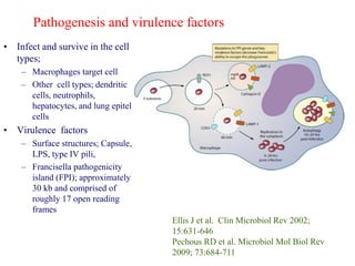
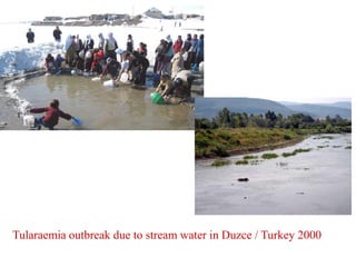
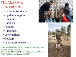
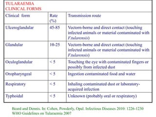
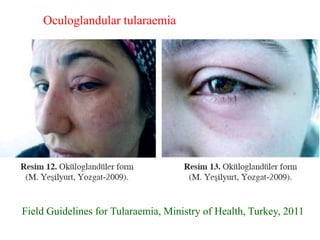
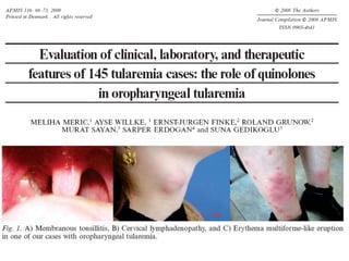
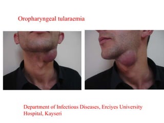
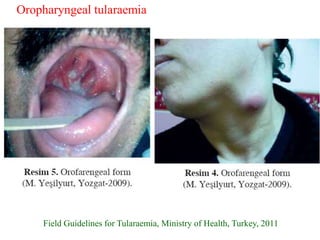
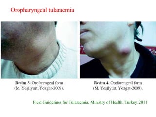
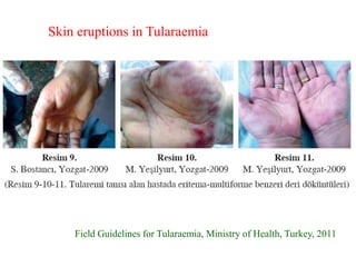
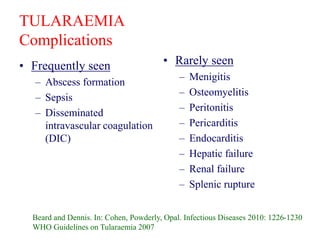
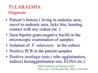
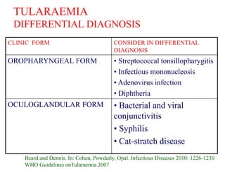
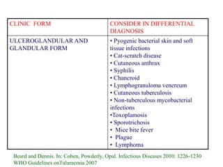
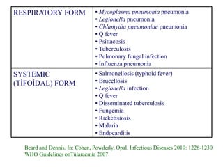
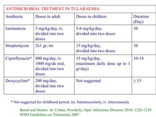
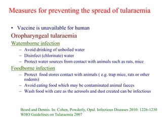
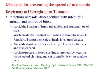
TULARAEMİA :
RE-EMERGING DISEASES
Mehmet Doganay
Department of Infectious Diseases,
Faculty of Medicine, Erciyes University
Kayseri / Turkey
Member of Zoonoses Working Group (International
Society of Chemotherapy )
Email. mdoganay@erciyes.edu.tr
mdoganay2003@yahoo.com
Outline of lecture
• Definition
• Etiologic agent
• Global epidemiology
• Transmission and pathogenesis
• Clinical presentation and diagnosis
• Treatment
• Prevention and control
• Conclusions
TULARAEMİA
Definition
• A bacterial zoonotic disease
• The etiologic agent: Francisella tularensis
• Highly virulent for humans and animals ( such as
rodents, hares, and rabbits)
• Cause epidemics and epizootics in humans and
animals
• A potential agent for bioterrorism, classified as a
Category A agent
MICROBIOLOGY :Francisella tularensis
• The bacteria;
– Gram-negative coccobacillus (0.2 - 0.5 in wide
and 0.7-1.0 mm in length),
– Capsulated ( in some strain)
– Facultative
– Intracellular
– Stain weakly
– Seen single and bipolar in microscopy
– Live in a long time in pooled water, mud and
animal carcass
• Not grow in routine media
• A fastidious microorganism which requires
enriched medium for growth ( cystein hearth
agar supplemented with 9 % heated sheep red
blood cells and nonselective buffered charchoal
yeast extract agar or Thayer- Martin agar may
be used for isolation)
• Grow in cystein enriched medium in aerobic
condition at 37oC within 2-4 days
WHO Guidelines on Tularaemia,
Roger D et al. Microbiol Mol Biol Rev 2009; 73:684-711
MICROBIOLOGY :Francisella tularensis
Subspecies Virulence character Found place
F.tularensis subsp.
tularensis (Type A)
Highly virulent USA
F.tularensis subsp.
holarchtica (Type B)
Less virulent Throughout the
northern hemisphere
F.tularensis subsp.
mediasiatica
Moderate virulent Kazakhstan,
Turkmenistan
F.tularensis novicida Low virulence and
water born
transmission
Australia, Spain,
USA
F.philomiragia Low virulence and
found in salty water
Atlantic and
Mediterranean
WHO Guidelines on Tularaemia,
Roger D et al. Microbiol Mol Biol 2009; 73:684-711
• Infect and survive in the cell
types;
– Macrophages target cell
– Other cell types; dendritic
cells, neutrophils,
hepatocytes, and lung epitel
cells
• Virulence factors
– Surface structures; Capsule,
LPS, type IV pili,
– Francisella pathogenicity
island (FPI); approximately
30 kb and comprised of
roughly 17 open reading
frames
Ellis J et al. Clin Microbiol Rev 2002;
15:631-646
Pechous RD et al. Microbiol Mol Biol Rev
2009; 73:684-711
Pathogenesis and virulence factors
TULARAEMİA
EPİDEMİOLOGY
• Reported in many countries of the Northern
hemisphere;
– Existed for a long time in the former Russian
federation, Kazakhstan and Turkmenistan, Finland
Sweden, Eastern Europe,
– Reported outbreaks from some European
Countries; Portugal, Spain, Sweden, (Kosovo)
Serbia and Turkey
– Also reported from Japan, China
– Regularly occur in USA and Canada
WHO Guidelines on Tularaemia 2007
Ellis J et al. Clin Microbiol Rev 2002; 15:631-634
World map showing the areas where tularemia is endemic
WHO Guidelines on Tularemia 2007, www.who.int
TULARAEMIA
Vectors and Reservoirs in the Environment
• Vectors
– Arthropods
• Ticks ( Ixodes spp.)
• Flies ( horse and deer flies)
• Mosquitoes (Aedes spp., Culex spp., Anopheles spp.)
• Flea ( their role is unclear)
– Mammals
• Outbreaks described in hares, prairie dogs and mink
• Mammals harbor and secrete the bacteria for a longer periods of time
• No evidence that they constitute a major natural reservoir for F.tularensis
• Voles and mice; accepting main source for the spread of F.tularensis to
humans
• Unclear the role of hares and rabbits for spreading the infection but infect
the hunters
– Reservoirs in Environment
• Field vole faeces are a source of human infection. Hay stored in barn is
contaminated with faeces and humans contract in the barns via inhalation
Beard and Dennis. In: Cohen, Powderly, Opal. Infectious Diseases 2010: 1226-1230
WHO Guidelines on Tularaemia 2007
Water / Soil Water / Soil
Voles
Voles
Mosquitoes
Ticks
Ticks
Lagomorphs
Lagomorphs
Deer flies
Life cycles of Francisella tularensis
A. Dominant in North America
B. Dominant in Eurasia
A B
TULARAEMIA
Transmission to Human
• Through insect bites (ticks, flies, mosquitoes
etc)
• Contact with infected animal or contaminated
animal products, water, mud
• Inhalation of aerosol droplets
• Consumption of contaminated water and food
( Outbreaks due to drinking contaminated water
have been seen in European countries such as
Kosovo, Poland and Turkey)
Beard and Dennis. In: Cohen, Powderly, Opal. Infectious Diseases 2010: 1226-1230
Pechous RD. Microbiol Mol Biol Rev 2009;73:684-711
WHO Guidelines on Tularaemia 2007
Meric M et al. APMIS 2008; 116:66-73
Tularaemia outbreak due to stream water in Duzce / Turkey 2000
TULARAEMIA
RISK GROUPS
• Living in rural area
in epidemic region
• Hunters
• Butchers
• Farmers
• Gardeners
• Veterinarians
• Cookers
• Laboratory workers
Beard and Dennis. In: Cohen, Powderly, Opal. Infectious
Diseases 2010: 1226-1230
Pechous RD. Microbiol Mol Biol Rev 2009;73:684-711
WHO Guidelines on Tularaemia 2007
TULARAEMİA
CINICAL PRESENTATION
• Severity of infection;
– Virulence
– Port of entry
– Systemic involvement
– Immune status of host
• An 3-5 days (1-21 days) incubation period
• Clinic pictures; asymptomatic
acute illness to sepsis
Beard and Dennis. In: Cohen, Powderly, Opal.
Infectious Diseases 2010: 1226-1230
Pechous RD. Microbiol Mol Biol Rev
2009;73:684-711
WHO Guidelines on Tularaemia 2007
TULARAEMIA
CLINICAL FORMS
Clinical form Rate
(%)
Transmission route
Ulceroglandular 45-85 Vectorn-borne and direct contact (touching
infected animals or material contaminated with
F.tularensis)
Glandular 10-25 Vectorn-borne and direct contact (touching
infected animals or material contaminated with
F.tularensis)
Oculoglandular < 5 Touching the eye with contaminated fingers or
possibly from infected dust
Oropharyngeal < 5 Ingestion contaminated food and water
Respiratory < 5 Inhaling contaminated dust or laboratory-
acquired infection
Typhoidal < 5 Unknown (probably oral or respiratory)
Beard and Dennis. In: Cohen, Powderly, Opal. Infectious Diseases 2010: 1226-1230
WHO Guidelines on Tularaemia 2007
Primary ulcer in a human case of
tularaemia. WHO Guidelines on
Tularemia. Geneva, 2007
Ulceroglandular tularaemia
• Appear a local papule at the site
of inoculation, then becomes
vesiculated and pustular and then
ulcerates within a few days
•Onset of fever and generalized
symptoms
• Around one or more painful
lymphadenetis
• Ulcer heals with leaving a scar
• In some cases, an abscessed
node may suppurate, create a sinus
tract and discharge purulent
material to the outside
Glandular tularaemia
Department of Infectious Diseases,
Erciyes University Hospital, Kayseri
Glandular tularaemia
•A similar clinical picture
as seen ulceroglandular
form
•Not having the local
cutaneous ulceration
• More likely follow
arthropod-borne
inoculation
Oculoglandular tularaemia
• Acquired by touching the eye
with contaminated finger or
possible by exposure to
F.tularensis containing dust
• Fever and unspecific symptoms
• Unilateral conjunctivitis,
together with swelling of eyelids,
excessive lacrimation,
photophobia, and mucopurulent
discharge
•A large , tender preauricular,
submandibular and cervical chain
lymph nodes
• Rarely leave vision loss
Oculoglandular tularaemia
Department of Infectious
Diseases, Erciyes University
Hospital, Kayseri
Field Guidelines for Tularaemia, Ministry of Health, Turkey, 2011
Oculoglandular tularaemia
Department of Infectious Diseases, Erciyes University
Hospital, Kayseri
Oropharyngeal tularaemia
Field Guidelines for Tularaemia, Ministry of Health, Turkey, 2011
Oropharyngeal tularaemia
Field Guidelines for Tularaemia, Ministry of Health, Turkey, 2011
Oropharyngeal tularaemia
Field Guidelines for Tularaemia, Ministry of Health, Turkey, 2011
Skin eruptions in Tularaemia
Typhoidal Tularaemia
• The most severe clinical form
• Seen pneumonia in 50 % of the cases
• Presents as an acute illness without localizing signs
• May be prominent a early systemic symptoms with
abdominal pain, diarrhea, vomiting and toxic
appearance
• May ensue the systemic inflammatory response
syndrome and sepsis
• May be complicated with acute respiratory distress
syndrome, disseminated intravascular coagulation and
bleeding, shock and organ failure
• Positive blood culture
Beard and Dennis. In: Cohen, Powderly, Opal. Infectious Diseases 2010: 1226-1230
WHO Guidelines on Tularaemia 2007
Pnömonik Tularemi
• Pathogenesis
– Inhalation of the bacteria
– Spread with hemothogenic
route
• Seen in 5-10 %
• Systemic symptoms (such as
fever, toxemia)
• Respiratory symptoms (such as
coughing, side pain in the chest)
• Chest X- Ray :
– Unilateral or bilateral infiltrations,
– Hilary adenophaty,
– Pleural effusion
Extended right-sided
pulmonary consolidation
Hilar lymphadenophaty en
WHO Guidelines on Tularemia. Geneva, 2007
TULARAEMIA
Complications
• Frequently seen
– Abscess formation
– Sepsis
– Disseminated
intravascular coagulation
(DIC)
• Rarely seen
– Menigitis
– Osteomyelitis
– Peritonitis
– Pericarditis
– Endocarditis
– Hepatic failure
– Renal failure
– Splenic rupture
Beard and Dennis. In: Cohen, Powderly, Opal. Infectious Diseases 2010: 1226-1230
WHO Guidelines on Tularaemia 2007
TULARAEMIA
Diagnosis
• Patient’s history ( living in endemic area,
travel to endemic area, ticks bite, hunting,
contact with any rodent etc. )
• Seen bipolar gram-negative bacilli in the
microscopic examination of samples
• Isolation of F. tularensis in the culture
• Positive PCR in the patient samples
• Positive serologic tests ( tube agglutination,
indirect hemagglutination test, ELISA etc.)
WHO Guidelines onTularaemia 2007
Ellis J et al. Clin Microbiol Rev 2002; 15:631-646
TULARAEMIA
DIFFERENTIAL DIAGNOSIS
CLINIC FORM CONSIDER IN DIFFERENTIAL
DIAGNOSIS
OROPHARYNGEAL FORM • Streptococcal tonsillopharygitis
• Infectious mononucleosis
• Adenovirus infection
• Diphtheria
OCULOGLANDULAR FORM • Bacterial and viral
conjunctivitis
• Syphilis
• Cat-stratch disease
Beard and Dennis. In: Cohen, Powderly, Opal. Infectious Diseases 2010: 1226-1230
WHO Guidelines onTularaemia 2007
CLINIC FORM CONSIDER IN DIFFERENTIAL
DIAGNOSIS
ULCEROGLANDULAR AND
GLANDULAR FORM
• Pyogenic bacterial skin and soft
tissue infections
• Cat-scratch disease
• Cutaneous anthrax
• Syphilis
• Chancroid
• Lymphogranuloma venereum
• Cutaneous tuberculosis
• Non-tuberculous mycobacterial
infections
•Toxoplamosis
• Sporotrichosis
• Mice bite fever
• Plague
• Lymphoma
Beard and Dennis. In: Cohen, Powderly, Opal. Infectious Diseases 2010: 1226-1230
WHO Guidelines onTularaemia 2007
RESPIRATORY FORM • Mycoplasma pneumonia pneumonia
• Legionella pneumonia
• Chlamydia pneumoniae pneumonia
• Q fever
• Psittacosis
• Tuberculosis
• Pulmonary fungal infection
• Influenza pneumonia
SYSTEMIC
(TİFOİDAL) FORM
• Salmonellosis (typhoid fever)
• Brucellosis
• Legionella infection
• Q fever
• Disseminated tuberculosis
• Fungemia
• Rickettsiosis
• Malaria
• Endocarditis
Beard and Dennis. In: Cohen, Powderly, Opal. Infectious Diseases 2010: 1226-1230
WHO Guidelines onTularaemia 2007
ANTIMICROBIAL TRETMENT IN TULARAEMIA
Antibiotic Doses in adult Doses in children Duration
(Day)
Gentamicin 5 mg/kg/day, iv,
divided into two
doses
5-6 mg/kg/day,
divided into two doses
10
Streptomycin 2x1 gr, im 15 mg/kg/day,
divided into two doses
10
Ciprofloxacin* 800 mg/day, iv
1000 mg/da oral,
divided into two
doses
15 mg/kg/day,
(maximum daily dose up to 1
gr/day)
10-14
Doxycycline* 200 mg/day,
divided into two
doses
Not suggested ≥ 15
* Not suggested for childhood period, im. Intramuscularly, iv. intravenously
Beard and Dennis. In: Cohen, Powderly, Opal. Infectious Diseases 2010: 1226-1230
WHO Guidelines on Tularaemia 2007
Measures for preventing the spread of tularaemia
• Vaccine is unavailable for human
Oropharyngeal tularaemia
Waternborne infection
– Avoid drinking of unboiled water
– Disinfect (chlorinate) water
– Protect water sources from contact with animals such as rats, mice
Foodborne infection
– Protect food stores contact with animals ( e.g. trap mice, rats or other
rodents)
– Avoid eating food which may be contaminated animal faeces
– Wash food with care as the aerosols and dust created can be infectious
Beard and Dennis. In: Cohen, Powderly, Opal. Infectious Diseases 2010: 1226-1230
WHO Guidelines on Tularaemia 2007
Measures for preventing the spread of tularaemia
Respiratory or Ulceroglandular Tularaemia
• Infectious aerosols, direct contact with infectious
animal, and arthropod bites:
– Avoid the hunting of hares and rabbits and consumption of
meat
– Wash hands after contact with wild and domestic animals
– Regularly inspect domestic animals for sign of disease
– Avoid dust and aerosols ( especially relevant for farmers
and landscapers)
– Avoid exposure to blood-sucking arthropods by wearing
long-sleeved clothing, and using repellents or mosquitoes
nets
Beard and Dennis. In: Cohen, Powderly, Opal. Infectious Diseases 2010: 1226-1230
WHO Guidelines on Tularaemia 2007
TULARAEMIA
Conclusions
• Tularaemia is a re-emerging disease
• The disease is prevalent in northern hemisphere
• Currently, some outbreaks have been occurred
• Franciesella tularensis subsp. tularensis is more virulent than others subspecies
• Franciesella tularensis subsp. tularensis is generally transmitted from animals (
rabbits, ticks and sheep) to human
• Franciesella tularensis subsp. Holarctica is often isolated in associated with
streams, ponds, lakes and riversd
• A wide range of arthropods ( ticks, mosquitoes, horse and deer flies) has been
identified in the transmission between mammalian hosts.
• Human-to-human transmission is not reported
• Varies clinical forms are seen
• Diagnose is based on history, clinical presentation, positive culture or /and serology
• The first line drugs are streptomycin, gentamicin, doxycyline and ciprofloxacin
• Vaccine is not available
• Applying to preventive measurements is very important for the control of tularemia
• We need a new vaccine for future and more researches for understanding of disease
transmission

Tularemia .ppt

  • 1.
    TULARAEMİA : RE-EMERGING DISEASES MehmetDoganay Department of Infectious Diseases, Faculty of Medicine, Erciyes University Kayseri / Turkey Member of Zoonoses Working Group (International Society of Chemotherapy ) Email. mdoganay@erciyes.edu.tr mdoganay2003@yahoo.com
  • 2.
    Outline of lecture •Definition • Etiologic agent • Global epidemiology • Transmission and pathogenesis • Clinical presentation and diagnosis • Treatment • Prevention and control • Conclusions
  • 3.
    TULARAEMİA Definition • A bacterialzoonotic disease • The etiologic agent: Francisella tularensis • Highly virulent for humans and animals ( such as rodents, hares, and rabbits) • Cause epidemics and epizootics in humans and animals • A potential agent for bioterrorism, classified as a Category A agent
  • 4.
    MICROBIOLOGY :Francisella tularensis •The bacteria; – Gram-negative coccobacillus (0.2 - 0.5 in wide and 0.7-1.0 mm in length), – Capsulated ( in some strain) – Facultative – Intracellular – Stain weakly – Seen single and bipolar in microscopy – Live in a long time in pooled water, mud and animal carcass • Not grow in routine media • A fastidious microorganism which requires enriched medium for growth ( cystein hearth agar supplemented with 9 % heated sheep red blood cells and nonselective buffered charchoal yeast extract agar or Thayer- Martin agar may be used for isolation) • Grow in cystein enriched medium in aerobic condition at 37oC within 2-4 days WHO Guidelines on Tularaemia, Roger D et al. Microbiol Mol Biol Rev 2009; 73:684-711
  • 5.
    MICROBIOLOGY :Francisella tularensis SubspeciesVirulence character Found place F.tularensis subsp. tularensis (Type A) Highly virulent USA F.tularensis subsp. holarchtica (Type B) Less virulent Throughout the northern hemisphere F.tularensis subsp. mediasiatica Moderate virulent Kazakhstan, Turkmenistan F.tularensis novicida Low virulence and water born transmission Australia, Spain, USA F.philomiragia Low virulence and found in salty water Atlantic and Mediterranean WHO Guidelines on Tularaemia, Roger D et al. Microbiol Mol Biol 2009; 73:684-711
  • 6.
    • Infect andsurvive in the cell types; – Macrophages target cell – Other cell types; dendritic cells, neutrophils, hepatocytes, and lung epitel cells • Virulence factors – Surface structures; Capsule, LPS, type IV pili, – Francisella pathogenicity island (FPI); approximately 30 kb and comprised of roughly 17 open reading frames Ellis J et al. Clin Microbiol Rev 2002; 15:631-646 Pechous RD et al. Microbiol Mol Biol Rev 2009; 73:684-711 Pathogenesis and virulence factors
  • 7.
    TULARAEMİA EPİDEMİOLOGY • Reported inmany countries of the Northern hemisphere; – Existed for a long time in the former Russian federation, Kazakhstan and Turkmenistan, Finland Sweden, Eastern Europe, – Reported outbreaks from some European Countries; Portugal, Spain, Sweden, (Kosovo) Serbia and Turkey – Also reported from Japan, China – Regularly occur in USA and Canada WHO Guidelines on Tularaemia 2007 Ellis J et al. Clin Microbiol Rev 2002; 15:631-634
  • 8.
    World map showingthe areas where tularemia is endemic WHO Guidelines on Tularemia 2007, www.who.int
  • 9.
    TULARAEMIA Vectors and Reservoirsin the Environment • Vectors – Arthropods • Ticks ( Ixodes spp.) • Flies ( horse and deer flies) • Mosquitoes (Aedes spp., Culex spp., Anopheles spp.) • Flea ( their role is unclear) – Mammals • Outbreaks described in hares, prairie dogs and mink • Mammals harbor and secrete the bacteria for a longer periods of time • No evidence that they constitute a major natural reservoir for F.tularensis • Voles and mice; accepting main source for the spread of F.tularensis to humans • Unclear the role of hares and rabbits for spreading the infection but infect the hunters – Reservoirs in Environment • Field vole faeces are a source of human infection. Hay stored in barn is contaminated with faeces and humans contract in the barns via inhalation Beard and Dennis. In: Cohen, Powderly, Opal. Infectious Diseases 2010: 1226-1230 WHO Guidelines on Tularaemia 2007
  • 10.
    Water / SoilWater / Soil Voles Voles Mosquitoes Ticks Ticks Lagomorphs Lagomorphs Deer flies Life cycles of Francisella tularensis A. Dominant in North America B. Dominant in Eurasia A B
  • 11.
    TULARAEMIA Transmission to Human •Through insect bites (ticks, flies, mosquitoes etc) • Contact with infected animal or contaminated animal products, water, mud • Inhalation of aerosol droplets • Consumption of contaminated water and food ( Outbreaks due to drinking contaminated water have been seen in European countries such as Kosovo, Poland and Turkey) Beard and Dennis. In: Cohen, Powderly, Opal. Infectious Diseases 2010: 1226-1230 Pechous RD. Microbiol Mol Biol Rev 2009;73:684-711 WHO Guidelines on Tularaemia 2007 Meric M et al. APMIS 2008; 116:66-73
  • 12.
    Tularaemia outbreak dueto stream water in Duzce / Turkey 2000
  • 13.
    TULARAEMIA RISK GROUPS • Livingin rural area in epidemic region • Hunters • Butchers • Farmers • Gardeners • Veterinarians • Cookers • Laboratory workers Beard and Dennis. In: Cohen, Powderly, Opal. Infectious Diseases 2010: 1226-1230 Pechous RD. Microbiol Mol Biol Rev 2009;73:684-711 WHO Guidelines on Tularaemia 2007
  • 14.
    TULARAEMİA CINICAL PRESENTATION • Severityof infection; – Virulence – Port of entry – Systemic involvement – Immune status of host • An 3-5 days (1-21 days) incubation period • Clinic pictures; asymptomatic acute illness to sepsis Beard and Dennis. In: Cohen, Powderly, Opal. Infectious Diseases 2010: 1226-1230 Pechous RD. Microbiol Mol Biol Rev 2009;73:684-711 WHO Guidelines on Tularaemia 2007
  • 15.
    TULARAEMIA CLINICAL FORMS Clinical formRate (%) Transmission route Ulceroglandular 45-85 Vectorn-borne and direct contact (touching infected animals or material contaminated with F.tularensis) Glandular 10-25 Vectorn-borne and direct contact (touching infected animals or material contaminated with F.tularensis) Oculoglandular < 5 Touching the eye with contaminated fingers or possibly from infected dust Oropharyngeal < 5 Ingestion contaminated food and water Respiratory < 5 Inhaling contaminated dust or laboratory- acquired infection Typhoidal < 5 Unknown (probably oral or respiratory) Beard and Dennis. In: Cohen, Powderly, Opal. Infectious Diseases 2010: 1226-1230 WHO Guidelines on Tularaemia 2007
  • 16.
    Primary ulcer ina human case of tularaemia. WHO Guidelines on Tularemia. Geneva, 2007 Ulceroglandular tularaemia • Appear a local papule at the site of inoculation, then becomes vesiculated and pustular and then ulcerates within a few days •Onset of fever and generalized symptoms • Around one or more painful lymphadenetis • Ulcer heals with leaving a scar • In some cases, an abscessed node may suppurate, create a sinus tract and discharge purulent material to the outside
  • 17.
    Glandular tularaemia Department ofInfectious Diseases, Erciyes University Hospital, Kayseri Glandular tularaemia •A similar clinical picture as seen ulceroglandular form •Not having the local cutaneous ulceration • More likely follow arthropod-borne inoculation
  • 18.
    Oculoglandular tularaemia • Acquiredby touching the eye with contaminated finger or possible by exposure to F.tularensis containing dust • Fever and unspecific symptoms • Unilateral conjunctivitis, together with swelling of eyelids, excessive lacrimation, photophobia, and mucopurulent discharge •A large , tender preauricular, submandibular and cervical chain lymph nodes • Rarely leave vision loss Oculoglandular tularaemia Department of Infectious Diseases, Erciyes University Hospital, Kayseri
  • 19.
    Field Guidelines forTularaemia, Ministry of Health, Turkey, 2011 Oculoglandular tularaemia
  • 21.
    Department of InfectiousDiseases, Erciyes University Hospital, Kayseri Oropharyngeal tularaemia
  • 22.
    Field Guidelines forTularaemia, Ministry of Health, Turkey, 2011 Oropharyngeal tularaemia
  • 23.
    Field Guidelines forTularaemia, Ministry of Health, Turkey, 2011 Oropharyngeal tularaemia
  • 24.
    Field Guidelines forTularaemia, Ministry of Health, Turkey, 2011 Skin eruptions in Tularaemia
  • 25.
    Typhoidal Tularaemia • Themost severe clinical form • Seen pneumonia in 50 % of the cases • Presents as an acute illness without localizing signs • May be prominent a early systemic symptoms with abdominal pain, diarrhea, vomiting and toxic appearance • May ensue the systemic inflammatory response syndrome and sepsis • May be complicated with acute respiratory distress syndrome, disseminated intravascular coagulation and bleeding, shock and organ failure • Positive blood culture Beard and Dennis. In: Cohen, Powderly, Opal. Infectious Diseases 2010: 1226-1230 WHO Guidelines on Tularaemia 2007
  • 26.
    Pnömonik Tularemi • Pathogenesis –Inhalation of the bacteria – Spread with hemothogenic route • Seen in 5-10 % • Systemic symptoms (such as fever, toxemia) • Respiratory symptoms (such as coughing, side pain in the chest) • Chest X- Ray : – Unilateral or bilateral infiltrations, – Hilary adenophaty, – Pleural effusion Extended right-sided pulmonary consolidation Hilar lymphadenophaty en WHO Guidelines on Tularemia. Geneva, 2007
  • 27.
    TULARAEMIA Complications • Frequently seen –Abscess formation – Sepsis – Disseminated intravascular coagulation (DIC) • Rarely seen – Menigitis – Osteomyelitis – Peritonitis – Pericarditis – Endocarditis – Hepatic failure – Renal failure – Splenic rupture Beard and Dennis. In: Cohen, Powderly, Opal. Infectious Diseases 2010: 1226-1230 WHO Guidelines on Tularaemia 2007
  • 28.
    TULARAEMIA Diagnosis • Patient’s history( living in endemic area, travel to endemic area, ticks bite, hunting, contact with any rodent etc. ) • Seen bipolar gram-negative bacilli in the microscopic examination of samples • Isolation of F. tularensis in the culture • Positive PCR in the patient samples • Positive serologic tests ( tube agglutination, indirect hemagglutination test, ELISA etc.) WHO Guidelines onTularaemia 2007 Ellis J et al. Clin Microbiol Rev 2002; 15:631-646
  • 29.
    TULARAEMIA DIFFERENTIAL DIAGNOSIS CLINIC FORMCONSIDER IN DIFFERENTIAL DIAGNOSIS OROPHARYNGEAL FORM • Streptococcal tonsillopharygitis • Infectious mononucleosis • Adenovirus infection • Diphtheria OCULOGLANDULAR FORM • Bacterial and viral conjunctivitis • Syphilis • Cat-stratch disease Beard and Dennis. In: Cohen, Powderly, Opal. Infectious Diseases 2010: 1226-1230 WHO Guidelines onTularaemia 2007
  • 30.
    CLINIC FORM CONSIDERIN DIFFERENTIAL DIAGNOSIS ULCEROGLANDULAR AND GLANDULAR FORM • Pyogenic bacterial skin and soft tissue infections • Cat-scratch disease • Cutaneous anthrax • Syphilis • Chancroid • Lymphogranuloma venereum • Cutaneous tuberculosis • Non-tuberculous mycobacterial infections •Toxoplamosis • Sporotrichosis • Mice bite fever • Plague • Lymphoma Beard and Dennis. In: Cohen, Powderly, Opal. Infectious Diseases 2010: 1226-1230 WHO Guidelines onTularaemia 2007
  • 31.
    RESPIRATORY FORM •Mycoplasma pneumonia pneumonia • Legionella pneumonia • Chlamydia pneumoniae pneumonia • Q fever • Psittacosis • Tuberculosis • Pulmonary fungal infection • Influenza pneumonia SYSTEMIC (TİFOİDAL) FORM • Salmonellosis (typhoid fever) • Brucellosis • Legionella infection • Q fever • Disseminated tuberculosis • Fungemia • Rickettsiosis • Malaria • Endocarditis Beard and Dennis. In: Cohen, Powderly, Opal. Infectious Diseases 2010: 1226-1230 WHO Guidelines onTularaemia 2007
  • 32.
    ANTIMICROBIAL TRETMENT INTULARAEMIA Antibiotic Doses in adult Doses in children Duration (Day) Gentamicin 5 mg/kg/day, iv, divided into two doses 5-6 mg/kg/day, divided into two doses 10 Streptomycin 2x1 gr, im 15 mg/kg/day, divided into two doses 10 Ciprofloxacin* 800 mg/day, iv 1000 mg/da oral, divided into two doses 15 mg/kg/day, (maximum daily dose up to 1 gr/day) 10-14 Doxycycline* 200 mg/day, divided into two doses Not suggested ≥ 15 * Not suggested for childhood period, im. Intramuscularly, iv. intravenously Beard and Dennis. In: Cohen, Powderly, Opal. Infectious Diseases 2010: 1226-1230 WHO Guidelines on Tularaemia 2007
  • 33.
    Measures for preventingthe spread of tularaemia • Vaccine is unavailable for human Oropharyngeal tularaemia Waternborne infection – Avoid drinking of unboiled water – Disinfect (chlorinate) water – Protect water sources from contact with animals such as rats, mice Foodborne infection – Protect food stores contact with animals ( e.g. trap mice, rats or other rodents) – Avoid eating food which may be contaminated animal faeces – Wash food with care as the aerosols and dust created can be infectious Beard and Dennis. In: Cohen, Powderly, Opal. Infectious Diseases 2010: 1226-1230 WHO Guidelines on Tularaemia 2007
  • 34.
    Measures for preventingthe spread of tularaemia Respiratory or Ulceroglandular Tularaemia • Infectious aerosols, direct contact with infectious animal, and arthropod bites: – Avoid the hunting of hares and rabbits and consumption of meat – Wash hands after contact with wild and domestic animals – Regularly inspect domestic animals for sign of disease – Avoid dust and aerosols ( especially relevant for farmers and landscapers) – Avoid exposure to blood-sucking arthropods by wearing long-sleeved clothing, and using repellents or mosquitoes nets Beard and Dennis. In: Cohen, Powderly, Opal. Infectious Diseases 2010: 1226-1230 WHO Guidelines on Tularaemia 2007
  • 35.
    TULARAEMIA Conclusions • Tularaemia isa re-emerging disease • The disease is prevalent in northern hemisphere • Currently, some outbreaks have been occurred • Franciesella tularensis subsp. tularensis is more virulent than others subspecies • Franciesella tularensis subsp. tularensis is generally transmitted from animals ( rabbits, ticks and sheep) to human • Franciesella tularensis subsp. Holarctica is often isolated in associated with streams, ponds, lakes and riversd • A wide range of arthropods ( ticks, mosquitoes, horse and deer flies) has been identified in the transmission between mammalian hosts. • Human-to-human transmission is not reported • Varies clinical forms are seen • Diagnose is based on history, clinical presentation, positive culture or /and serology • The first line drugs are streptomycin, gentamicin, doxycyline and ciprofloxacin • Vaccine is not available • Applying to preventive measurements is very important for the control of tularemia • We need a new vaccine for future and more researches for understanding of disease transmission