This document provides an overview of dengue fever in children. It discusses the epidemiology, transmission, pathophysiology, classification, clinical presentation, investigations, differential diagnosis, management, prognosis, and prevention of dengue fever in children. Some key points include:
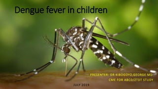
- Dengue is caused by one of four serotypes of dengue virus and is transmitted by Aedes mosquitoes.
- It is a major public health problem in many tropical and subtropical countries.
- Clinical presentation varies from mild fever to severe dengue with hemorrhage, plasma leakage, or organ involvement.
- Diagnosis involves IgM/IgG detection, NS1 antigen detection, PCR, or viral isolation from blood samples.
A mosquito-borne viral disease occurring in tropical and subtropical areas.
Spreads by animals or insects
Requires a medical diagnosis
Lab tests or imaging often required
Short-term: resolves within days to weeks
Those who become infected with the virus a second time are at a significantly greater risk of developing severe disease.
Symptoms include high fever, headache, rash and muscle and joint pain. In severe cases there is serious bleeding and shock, which can be life threatening.
Treatment includes fluids and pain relievers. Severe cases require hospital care.
The lecture gives concise review about the main four groups of viruses causing hemorrhagic fever i.e. Flavivirues, Filoviruses, Arenaviruses and Bunyaviruses.
A basic description of Leishmania spp. along with Old and New world Leishmaniasis regarding Parasite morphology, Life Cycle, Pathogenesis, Clinical manifestations, Laboratory Diagnosis and Treatment.
A basic description of Leishmania spp. along with Old and New world Leishmaniasis regarding Parasite morphology, Life Cycle, Pathogenesis, Clinical manifestations, Laboratory Diagnosis and Treatment.
Rainbow Hospital is the No. 1 super specialty hospital offering the best NICU care in Hyderabad, a state-of-the-art neonatal care, rated as best intensive units for children and neonatal intensive care unit.
Dengue fever is the fastest emerging arboviral infection spread
by Aedes mosquitoes with major public health consequences in
over 100 tropical and sub-tropical countries in South-East Asia,
the Western Pacific, and South and Central America. Up to 2.5
billion people globally live under the threat of dengue fever and its
severe forms—dengue hemorrhagic fever (DHF) or dengue shock
syndrome (DSS). More than 75% of these people, or approximately
1.8 billion, live in the Asia-Pacific Region. As the disease spreads to
new geographical areas, the frequency of the outbreaks is increasing
along with changing disease epidemiology. It is estimated that 50
a million cases of dengue fever occur worldwide annually and half a
million people suffering from DHF require hospitalization each year,
a very large proportion of whom (approximately 90%) are children
less than five years old. About 2.5% of those affected with dengue
die of the disease.
As an intern house officer, I prepared this presentation after I came across a rare case of dengue fever complicated by hemophagocytic lymphohistiocytosis (HLH). Dengue fever itself is a rare disease entity in the UAE, as a developed country; and the presence of such a complication merely added to the complexity of the diagnosis. Therefore, I am delighted to share this lively PowerPoint Presentation about dengue, which was initially supplemented with an interesting case presentation but was removed for confidentiality purposes when sharing the document. I hope you enjoy it!
PS: Use the slideshow button in Microsoft PowerPoint for the best experience.
Similar to Dengue fever in children 2019 by Dr Kibogoyo (20)
The prostate is an exocrine gland of the male mammalian reproductive system
It is a walnut-sized gland that forms part of the male reproductive system and is located in front of the rectum and just below the urinary bladder
Function is to store and secrete a clear, slightly alkaline fluid that constitutes 10-30% of the volume of the seminal fluid that along with the spermatozoa, constitutes semen
A healthy human prostate measures (4cm-vertical, by 3cm-horizontal, 2cm ant-post ).
It surrounds the urethra just below the urinary bladder. It has anterior, median, posterior and two lateral lobes
It’s work is regulated by androgens which are responsible for male sex characteristics
Generalised disease of the prostate due to hormonal derangement which leads to non malignant enlargement of the gland (increase in the number of epithelial cells and stromal tissue)to cause compression of the urethra leading to symptoms (LUTS
Anti ulcer drugs and their Advance pharmacology ||
Anti-ulcer drugs are medications used to prevent and treat ulcers in the stomach and upper part of the small intestine (duodenal ulcers). These ulcers are often caused by an imbalance between stomach acid and the mucosal lining, which protects the stomach lining.
||Scope: Overview of various classes of anti-ulcer drugs, their mechanisms of action, indications, side effects, and clinical considerations.
Report Back from SGO 2024: What’s the Latest in Cervical Cancer?bkling
Are you curious about what’s new in cervical cancer research or unsure what the findings mean? Join Dr. Emily Ko, a gynecologic oncologist at Penn Medicine, to learn about the latest updates from the Society of Gynecologic Oncology (SGO) 2024 Annual Meeting on Women’s Cancer. Dr. Ko will discuss what the research presented at the conference means for you and answer your questions about the new developments.
Factory Supply Best Quality Pmk Oil CAS 28578–16–7 PMK Powder in Stockrebeccabio
Factory Supply Best Quality Pmk Oil CAS 28578–16–7 PMK Powder in Stock
Telegram: bmksupplier
signal: +85264872720
threema: TUD4A6YC
You can contact me on Telegram or Threema
Communicate promptly and reply
Free of customs clearance, Double Clearance 100% pass delivery to USA, Canada, Spain, Germany, Netherland, Poland, Italy, Sweden, UK, Czech Republic, Australia, Mexico, Russia, Ukraine, Kazakhstan.Door to door service
Hot Selling Organic intermediates
Title: Sense of Smell
Presenter: Dr. Faiza, Assistant Professor of Physiology
Qualifications:
MBBS (Best Graduate, AIMC Lahore)
FCPS Physiology
ICMT, CHPE, DHPE (STMU)
MPH (GC University, Faisalabad)
MBA (Virtual University of Pakistan)
Learning Objectives:
Describe the primary categories of smells and the concept of odor blindness.
Explain the structure and location of the olfactory membrane and mucosa, including the types and roles of cells involved in olfaction.
Describe the pathway and mechanisms of olfactory signal transmission from the olfactory receptors to the brain.
Illustrate the biochemical cascade triggered by odorant binding to olfactory receptors, including the role of G-proteins and second messengers in generating an action potential.
Identify different types of olfactory disorders such as anosmia, hyposmia, hyperosmia, and dysosmia, including their potential causes.
Key Topics:
Olfactory Genes:
3% of the human genome accounts for olfactory genes.
400 genes for odorant receptors.
Olfactory Membrane:
Located in the superior part of the nasal cavity.
Medially: Folds downward along the superior septum.
Laterally: Folds over the superior turbinate and upper surface of the middle turbinate.
Total surface area: 5-10 square centimeters.
Olfactory Mucosa:
Olfactory Cells: Bipolar nerve cells derived from the CNS (100 million), with 4-25 olfactory cilia per cell.
Sustentacular Cells: Produce mucus and maintain ionic and molecular environment.
Basal Cells: Replace worn-out olfactory cells with an average lifespan of 1-2 months.
Bowman’s Gland: Secretes mucus.
Stimulation of Olfactory Cells:
Odorant dissolves in mucus and attaches to receptors on olfactory cilia.
Involves a cascade effect through G-proteins and second messengers, leading to depolarization and action potential generation in the olfactory nerve.
Quality of a Good Odorant:
Small (3-20 Carbon atoms), volatile, water-soluble, and lipid-soluble.
Facilitated by odorant-binding proteins in mucus.
Membrane Potential and Action Potential:
Resting membrane potential: -55mV.
Action potential frequency in the olfactory nerve increases with odorant strength.
Adaptation Towards the Sense of Smell:
Rapid adaptation within the first second, with further slow adaptation.
Psychological adaptation greater than receptor adaptation, involving feedback inhibition from the central nervous system.
Primary Sensations of Smell:
Camphoraceous, Musky, Floral, Pepperminty, Ethereal, Pungent, Putrid.
Odor Detection Threshold:
Examples: Hydrogen sulfide (0.0005 ppm), Methyl-mercaptan (0.002 ppm).
Some toxic substances are odorless at lethal concentrations.
Characteristics of Smell:
Odor blindness for single substances due to lack of appropriate receptor protein.
Behavioral and emotional influences of smell.
Transmission of Olfactory Signals:
From olfactory cells to glomeruli in the olfactory bulb, involving lateral inhibition.
Primitive, less old, and new olfactory systems with different path
Couples presenting to the infertility clinic- Do they really have infertility...Sujoy Dasgupta
Dr Sujoy Dasgupta presented the study on "Couples presenting to the infertility clinic- Do they really have infertility? – The unexplored stories of non-consummation" in the 13th Congress of the Asia Pacific Initiative on Reproduction (ASPIRE 2024) at Manila on 24 May, 2024.
micro teaching on communication m.sc nursing.pdfAnurag Sharma
Microteaching is a unique model of practice teaching. It is a viable instrument for the. desired change in the teaching behavior or the behavior potential which, in specified types of real. classroom situations, tends to facilitate the achievement of specified types of objectives.
Lung Cancer: Artificial Intelligence, Synergetics, Complex System Analysis, S...Oleg Kshivets
RESULTS: Overall life span (LS) was 2252.1±1742.5 days and cumulative 5-year survival (5YS) reached 73.2%, 10 years – 64.8%, 20 years – 42.5%. 513 LCP lived more than 5 years (LS=3124.6±1525.6 days), 148 LCP – more than 10 years (LS=5054.4±1504.1 days).199 LCP died because of LC (LS=562.7±374.5 days). 5YS of LCP after bi/lobectomies was significantly superior in comparison with LCP after pneumonectomies (78.1% vs.63.7%, P=0.00001 by log-rank test). AT significantly improved 5YS (66.3% vs. 34.8%) (P=0.00000 by log-rank test) only for LCP with N1-2. Cox modeling displayed that 5YS of LCP significantly depended on: phase transition (PT) early-invasive LC in terms of synergetics, PT N0—N12, cell ratio factors (ratio between cancer cells- CC and blood cells subpopulations), G1-3, histology, glucose, AT, blood cell circuit, prothrombin index, heparin tolerance, recalcification time (P=0.000-0.038). Neural networks, genetic algorithm selection and bootstrap simulation revealed relationships between 5YS and PT early-invasive LC (rank=1), PT N0—N12 (rank=2), thrombocytes/CC (3), erythrocytes/CC (4), eosinophils/CC (5), healthy cells/CC (6), lymphocytes/CC (7), segmented neutrophils/CC (8), stick neutrophils/CC (9), monocytes/CC (10); leucocytes/CC (11). Correct prediction of 5YS was 100% by neural networks computing (area under ROC curve=1.0; error=0.0).
CONCLUSIONS: 5YS of LCP after radical procedures significantly depended on: 1) PT early-invasive cancer; 2) PT N0--N12; 3) cell ratio factors; 4) blood cell circuit; 5) biochemical factors; 6) hemostasis system; 7) AT; 8) LC characteristics; 9) LC cell dynamics; 10) surgery type: lobectomy/pneumonectomy; 11) anthropometric data. Optimal diagnosis and treatment strategies for LC are: 1) screening and early detection of LC; 2) availability of experienced thoracic surgeons because of complexity of radical procedures; 3) aggressive en block surgery and adequate lymph node dissection for completeness; 4) precise prediction; 5) adjuvant chemoimmunoradiotherapy for LCP with unfavorable prognosis.
Pulmonary Thromboembolism - etilogy, types, medical- Surgical and nursing man...VarunMahajani
Disruption of blood supply to lung alveoli due to blockage of one or more pulmonary blood vessels is called as Pulmonary thromboembolism. In this presentation we will discuss its causes, types and its management in depth.
3. Introduction
Dengue is the most common and important arthropod-borne viral (arboviral) illness in humans
It is caused by infection with 1 of the 4 serotypes of dengue virus, which is a Flavivirus (a genus of single-
stranded nonsegmented RNA viruses). DEN 1, DEN 2, DEN 3 and DEN 4
Infection with one dengue serotype confers lifelong homotypic immunity to that serotype and a brief period
(approximately 2 years) of partial heterotypic immunity to other serotypes, but an individual can eventually be
infected by all 4 serotypes.
Dengue virus is transmitted by female mosquitoes mainly of the species Aedes aegypti and to a lesser extent,
Aedes albopictus.
Dengue is widespread throughout the tropics, with local variations in risk influenced by rainfall, temperature
and unplanned rapid urbanization.
Today, severe dengue affects most Asian and Latin American countries and has become a leading cause of
hospitalization and death among children and adults in these regions.
4. Epidermiology
Before 1970, only 9 countries had experienced severe dengue epidemics. The disease is now
endemic in more than 100 countries in the WHO regions of Africa, the Americas, the Eastern
Mediterranean, South-East Asia and the Western Pacific.
Severe dengue is a leading cause of serious illness and death among children in some Asian and
Latin American countries
The prevalence of dengue estimates that 3.9 billion people, in 128 countries, are at risk of
infection with dengue viruses.
Each year, an estimated 390 million dengue infections occur around the world. Of these,
500,000 cases develop into dengue haemorrhagic fever, a more severe form of the disease,
which results in up to 25,000 deaths annually worldwide. (WHO)
The World Health Organization (WHO) ranked dengue as one of the top ten threats to global
health in 2019
5. Epidermiology in Tanzania recent Outbreak
Tanzania continue to report dengue fever cases to WHO. It was notified to WHO on 31st jan 2019
As of week 25 (week ending on 23 June 2019), 229 new dengue cases were reported, from Dar es
Salaam (203 cases), Tanga (23) and Morogoro (3 cases). The total confirmed cases reported since 01st
Aug 2018 (the beginning of the outbreak) was 4456 cases including four deaths. (Source: WHO
Outbreak and Emergencies Bulletin - Week 25: 17-23 June 2019)
According to the Ministry of Health about 1,222 people have been diagnosed with dengue fever in Dar
es Salaam, Dodoma and Singida Regions, a steep rise from the 304 cases reported in April. Two people
have since died. (May 16th 2019)
Deputy Minister of Health reported that Dar es Salaam leads with 1,145 patients, Tanga follows with
75 patients and Singida so far has registered one patient with the fever.
This year is the worst dengue outbreak compared with 2014 when more than 400 patients in Dar es
Salaam were diagnosed with the disease.
8. Transmission
The Aedes aegypti mosquito is the primary vector of dengue. Human is a primary host.
The virus is transmitted to humans through the bites of infected female mosquitoes. After virus incubation
for 4–10 days, an infected mosquito is capable of transmitting the virus for the rest of its life.
Infected symptomatic or asymptomatic humans are the main carriers and multipliers of the virus, serving as
a source of the virus for uninfected mosquitoes. Patients who are already infected with the dengue virus can
transmit the infection (for 4–5 days; maximum 12) via Aedes mosquitoes after their first symptoms appear.
The Aedes aegypti mosquito lives in urban habitats and breeds mostly in man-made containers. Unlike other
mosquitoes Ae. aegypti is a day-time feeder; its peak biting periods are early in the morning and in the
evening before dusk. Female Ae. aegypti bites multiple people during each feeding period. Aedes eggs can
remain dry for over a year in their breeding habitat and hatch when in contact with water.
Aedes albopictus, a secondary dengue vector in Asia, has spread to North America and more than 25
countries in the European Region, largely due to the international trade in used tyres (a breeding habitat).
Ae. albopictus is highly adaptive and, therefore, can survive in cooler temperate regions of Europe. Its spread
is due to its tolerance to temperatures below freezing, hibernation, and ability to shelter in microhabitats.
16. Tourniquet Test for Dengue
The tourniquet test is part of the new WHO case definition for dengue. The test is a marker of capillary fragility and it can be used as a
triage tool to differentiate patients with acute gastroenteritis, for example, from those with dengue.
Even if a tourniquet test was previously done, it should be repeated if
It was previously negative
There is no bleeding manifestation
How to do a Tourniquet Test
Take the patient's blood pressure and record it, for example, 100/70.
Inflate the cuff to a point midway between SBP and DBP and maintain for 5 minutes.
(100 + 70) ÷ 2 = 85 mm Hg
Reduce and wait 2 minutes.
Count petechiae below antecubital fossa.
A positive test is 10 or more petechiae per 1 square inch.
18. Febrile Phase
Findings on physical examination during the febrile phase can include
Signs of dehydration, such as dry, cracked lips
Erythema on face
Hemorrhage manifestations
Transient macular or maculopapular rash that spreads to the face and
extremities
19. Critical phase
Most dengue patients will improve during the critical phase and they will not develop manifestations of severe disease.
Patients who do not improve but report warning signs or worsening of their illness during defervescence need to be evaluated.
Around time of defervescence, petechiae can appear, especially on lower extremities.
Warning signs might develop, such as
•Severe abdominal pain
•Persistent vomiting
•Ascites, pleural effusion
•Mucosal bleed
•Lethargy, restlessness
•Liver enlargement > 2 cm
•Drop in PLT with increase in HCT
20. Recovery phase
After defervescence, a confluent erythematous rash with round islands of normal skin might
appear. The rash can be very pruritic and desquamate.
Some patients also have petechiae. This rash is known as the convalescent rash of dengue,
commonly described as "islands of white in a sea of red." Patients can present complaining of
being unable to sleep because their skin itches.
23. Clinical and laboratory features that distinguish dengue from other febrile illnesses in endemic populations.
Published Sept 2008
Author: Potts JA1, Rothman AL.
OBJECTIVE:
Clinicians in resource-poor countries need to identify patients with dengue using readily-available data. The objective of this systematic
review was to identify clinical and laboratory features that differentiate dengue fever (DF) and/or dengue haemorrhagic fever (DHF)
from other febrile illnesses (OFI) in dengue-endemic populations.
METHOD: Systematic review of the literature from 1990 to 30 October 2007 including English publications comparing dengue and OFI.
RESULTS:
Among 49 studies reviewed, 34 did not meet our criteria for inclusion. Of the 15 studies included, 10 were prospective cohort studies
and five were case-control studies. Seven studies assessed all ages, four assessed children only, and four assessed adults only. Patients
with dengue had significantly lower platelet, white blood cell (WBC) and neutrophil counts, and a higher frequency of petechiae than
OFI patients. Higher frequencies of myalgia, rash, haemorrhagic signs, lethargy/prostration, and arthralgia/joint pain and higher
haematocrits were reported in adult patients with dengue but not in children. Most multivariable models included platelet count,
WBC, rash, and signs of liver damage; however, none had high statistical validity and none considered changes in clinical features over
the course of illness.
CONCLUSIONS:
Several individual clinical and laboratory variables distinguish dengue from OFI; however, some variables may be dependent on age.
Study design, populations, diagnostic criteria, and data collection methods differed widely across studies, and the majority of studies did
not identify specific aetiologies of OFIs. More prospective studies are needed to construct a valid and generalizable algorithm to guide
the differential diagnosis of dengue in endemic countries.
24. Lab Findings
Clinical laboratory findings in the febrile phase include
Mild-to-moderate thrombocytopenia
Normal or slightly increased HCT
Elevated AST and ALT
25. Lab Findings
The critical phase usually can be identified by
Onset of defervescence
A rapid decline in platelet count but with a rise in HCT
Leukopenia for about 24 hours, before platelet drop
Warning signs
26. Laboratory-confirmedDengueinChildreninThreeRegionalHospitalsinthePhilippinesin2009−2010
Capeding, MariaRosario Z.MD*;L’Azou,Maïna MSc†;Manalaysay,MichaelMD‡;Vince-Woo,CristinaR.MD§;Rivera,Religaya
G.MD¶;KristySy,AvaMSc*;Mercado,EdelwisaSegubre MSc*;Inobaya,Marianette T.MSc*;Tayag,Enrique G.MD‖
ThePediatric InfectiousDiseaseJournal:November2015-Volume34 - Issue11 -p 1145–1151
Background: The burden of dengue is high in the Philippines but the prevalence of confirmed cases is unknown, and the
disease is subject to underreporting because surveillance of suspected cases is passive. We conducted a prospective
epidemiological study to estimate the proportion of laboratory-confirmed dengue among clinically suspected hospitalized
cases in the pediatric wards of 3 regional hospitals in the Philippines and to describe the clinical and laboratory features, age
distributions, case fatality rates and serotype distributions of these hospitalized cases.
José B. Lingad Memorial Regional Hospital (JBL); Davao Medical Center (DMC; now the Southern Philippines Medical Center)
and Western Visayas Medical Center (WVMC)
Methods: Patients ≤18 years and hospitalized for suspected dengue were included if they had an axillary temperature ≥38°C
for 2−7 days and 2 or more dengue-associated symptoms. Dengue infection was confirmed in acute blood samples by
serotype-specific reverse transcription-polymerase chain reaction and IgM immunoassay.
Results: We confirmed dengue infection in 1809 (86.1%) cases of 2103 suspected cases between November 2009 and
November 2010. The 6- to 10-year-old age group had the highest proportion of cases overall (36.7%).
Fever, anorexia, myalgia, abdominal pain and headache were the most common symptoms at admission. Hemorrhagic
manifestations, signs of plasma leakage, thrombocytopenia and leucopenia were all significantly more common in
confirmed than in nonconfirmed cases.
Most cases (76.5%) developed dengue hemorrhagic fever or dengue shock syndrome, and the overall case fatality rate was
0.94%. Distributions of all 4 virus serotypes varied at each hospital.
Conclusions: The clinical burden of pediatric dengue continues to be substantial in the Philippines. Most hospitalized cases of
suspected pediatric dengue can be laboratory confirmed and most develop severe disease.
27. Diagnosis
For Dengue Fever
Diagnosis Suspect dengue fever in an area of risk for dengue if the child has fever lasting > 2
days.
Headache
pain behind the eyes
joint and muscle pain
abdominal pain
vomiting and/or a rash may occur but are not always present.
It can be difficult to distinguish dengue from other common childhood infections
28. Diagnosis
For Severe Dengue Fever
Suspect severe dengue in an area of risk for dengue if the child has fever lasting > 2 days, and any of the following features:
evidence of plasma leakage
high or progressively rising EVF (erythrocyte volume fraction /haematocrit)
pleural effusions or ascites
circulatory compromise or shock – cold, clammy extremities
prolonged capillary refill time (> 3 s)
weak pulse (fast pulse may be absent even with significant volume depletion)
narrow pulse pressure (see above)
spontaneous bleeding
from the nose or gums
black stools or coffee-ground vomit
skin bruising or extensive petaechiae
29. Severe dengue fever….
altered level of consciousness
lethargy or restlessness
coma
convulsions
severe gastrointestinal involvement
persistent vomiting
increasing abdominal pain with tenderness in the right upper quadrant
jaundice
30. A Screening Tool for Dengue Fever in Children
Wen-Pin Lai, MD; Tsair-Wei Chien, MBA; Hung-Jung Lin, MD, MBA; Shih-Bin Su, MD, PhD; Chih-Hung Chang, PhD
PUBLISHED 2013
Main Findings
WBC and platelet counts were the 2 items in the DF-14 scale that had the most power to predict DF in
children.
The DF-14, DF-11 and DF-2 scales, despite their having a different number of items, were found to be measuring
the same "tendency for DF" construct.
The DF-2 scale is considered acceptable for its simplicity in practice, even though it is less accurate than the DF-
11 and DF-14 scales.
This suggests that using signs or symptoms only, to distinguish DF from non-DF is insufficient. Considering and
evaluating the symptom characteristics and the laboratory characteristics together (DF-11 and DF-14) are more
effective for the early detection of DF.
This is consistent with other study findings that used different variables together to distinguish DF from non-DF.
Similar to other studies, we found that thrombocytopenia and leukopenia were highly associated with DF.
WBC and platelet counts are easily obtainable in primary care settings; thus, a combination of laboratory
variables (low platelet or WBC counts) for early prediction of DENV infection is feasible.
32. OTHER INVESTIGATIONS
FBP
MRDT (if negative and dengue symptoms persist esp fever Do BS there is high prevalence of co
mobidity in our set up hence having dengue fever doesn’t rule out Malaria)
PT PTT
RFT
LFT
RBG
SERUM ELECTROLYTES
CXR
USS
33. Management
Dengue Fever
Most children can be managed at home, provided the parents have good access to a hospital.
Counsel the parents to bring the child back for daily follow-up and to return immediately if any of the
following occur: severe abdominal pain, persistent vomiting, cold, clammy extremities, lethargy or
restlessness, bleeding, e.g. black stools or coffee-ground vomit.
Encourage oral fluid intake with clean water or ORS solution to replace losses during fever and vomiting.
Give paracetamol for high fever if the child is uncomfortable. Do not give aspirin or NSAIDs such as
ibuprofen, as these drugs may aggravate bleeding.
Follow-up the child daily until the temperature is normal. Check the EVF daily if possible. Check for signs
of severe disease.
Admit any child with signs of severe disease (mucosal or severe skin bleeding, shock, altered mental
status, convulsions or jaundice) or with a rapid or marked rise in EVF(erythrocyte volume fraction or
haematocrit)
34. Severe Dengue Fever
Admit all patients with severe dengue to a hospital with facilities for urgent IV fluid treatment and
blood pressure and EVF monitoring.
Fluid management: patients without shock (pulse pressure > 20 mm Hg)
Give IV fluids for repeated vomiting or a high or rapidly rising EVF.
Give only isotonic solutions such as normal saline and Ringer’s lactate (Hartmann’s solution) or 5%
glucose in Ringer’s lactate.
Start with 6 ml/kg per h for 2 h, and then reduce to 2–3 ml/kg per h as soon as possible, depending
on the clinical response.
Give the minimum volume required to maintain good perfusion and urine output. IV fluids are usually
needed only for 24–48 h, as the capillary leak resolves spontaneously after that time.
35. Fluid management: patients in shock (pulse pressure ≤ 20 mm Hg)
Treat as an emergency.
Give 10–20 ml/kg of an isotonic crystalloid solution such as Ringer’s lactate (Hartmann’s solution) or normal saline over 1 h.
If the child responds (capillary refill and peripheral perfusion start to improve, pulse pressure widens), reduce to 10 ml/kg
for 1 h and then gradually to 2–3 ml/kg per h over the next 6–8 h.
If the child does not respond (continuing signs of shock), give a further 20 ml/kg of the crystalloid over 1 h, or consider
giving 10 ml/kg of a colloid solution such as 6% dextran 70 over 1 h.
Further small boluses of extra fluid (5–10 ml/kg over 1 h) may be required during the next 24–48 h.
Decide on fluid treatment on the basis of clinical response, i.e. review vital signs hourly, EVF and monitor urine output
closely.
Changes in the EVF can be a useful guide to treatment but must be interpreted with the clinical response. In these
circumstances, continue to monitor frequently. The EVF is likely to start falling within the next 24 h as the reabsorptive
phase of the disease begins.
In most cases, IV fluids can be stopped after 36–48 h. Remember that too much fluid can result into death due to fluid
overload.
36. Supportive care
Treat high fever with paracetamol if the child is uncomfortable.
Do not give aspirin or NSAIDs such as ibuprofen, as they aggravate the bleeding.
Do not give steroids.
Children in shock or with respiratory distress should receive oxygen, if possible with nasal
continuous positive airway pressure .
Hypoglycaemia (blood glucose < 2.5 mmol/litre or < 45 mg/dl) is unusual. If present, give IV
glucose
37. MONITORING
For children in shock, monitor the vital signs hourly (particularly the pulse pressure, if possible)
until the patient is stable, and check the EVF three or four times a day.
Review the patient at least four times a day and prescribe IV fluids for a maximum of 6 h at a
time.
For children without shock, a nurse should check the child’s vital signs (temperature, pulse and
blood pressure) at least four times a day and the EVF once daily, and a doctor should review the
patient at least once daily.
Check the platelet count daily, when possible in the acute phase.
Keep a detailed record of all fluid intake and output
38.
39. Turning papaya leaf into a cure for dengue fever
A traditional herbal remedy for the dangerous tropical disease ‘dengue fever’
could be turned into a pill to treat patients thanks to groundbreaking research by
scientists at the University of Nottingham Malaysia.
Papaya leaf juice has for a long time been used in some areas of India and South
East Asia as a treatment for dengue fever. A compound in the juice is known to
help with blood clotting and can restrict the internal bleeding caused by the
disease.
Now, thanks to funding from the Malaysian Ministry of Higher Education, a team
of chemical engineers in the University’s Food and Pharmaceutical Engineering
Group in Malaysia are tackling the challenge of extracting the bioactive
compound ‘carpaine’ for use in a pill for dengue
40. Video below showing how papaya leaf can be a cure to dengue fever
click link below
https://youtu.be/5yKFMJnWKrk
41. WHEN TO DISCHARGE
Patients can be discharged from the hospital when all of the following occur:
No fever for 24–48 hours
Improvement in clinical status
• General well being
• Appetite
• Hemodynamic status
• Urine output
• No respiratory distress
Increasing trend of platelet count (usually preceded by rising WBC)
Stable HCT with adequate oral intake while off IVFs
44. Prognosis
Prognosis depends on early diagnosis, recognition of warning signs and early signs of shock, and
timely initiation of correct management.
Patients with severe dengue can deteriorate quickly during defervescence, with rapid onset of shock
due to increase in vascular permeability during the critical phase.
Prolonged shock is associated with poor outcomes, including hemorrhage
Dengue fever is typically a self-limiting disease with a mortality rate of less than 1%.
When treated, dengue hemorrhagic fever has a mortality rate of 2-5%. When left untreated, dengue
hemorrhagic fever has a mortality rate as high as 50%.
Survivors usually recover without sequelae and develop immunity to the infecting serotype
45. PREVENTATION AND CONTROL
At present, the main method to control or prevent the transmission of dengue virus is to combat vector mosquitoes through:
preventing mosquitoes from accessing egg-laying habitats by environmental management and modification;
disposing of solid waste properly and removing artificial man-made habitats;
covering, emptying and cleaning of domestic water storage containers on a weekly basis;
applying appropriate insecticides to water storage outdoor containers;
using of personal household protection measures, such as window screens, long-sleeved clothes, repellents, insecticide treated
materials, coils and vaporizers (These measures have to be observed during the day both at home and place of work since the
mosquito bites during the day);
improving community participation and mobilization for sustained vector control;
applying insecticides as space spraying during outbreaks as one of the emergency vector-control measures;
active monitoring and surveillance of vectors should be carried out to determine effectiveness of control interventions.
46. Preventation
The World Mosquito Program’s Wolbachia method
Wolbachia are natural bacteria present in up to 60% of insect species, including some mosquitoes. However,
Wolbachia is not usually found in the Aedes aegypti mosquito.
Wolbachia can help to reduce the transmission of these viruses to people. This important discovery has the
potential to transform the fight against life-threatening mosquito-borne diseases
The WMP's field teams release male and female Aedes aegypti mosquitoes with Wolbachia over a number of
weeks. These mosquitoes then breed with the wild mosquito population. Over time, the percentage of
mosquitoes carrying Wolbachia grows until it remains high without the need for further releases
47. Video below showing Wolbachia method of preventing mosquito
from spreading dengue virus
https://youtu.be/ut2UxF5gEDI
48. IMMUNIZATION
The first dengue vaccine, Dengvaxia® (CYD-TDV) developed by Sanofi Pasteur was licensed in
December 2015 and has now been approved by regulatory authorities in 20 countries for use in
endemic areas in persons ranging from 9-45 years of age.
In April 2016, WHO issued a conditional recommendation on the use of the vaccine for areas in
which dengue is highly endemic as defined by seroprevalence of 70% or higher.
In November 2017, the results of an additional analysis to retrospectively determine serostatus
at the time of vaccination were released. The analysis showed that the subset of trial
participants who were inferred to be seronegative at time of first vaccination had a higher risk of
more severe dengue and hospitalizations from dengue compared to unvaccinated participants
49. References
1. World Health Organization. Global Strategy for Dengue Prevention and
Control. 2012 Geneva WHO Press
2. . World Health Organization. Dengue and severe dengue. 2019
Available at: https://www.who.int/news-room/fact-sheets/detail/dengue-
and-severe-dengue. Accessed April 15, 2019
3. https://www.afro.who.int/publications/outbreaks-and-emergencies-
bulletin-week-26-24-30-june-2019.
4. Brady OJ, Gething PW, Bhatt S, Messina JP, Brownstein JS, Hoen AG
et al. Refining the global spatial limits of dengue virus transmission by
evidence-based consensus.
5. https://www.cdc.gov/dengue/training/cme/ccm/page53590.html
6. https://emedicine.medscape.com/article/215840
50. Thank for your Attention
This presentation is dedicated to my
daughter Joycefelicia
DENGUE DAY 2019 (16th May).
THEME: “End Dengue: Starts With Me.
51. CASE SCENARIO
A 3 years male child came to round table diarrhea clinic with complaint of severe vomiting,
diarrhea, fever, history of passing black stool and running nose for 2 days. On examination
finding were positive tourniquet test, no signs of shock ,jaundiced and hepatomegaly. On lab
findings mrdt was negative and low WBC count.
1. what are your differential diagnosis
2. What other investigation would you order
3. How will you manage the child
52. Lab results for dengue case showing
marked decrease in WBC and platelets