This document provides guidance on evaluating and categorizing thrombocytopenia in hospitalized patients. It defines thrombocytopenia as a platelet count below 150,000/mL and discusses the life cycle of platelets. Thrombocytopenia can be categorized as pseudothrombocytopenia, decreased production, increased destruction, consumption, or sequestration. A detailed history and physical exam are important for determining the cause. Initial testing should include a CBC, peripheral smear, and tests for HIV and hepatitis C. Common causes of drug-induced thrombocytopenia include antibiotics and heparin.
When your blood has too few platelets, mild
to serious bleeding can occur. Bleeding can occur inside your body (internal
bleeding) or underneath your skin or from the surface of your skin (external
bleeding).
A normal platelet count in adults ranges
from 150,000 to 450,000 platelets per microliter of blood. A platelet count of
less than 150,000 platelets per microliter is lower than normal. If your blood
platelet count falls below normal, you have thrombocytopenia.
However, the risk for serious bleeding
doesn't occur until the count becomes very low—less than 10,000 or 20,000
platelets per microliter. Mild bleeding sometimes occurs when the count is less
than 50,000 platelets per microliter.
Many factors can cause a low platelet
count, such as:
-- The body's bone marrow doesn't make enough
platelets.
-- The bone marrow makes enough platelets, but
the body destroys them or uses them up.
-- The spleen holds on to too many platelets.
The spleen is an organ that normally stores about one-third of the body's
platelets. It also helps your body fight infection and remove unwanted cell
material.
-- A combination of the above factors.
-- How long thrombocytopenia lasts depends on
its cause. It can last from days to years.
The treatment for this condition also
depends on its cause and severity. Mild thrombocytopenia often doesn't require
treatment. If the condition causes or puts you at risk for serious bleeding,
you may need medicines or blood or
platelet transfusions. Rarely, the spleen may need to be removed.
Thrombocytopenia is frequently encountered in the ICU. It is important to have an understanding of the common and important causes of TP as well as to have a simple framework to approach this problem. A simple approach is to identify if TP is expected or unexpected and work through the management that way. Platelet transfusions or repeating the FBC is not always the right approach. Heparin induced thrombocytopenia is frequently considered as the cause of TP. This leads to excessive HITS screen being ordered and risks false positive results.
Thrombotic Microangiopathies are diverse group of disorders wherein thrombocytopenia, hemolytic anemia and organ dysfunction such as Kidney and brain occur . Major recent advances in this field have occurred which opens up oppurtunities to effectively manage its clinical challenges .
Thrombocytopenia is most frequently encountered Hematological problem in hospitalized patients. The most common causes and differential diagnosis of In-patient and Outpatient presentations of Thrombocytopenia is discussed here. Useful for Internal Medicine Boards . Archer Internal Medicine Board review lectures will be released soon.
Hemostasis, Coagulation, Intrinsic, Extrinsic & common Pathways of Clotting, Common bleeding disorders & their investigations, BT, CT, PT, APTT, TT, Blood & its products, Blood transfusion & its complication.
Hemostasis
Seminar Prepared by :-
Mohammed Saadi
Mohammed Musa
Hussein Jassam
Mahmoud Ahmed
Internal Medicine
College of Medicine - University of Kirkuk
Pulmonary Thromboembolism - etilogy, types, medical- Surgical and nursing man...VarunMahajani
Disruption of blood supply to lung alveoli due to blockage of one or more pulmonary blood vessels is called as Pulmonary thromboembolism. In this presentation we will discuss its causes, types and its management in depth.
Lung Cancer: Artificial Intelligence, Synergetics, Complex System Analysis, S...Oleg Kshivets
RESULTS: Overall life span (LS) was 2252.1±1742.5 days and cumulative 5-year survival (5YS) reached 73.2%, 10 years – 64.8%, 20 years – 42.5%. 513 LCP lived more than 5 years (LS=3124.6±1525.6 days), 148 LCP – more than 10 years (LS=5054.4±1504.1 days).199 LCP died because of LC (LS=562.7±374.5 days). 5YS of LCP after bi/lobectomies was significantly superior in comparison with LCP after pneumonectomies (78.1% vs.63.7%, P=0.00001 by log-rank test). AT significantly improved 5YS (66.3% vs. 34.8%) (P=0.00000 by log-rank test) only for LCP with N1-2. Cox modeling displayed that 5YS of LCP significantly depended on: phase transition (PT) early-invasive LC in terms of synergetics, PT N0—N12, cell ratio factors (ratio between cancer cells- CC and blood cells subpopulations), G1-3, histology, glucose, AT, blood cell circuit, prothrombin index, heparin tolerance, recalcification time (P=0.000-0.038). Neural networks, genetic algorithm selection and bootstrap simulation revealed relationships between 5YS and PT early-invasive LC (rank=1), PT N0—N12 (rank=2), thrombocytes/CC (3), erythrocytes/CC (4), eosinophils/CC (5), healthy cells/CC (6), lymphocytes/CC (7), segmented neutrophils/CC (8), stick neutrophils/CC (9), monocytes/CC (10); leucocytes/CC (11). Correct prediction of 5YS was 100% by neural networks computing (area under ROC curve=1.0; error=0.0).
CONCLUSIONS: 5YS of LCP after radical procedures significantly depended on: 1) PT early-invasive cancer; 2) PT N0--N12; 3) cell ratio factors; 4) blood cell circuit; 5) biochemical factors; 6) hemostasis system; 7) AT; 8) LC characteristics; 9) LC cell dynamics; 10) surgery type: lobectomy/pneumonectomy; 11) anthropometric data. Optimal diagnosis and treatment strategies for LC are: 1) screening and early detection of LC; 2) availability of experienced thoracic surgeons because of complexity of radical procedures; 3) aggressive en block surgery and adequate lymph node dissection for completeness; 4) precise prediction; 5) adjuvant chemoimmunoradiotherapy for LCP with unfavorable prognosis.
ARTIFICIAL INTELLIGENCE IN HEALTHCARE.pdfAnujkumaranit
Artificial intelligence (AI) refers to the simulation of human intelligence processes by machines, especially computer systems. It encompasses tasks such as learning, reasoning, problem-solving, perception, and language understanding. AI technologies are revolutionizing various fields, from healthcare to finance, by enabling machines to perform tasks that typically require human intelligence.
Anti ulcer drugs and their Advance pharmacology ||
Anti-ulcer drugs are medications used to prevent and treat ulcers in the stomach and upper part of the small intestine (duodenal ulcers). These ulcers are often caused by an imbalance between stomach acid and the mucosal lining, which protects the stomach lining.
||Scope: Overview of various classes of anti-ulcer drugs, their mechanisms of action, indications, side effects, and clinical considerations.
MANAGEMENT OF ATRIOVENTRICULAR CONDUCTION BLOCK.pdfJim Jacob Roy
Cardiac conduction defects can occur due to various causes.
Atrioventricular conduction blocks ( AV blocks ) are classified into 3 types.
This document describes the acute management of AV block.
Flu Vaccine Alert in Bangalore Karnatakaaddon Scans
As flu season approaches, health officials in Bangalore, Karnataka, are urging residents to get their flu vaccinations. The seasonal flu, while common, can lead to severe health complications, particularly for vulnerable populations such as young children, the elderly, and those with underlying health conditions.
Dr. Vidisha Kumari, a leading epidemiologist in Bangalore, emphasizes the importance of getting vaccinated. "The flu vaccine is our best defense against the influenza virus. It not only protects individuals but also helps prevent the spread of the virus in our communities," he says.
This year, the flu season is expected to coincide with a potential increase in other respiratory illnesses. The Karnataka Health Department has launched an awareness campaign highlighting the significance of flu vaccinations. They have set up multiple vaccination centers across Bangalore, making it convenient for residents to receive their shots.
To encourage widespread vaccination, the government is also collaborating with local schools, workplaces, and community centers to facilitate vaccination drives. Special attention is being given to ensuring that the vaccine is accessible to all, including marginalized communities who may have limited access to healthcare.
Residents are reminded that the flu vaccine is safe and effective. Common side effects are mild and may include soreness at the injection site, mild fever, or muscle aches. These side effects are generally short-lived and far less severe than the flu itself.
Healthcare providers are also stressing the importance of continuing COVID-19 precautions. Wearing masks, practicing good hand hygiene, and maintaining social distancing are still crucial, especially in crowded places.
Protect yourself and your loved ones by getting vaccinated. Together, we can help keep Bangalore healthy and safe this flu season. For more information on vaccination centers and schedules, residents can visit the Karnataka Health Department’s official website or follow their social media pages.
Stay informed, stay safe, and get your flu shot today!
HOT NEW PRODUCT! BIG SALES FAST SHIPPING NOW FROM CHINA!! EU KU DB BK substit...GL Anaacs
Contact us if you are interested:
Email / Skype : kefaya1771@gmail.com
Threema: PXHY5PDH
New BATCH Ku !!! MUCH IN DEMAND FAST SALE EVERY BATCH HAPPY GOOD EFFECT BIG BATCH !
Contact me on Threema or skype to start big business!!
Hot-sale products:
NEW HOT EUTYLONE WHITE CRYSTAL!!
5cl-adba precursor (semi finished )
5cl-adba raw materials
ADBB precursor (semi finished )
ADBB raw materials
APVP powder
5fadb/4f-adb
Jwh018 / Jwh210
Eutylone crystal
Protonitazene (hydrochloride) CAS: 119276-01-6
Flubrotizolam CAS: 57801-95-3
Metonitazene CAS: 14680-51-4
Payment terms: Western Union,MoneyGram,Bitcoin or USDT.
Deliver Time: Usually 7-15days
Shipping method: FedEx, TNT, DHL,UPS etc.Our deliveries are 100% safe, fast, reliable and discreet.
Samples will be sent for your evaluation!If you are interested in, please contact me, let's talk details.
We specializes in exporting high quality Research chemical, medical intermediate, Pharmaceutical chemicals and so on. Products are exported to USA, Canada, France, Korea, Japan,Russia, Southeast Asia and other countries.
These lecture slides, by Dr Sidra Arshad, offer a quick overview of physiological basis of a normal electrocardiogram.
Learning objectives:
1. Define an electrocardiogram (ECG) and electrocardiography
2. Describe how dipoles generated by the heart produce the waveforms of the ECG
3. Describe the components of a normal electrocardiogram of a typical bipolar leads (limb II)
4. Differentiate between intervals and segments
5. Enlist some common indications for obtaining an ECG
Study Resources:
1. Chapter 11, Guyton and Hall Textbook of Medical Physiology, 14th edition
2. Chapter 9, Human Physiology - From Cells to Systems, Lauralee Sherwood, 9th edition
3. Chapter 29, Ganong’s Review of Medical Physiology, 26th edition
4. Electrocardiogram, StatPearls - https://www.ncbi.nlm.nih.gov/books/NBK549803/
5. ECG in Medical Practice by ABM Abdullah, 4th edition
6. ECG Basics, http://www.nataliescasebook.com/tag/e-c-g-basics
micro teaching on communication m.sc nursing.pdfAnurag Sharma
Microteaching is a unique model of practice teaching. It is a viable instrument for the. desired change in the teaching behavior or the behavior potential which, in specified types of real. classroom situations, tends to facilitate the achievement of specified types of objectives.
Title: Sense of Smell
Presenter: Dr. Faiza, Assistant Professor of Physiology
Qualifications:
MBBS (Best Graduate, AIMC Lahore)
FCPS Physiology
ICMT, CHPE, DHPE (STMU)
MPH (GC University, Faisalabad)
MBA (Virtual University of Pakistan)
Learning Objectives:
Describe the primary categories of smells and the concept of odor blindness.
Explain the structure and location of the olfactory membrane and mucosa, including the types and roles of cells involved in olfaction.
Describe the pathway and mechanisms of olfactory signal transmission from the olfactory receptors to the brain.
Illustrate the biochemical cascade triggered by odorant binding to olfactory receptors, including the role of G-proteins and second messengers in generating an action potential.
Identify different types of olfactory disorders such as anosmia, hyposmia, hyperosmia, and dysosmia, including their potential causes.
Key Topics:
Olfactory Genes:
3% of the human genome accounts for olfactory genes.
400 genes for odorant receptors.
Olfactory Membrane:
Located in the superior part of the nasal cavity.
Medially: Folds downward along the superior septum.
Laterally: Folds over the superior turbinate and upper surface of the middle turbinate.
Total surface area: 5-10 square centimeters.
Olfactory Mucosa:
Olfactory Cells: Bipolar nerve cells derived from the CNS (100 million), with 4-25 olfactory cilia per cell.
Sustentacular Cells: Produce mucus and maintain ionic and molecular environment.
Basal Cells: Replace worn-out olfactory cells with an average lifespan of 1-2 months.
Bowman’s Gland: Secretes mucus.
Stimulation of Olfactory Cells:
Odorant dissolves in mucus and attaches to receptors on olfactory cilia.
Involves a cascade effect through G-proteins and second messengers, leading to depolarization and action potential generation in the olfactory nerve.
Quality of a Good Odorant:
Small (3-20 Carbon atoms), volatile, water-soluble, and lipid-soluble.
Facilitated by odorant-binding proteins in mucus.
Membrane Potential and Action Potential:
Resting membrane potential: -55mV.
Action potential frequency in the olfactory nerve increases with odorant strength.
Adaptation Towards the Sense of Smell:
Rapid adaptation within the first second, with further slow adaptation.
Psychological adaptation greater than receptor adaptation, involving feedback inhibition from the central nervous system.
Primary Sensations of Smell:
Camphoraceous, Musky, Floral, Pepperminty, Ethereal, Pungent, Putrid.
Odor Detection Threshold:
Examples: Hydrogen sulfide (0.0005 ppm), Methyl-mercaptan (0.002 ppm).
Some toxic substances are odorless at lethal concentrations.
Characteristics of Smell:
Odor blindness for single substances due to lack of appropriate receptor protein.
Behavioral and emotional influences of smell.
Transmission of Olfactory Signals:
From olfactory cells to glomeruli in the olfactory bulb, involving lateral inhibition.
Primitive, less old, and new olfactory systems with different path
The prostate is an exocrine gland of the male mammalian reproductive system
It is a walnut-sized gland that forms part of the male reproductive system and is located in front of the rectum and just below the urinary bladder
Function is to store and secrete a clear, slightly alkaline fluid that constitutes 10-30% of the volume of the seminal fluid that along with the spermatozoa, constitutes semen
A healthy human prostate measures (4cm-vertical, by 3cm-horizontal, 2cm ant-post ).
It surrounds the urethra just below the urinary bladder. It has anterior, median, posterior and two lateral lobes
It’s work is regulated by androgens which are responsible for male sex characteristics
Generalised disease of the prostate due to hormonal derangement which leads to non malignant enlargement of the gland (increase in the number of epithelial cells and stromal tissue)to cause compression of the urethra leading to symptoms (LUTS
1. An Approach to
Thrombocytopenia in the Hospital
Estebes Hernandez, MD
a,
*, Efrain Talamanates, MD, MBA
a,
*,
Evangelia Kirimis, MD
b
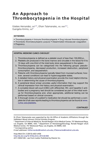
HOSPITAL MEDICINE CLINICS CHECKLIST
1. Thrombocytopenia is defined as a platelet count of less than 150,000/mL.
2. Platelets are produced in the bone marrow and circulate in the blood for 8 to
10 days with one-third of the total body store sequestered in the spleen.
3. Thrombocytopenia can be categorized into the following groups: pseudo-
thrombocytopenia, decreased production, increased destruction, peripheral
consumption, and sequestration.
4. Patients with thrombocytopenia typically bleed from mucosal surfaces; how-
ever, several conditions can lead to hypercoagulable states.
5. A detailed history and physical examination provide the most helpful informa-
tion in determining the cause of thrombocytopenia.
6. A peripheral blood smear provides useful information that may support the
clinical impression from the history and physical examination.
7. A complete blood cell count (CBC) with differential, HIV, and hepatitis C anti-
bodies and a pregnancy test should be considered as part of the initial work-
up for thrombocytopenia and, when appropriate, additional testing can be
performed based on clinical concerns.
8. A thorough review of a patient’s medication list should be performed; a com-
plete list of all case reports leading to thrombocytopenia can be found at www.
ouhsc.edu/platelets.
CONTINUED
Dr Efrain Talamanates was supported by the VA Office of Academic Affiliations through the
VA/Robert Wood Johnson Clinical Scholars Program.
a
Department of Internal Medicine, Ronald Reagan UCLA Medical Center, 757 Westwood Plaza,
Los Angeles, CA 90095, USA; b
Department of Hematology/Oncology, Ronald Reagan UCLA
Medical Center, 757 Westwood Plaza, Los Angeles, CA 90095, USA
* Corresponding author.
E-mail addresses: EAHernandez@mednet.ucla.edu; ETalamantes@mednet.ucla.edu
KEYWORDS
Thrombocytopenia Immune thrombocytopenia Drug induced thrombocytopenia
Thrombotic thromobocytopenic purpura Disseminated intravascular coagulation
Pregnancy
Hosp Med Clin 2 (2013) e422–e436
http://dx.doi.org/10.1016/j.ehmc.2013.02.003
2211-5943/13/$ – see front matter Published by Elsevier Inc.
2. CONTINUED
9. Hematology consultation should be considered when trying to determine if a
patient has thrombotic thromobocytopenic purpura (TTP) or disseminated
intravascular coagulation (DIC).
10. A bone marrow biopsy should be considered in all patients with severe unex-
plained thrombocytopenia and high risk for bleeding or, alternatively, if the re-
sults alter management.
11. Follow-up of minimally asymptomatic patients with thrombocytopenia should
occur within 1 to 2 weeks of diagnosis, and further work-up versus close
follow-up is advised based on the clinical context.
12. In the setting of multisystem illness, management of thrombocytopenia is
directed at treating the underlying cause as well as managing bleeding and
thrombotic risk.
13. The threshold for considering treatment of immune-mediated thrombocyto-
penia should be platelet counts less than 30,000/mL, with the goal of prevent-
ing serious bleeding.
14. To evaluate a patient’s risk of heparin-induced thrombocytopenia (HIT), the
4 Ts (thrombocytopenia, timing, thrombosis, and other cause of thrombocyto-
penia) scoring system can be used.
15. Therapeutic anticoagulation likely is safe for most patients with platelet counts
above 30,000/mL, but higher platelet count thresholds are advised based on
bleeding risk.
16. Safe platelet thresholds vary depending on type of invasive procedure
(30,000/mL–100,000/mL).
DEFINITION
1. What criteria constitute thrombocytopenia?
Thrombocytopenia is defined as a platelet count lower than the normal range (typically
150,000/mL–450,000/mL), which is calculated by laboratories as 2 standard deviations
below the mean. The lower limit of normal, however, far exceeds the platelet count that
leads to pathologic bleeding. Bleeding during surgery does not occur until the platelet
count drops below 50,000/mL and spontaneous bleeding can be observed with counts
of 10,000/mL to 20,000/mL.
2. What is the life cycle of a platelet?
Hematopoietic stems cells in the bone marrow (hemocytoblasts) are precursors of
megakaryocytes, which produce platelets by cytoplasmic shedding into bone marrow
sinusoids. Thrombopoietin is the major hormone that stimulates megarkaryocyte pro-
duction and is produced by the liver. The typical lifespan of platelets ranges from 8 to
9 days and, thereafter, most platelets become senescent and are cleared by the retic-
uloendothelial system in the liver and spleen.1
Approximately one-third of platelets are
sequestered in the spleen.2
3. How is thrombocytopenia categorized?
When considering the cause of thrombocytopenia, clinicians must first determine
whether the laboratory value is real. Pseudothrombocytopenia, or a falsely low platelet
An Approach to Thrombocytopenia in the Hospital e423
3. count due to platelet clumping, can occur in association with the anticoagulant, EDTA
(Box 1). In this setting, platelet clumps can be counted as a leukocyte, leading to an
erroneous report on the CBC. Once the platelet count is confirmed as low, thrombocy-
topenia can be categorized into (1) decreased production, (2) peripheral destruction, (3)
consumption, or (4) splenic sequestration. Patients with thrombocytopenia due to
decreased production have low platelet counts because the body’s fixed daily con-
sumption of platelets accounts for a larger fraction of the reduced total daily production.
Patients with thrombocytopenia because of platelet destruction or consumption have a
markedly decreased platelet survival. In patients with thrombocytopenia due to splenic
sequestration, up to 90% of circulating platelets can be sequestered in the spleen.3
HISTORY AND EXAMINATION
4. How do patients with thrombocytopenia present?
Often, thrombocytopenia comes to clinicians’ attention in the hospital setting due to
review of the admission or daily CBC and is not the primary reason for admission.
Consequently, most patients with thrombocytopenia are asymptomatic despite the
degree of decline. Symptomatic patients with thrombocytopenia may present with
mucosal bleeding (epistaxis, menorrhagia, or gingival) or cutaneous bleeding (pete-
chiae and superficial ecchymoses). Petechiae occur most frequently in the lower ex-
tremities, around the mouth, or where there is constriction (blood pressure cuff or tight
clothing) causing pressure on small vessels. Patients may be predisposed to gastro-
intestinal bleeding, which may manifest as hematochezia and melena. Patients may
also have persistent bleeding at sites of surgery and central venous catheters,
depending on the severity of thrombocytopenia. Although bleeding in the central ner-
vous system (CNS) is rare, any trauma to the head should be evaluated promptly
because it remains the most common cause of death due to thrombocytopenia. If
the underlying cause of thrombocytopenia is HIT or antiphospholipid syndrome
(APS), thrombosis (both arterial and venous) may be the presenting symptom, instead
of bleeding. A seizure in a pregnant patient with thrombocytopenia could suggest TTP
or eclampsia if a CNS bleed has been ruled out.
5. What are the most important aspects of the history and physical examination?
The history often provides the most helpful information in determining the cause of the
thrombocytopenia. Clinicians should consider the time course of the problem (present
before admission or developed during the admission). A history of epistaxis, gingival
bleeding, cutaneous bleeding (petechiae and ecchymoses), and menorrhagia in
women should be obtained along with a history of past challenges in obtaining hemo-
stasis (during previous surgeries, child birth, and dental procedures). Clinicians should
inquire about current and previous alcohol use, dietary restrictions, intravenous drug
use, high-risk sexual behavior, and previous HIV and viral hepatitis testing. Medication
history, including over-the-counter medications, herbal supplements, and tonic water
(which contains quinine), is also important to obtain when considering a diagnosis of
drug-induced idiopathic thrombycytopenic purpura (ITP) (Box 2). Recent infections
(eg, parvovirus, cytomegalovirus, and HIV), previously diagnosed hematologic dis-
eases (eg, leukemias and myelodysplastic syndrome), autoimmune conditions
(eg, lupus), and pregnancy are among other important elements of the history (see
Box 2). Intermittent, acute purpura suggests a drug, herbal, or food-induced thrombo-
cytopenia. Recent travel to tropical regions of the world may alert clinicians to suspect
Hernandez et al
e424
5. Box 2
Important aspects of the history
Bleeding symptoms
Epistaxis
Gingival bleeding
Menorrhagia
Petechiae
Bruising
Hematochezia/melena
Hemostatic challenges
Surgeries
Dental
Trauma
Childbirth
Medical history
Recent infections (infectious symptoms, sick contacts, travel)
Previously diagnosed hematologic condition
Autoimmune conditions (rash, arthritis, Raynaud’s phenomenon)
Last menstrual period
Medication history
Platelet function inhibitors
Recent hospitalizations/exposure to heparin
Over-the-counter medications/herbal supplements
Tonic water—quinine
Social history
Alcohol, intravenous drug use, sexual history, blood transfusions, tattoos
Family history of bleeding
Modified from Wong EY, Rose MG. Why does my patient have thrombocytopenia? Hematol
Oncol Clin North Am. 2012;26(2):231–52; with permission.
Sequestration
Splenomegaly
Hypersplenism
Hypothermia
Modified from Wong EY, Rose MG. Why does my patient have thrombocytopenia? Hematol
Oncol Clin North Am 2012;26(2):231–52; with permission.
Hernandez et al
e426
6. a diagnosis of malaria as a cause of the thrombocytopenia. A family history of throm-
bocytopenia could suggest a congenital disorder.
The physical examination should first focus on evidence of bleeding, because it
is critical to determine if patients are symptomatic. Evaluation of heart rate and
blood pressure are important in assessing hemodynamic status in light of possible
internal bleeding. The skin examination may be significant for petechiae, purpura,
and ecchymoses. Inspection of the oral mucosa may reveal blood blisters and
the nasal cavity may have dried blood. If there is suspicion of gastrointestinal
bleeding, a rectal examination should be performed and a stool sample tested
for occult blood. The head should be inspected for any evidence of trauma and,
if present, a comprehensive neurologic and fundoscopic examination should be
performed.
Presence of fever may heighten suspicion of an infection, malignancy, or autoim-
mune disease. Careful evaluation of the lymph nodes, liver, and spleen may suggest
a lymphoproliferative disorder or chronic infection as the underlying cause. Stigmata
of chronic liver disease, such as asterixis, jaundice, spider telangiectasias, or palmar
erythema, may be apparent in patients with thrombocytopenia due to cirrhosis. A new
diastolic murmur in patients with a prosthetic valve might suggest that platelets are
being activated improperly on the valve surface, causing nonimmune destruction.
Asymmetric swelling of an extremity could be a sign of deep vein thrombosis in a pa-
tient with HIT or APS. Skin necrosis on the lower abdominal wall could be another sign
of HIT, which occurs where heparin products are injected.
DIAGNOSIS
6. What is the role of CBC and peripheral smear in diagnosing thrombocytopenia?
The CBC confirms a clinical suspicion of thrombocytopenia. A review of the rest of the
CBC, including the differential for the white blood cell count, is essential in determining
whether a patient has other cytopenias or abnormal circulating cells. The mean
platelet value may be helpful because a low value suggests old platelets and poor pro-
duction, whereas a large value suggests newer platelets and increased destruction.
The peripheral blood smear can be reviewed to determine if platelet clumping is pre-
sent (routinely reviewed by a laboratory technologist although the physician may only
receive report of a falsely low platelet count). Particular attention should be made to
note any morphologic abnormalities in all 3 cell lines (ie, schistocytes, dysmorphic
cells, and echinocytes) (Table 1). Certainly, trending of platelet counts from day to
day in the hospital also provides valuable information. A platelet count that has
decreased by more than 50% from a previous value could suggest HIT. A platelet
count that has started downtrending approximately 1 week after starting a new medi-
cation may bring to mind drug-induced thrombocytopenia.
7. What tests should be ordered in the initial work-up of a hospitalized patient who has
developed thrombocytopenia?
All patients should have a CBC repeated to confirm the diagnosis and a peripheral
blood smear, for reasons discussed previously. Patients should be offered HIV and
hepatitis C testing. Further testing can be considered depending on findings from
the history, physical, CBC, and peripheral smear (Table 2). Pregnancy tests should
be considered in all women of childbearing age. The test for antiplatelet antibodies
has not been standardized and routine testing is not recommended.
An Approach to Thrombocytopenia in the Hospital e427
7. 8. Which drugs are most commonly implicated in thrombocytopenia?
Drug-induced thrombocytopenia can be caused by both immune-mediated and
nonimmune-mediated mechanisms (eg, chemotherapy, which causes myelosuppres-
sion). Immune mechanisms include autoantibody production, ligand-induced binding,
and hapten-induced binding. Antibiotics and heparin are among the more common
causes of thrombocytopenia in hospitalized patients. Heparin causes thrombocyto-
penia by a different mechanism than other drugs whereby an antibody to the
heparin-platelet factor 4 complex causes platelet activation and clot formation, result-
ing in thrombosis.4
Although many drugs have been reported to have an association
with thrombocytopenia, 24 drugs have the strongest evidence (Box 3).5
A complete
list of all case reports describing drug-induced thrombocytopenia is on the Web
site, www.ouhsc.edu/platelets. Quinine (present in tonic water) is an often missed
cause of thrombocytopenia because patients believe it is a safe product without
side effects and often fail to report intake to physicians.
Table 2
Further testing considerations
Tests Conditions to Consider
PT, aPTT, fibrinogen, D-dimer DIC
HIT antibody and serotonin release assay HIT
HIV and hepatitis C serology Secondary ITP
ANA, anti–double-stranded DNA Systemic lupus erythematosus
Anticardiolipin antibody, lupus anticoagulant, anti–b2-
glycoprotein antibody
Antiphospholipid syndrome
Peripheral blood flow cytometry Lymphoproliferative disorders
Ultrasound of liver/spleen Cirrhosis/splenomegaly
Modified from Wong EY, Rose MG. Why does my patient have thrombocytopenia? Hematol Oncol
Clin North Am 2012;26(2):231–52; with permission.
Table 1
Findings on peripheral smear
Platelet clumps Evaluate for pseudothrombocytopenia
Abnormal morphology of platelets Large platelets suggest ITP
Giant platelets suggest congenital disorder
Red blood cell fragments (eg, schistocytes,
helmet cells)
Microangiopathic process
Toxic granulation Sepsis
Abnormal lymphocytes Viral infection or lymphoproliferative disorder
Intracellular organisms Malaria, ehrlichiosis, babesiosis
Circulating blast cells Acute leukemia
Leukoerythroblastic Response (tear drop cells,
nucleated red blood cells, early myeloid
forms in blood)
Marrow invasion with tumor, fibrosis,
granulomatous disease
Macrocytic anemia Vitamin B12 or folate deficiency, liver disease
Modified from Wong EY, Rose MG. Why does my patient have thrombocytopenia? Hematol Oncol
Clin North Am 2012;26(2):231–52; with permission.
Hernandez et al
e428
8. 9. What are the distinguishing features between thrombotic microangiopathy and
disseminated intravascular coagulation?
Thrombotic microangiopathy (TTP–hemolytic-uremic syndrome [HUS]) can be differ-
entiated from DIC by clinical history and laboratory studies. Although both conditions
can have a febrile component, microangiopthic hemolytic anemia, thrombocytopenia,
and renal failure, DIC is associated most commonly with sepsis, whereas TTP-HUS
may be preceded by a bloody diarrheal syndrome or initiation of certain medications
(eg, clopidogrel). Both can occur in pregnant women and in patients with malignancy.
In DIC, there is uncontrolled activation of the clotting cascade, which results in
increased fibrin degradation products, decreased fibrinogen, and increased clotting
times.6
In TTP, the condition is caused by a congenital or acquired deficiency in
ADAMTS13, a von Willebrand factor protease.7
The end result is that the uncleaved
Box 3
Drugs commonly associated with thrombocytopenia
Abciximab
Acetaminophen
Ampicillin
Carbamazepine
Eptifibatide
Ethambutol
Haloperidol
Ibuprofen
Irinotecan
Naproxen
Oxaliplatin
Phenytoin
Piperacillin
Quinidine
Quinine
Ranitidine
Rifampin
Simvastatin
Sulfisoxazole
Tirofiban
Trimethoprim-sulfamethoxazole
Valproic acid
Vancomycin
Data from Reese JA, Li X, Hauben M, et al. Identifying drugs that cause acute thrombocyto-
penia: an analysis using 3 distinct methods. Blood 2010;116(12):2127–33.
An Approach to Thrombocytopenia in the Hospital e429
9. von Willebrand factor activates endothelial cells, which leads to platelet aggregation
and clotting. In the latter condition, D-dimer, fibrinogen, and prothrombin time (PT)
and activated partial thromboplastin time (aPTT) are often normal. The peripheral
smear in both conditions show microangiopathic hemolysis and thrombocytopenia.
The conditions are managed differently (generally supportive care for DIC and plasma
exchange for TTP); therefore, hematologic consultation should be considered in these
situations.
10. How should thrombocytopenia be approached during pregnancy?
Thrombocytopenias in pregnancy should be evaluated similarly to nonpregnant pa-
tients although several other conditions should be considered in the differential diag-
nosis. Thrombocytopenia in pregnant patients is most likely a result of gestational
thrombocytopenia and ITP. Gestational thrombocytopenia occurs in approximately
5% of pregnancies and, if this is the suspected diagnosis, the platelet count can be
monitored every trimester.8
Platelet counts with testational thrombocytopenia rarely
decline below 70,000 and remit quickly on delivery.8,9
ITP is more likely the diagnosis
if the platelet count is less than 50,000/mL and if it is noted early in pregnancy.9
Other
diagnostic considerations in the third trimester include preeclampsia/eclampsia and
hemolysis, elevated liver enzymes, and low platelet count (HELLP) syndrome. Pre-
eclampsia as a cause of thrombocytopenia should be entertained if a patient has
new-onset hypertension and proteinuria. If a patient develops seizures, the clinical
syndrome of eclampsia may be difficult to distinguish from TTP-HUS. Progression
of hematologic abnormalities for 3 days after delivery may favor a diagnosis of
TTP-HUS.10
11. When do you obtain a hematological consultation and consider a bone marrow
biopsy?
When the cause of the thrombocytopenia (of any degree of severity) is not clear after
retesting and initial evaluation, hematologic consultation is warranted. Evaluating the
CBC, white cell differential, and description of the peripheral blood smear is of para-
mount importance in assuring that disorders, such as myelodysplastic syndrome,
acute leukemia, and microangiopathy (eg, DIC and TTP), are not present.
A bone marrow biopsy is indicated in all patients with severe unexplained thrombo-
cytopenia with high risk for bleeding or if the results alter management (eg, confirm the
diagnosis of a malignancy or infection). Increased megakaryocytes noted in the bone
marrow suggest increased destruction or peripheral consumption whereas a de-
creased number suggest a hypoproliferative process. Graunulomas, fibrosis, and ma-
lignant cells may be seen in patients with findings of leukoerythroblastic response on
the peripheral smear. If patients are less than 60 years old and a diagnosis of ITP is
entertained, forgoing biopsy could be considered given the low risk of having myelo-
dysplastic syndrome in this age group. In the inpatient setting, given that there may be
different factors influencing the platelet count (active infection, drugs, and so forth), it
is reasonable to defer biopsy after repeat testing in the outpatient setting unless it has
an impact on management.
12. When can you be certain that the cause of thrombocytopenia is immune mediated?
A diagnosis of immune-mediated thrombocytopenia can never be made with absolute
certainty given that it is a diagnosis of exclusion. However, a presumption diagnosis
of ITP can be made after history, physical examination, CBC, and peripheral blood
Hernandez et al
e430
10. smear do not identify another cause for patients with isolated thrombocytopenia. HIV
and hepatitis C testing are recommended, because management of these chronic in-
fections may result in improvement of a patient’s thrombocytopenia. Bone marrow
aspiration should be considered if patients are over 60 years old to rule out myelodys-
plastic syndrome.
13. What is the appropriate follow-up of minimal asymptomatic thrombocytopenia?
Asymptomatic and nonbleeding patients with modest reductions in platelet count (eg,
75,000/mL to 100,000/mL) need follow-up testing in 1 to 2 weeks after initial diagnosis,
provided patients report any changes in clinical status or bleeding.
For those with minimal degrees of thrombocytopenia (eg, 100,000/mL to 150,000/mL),
there is much less urgency to follow-up frequently; testing may be repeated in 1 or more
months, because a small percent of these patients develop a normal platelet count.
There is a 6.9% chance of developing a persistent platelet count of less than
100,000/mL over 10 years of follow-up.11
Inoncology patients,onlyseverethrombocytopenia,a plateletcountbelow10,000/mL
(and maybe 5000/mL), is associated with an increased risk of bleeding.12–14
Based on
thesestudies,aplatelettriggerof10,000/mLhasbeengenerallyadoptedforprophylactic
platelet transfusions in stable hematology/oncology patients.15
In nononcology patients, there are few studies that have examined the relationship
between thrombocytopenia and bleeding. In patients with ITP, observational data sug-
gest that bleeding rarely occurs with platelet counts above 30,000/mL.16
Evidence-
based guidelines suggest that platelet-raising therapies are generally not necessary
when platelet counts are above 20,000/mL to 30,000/mL.17
14. What are the management goals for thrombocytopenia in the setting of multisystem
illness?
Management of thrombocytopenia is directed at treating the underlying cause as well
as managing bleeding and thrombotic risk. Maintenance of safe platelet counts
greater than 30,000/mL to 50,000/mL using platelet transfusions is usually a small
part of the overall treatment plan unless there is active bleeding.
15. What are the indications for treatment of immune-mediated thrombocytopenia?
The goal of treating immune-mediated thrombocytopenia is to prevent serious
bleeding. There is no evidence that supports a minimum platelet count threshold or
specific age at which a typical patient with ITP should be treated. Based on the
American Society of Hematology 2011 evidence-based practice guideline, it is recom-
mended that treatment with glucocorticoids should be initiated when platelet counts
are less than less than 30,000/mL. If platelets remain less than 30,000/mL despite treat-
ment with glucocorticoids and there is no significant bleeding, toxic side effects of
intravenous immunoglobulin, splenectomy, rituximab, or immunosuppressive agents
for additional therapy options must be considered.
16. Which conditions require ICU level of care?
The clinical picture and context of thrombocytopenia are crucial in identifying patients
who need a higher level of care. Evolving thrombocytopenia in critically ill patients may
be an early warning sign of sepsis. Many patients may have more than 1 cause, however;
Box 4 lists some of the most common conditions contributing to thrombocytopenia.
An Approach to Thrombocytopenia in the Hospital e431
11. PERFORMANCE IMPROVEMENT
17. When do you use the heparin-induced thrombocytopenia 4 Ts scoring system?
There can be a significant delay before the results of laboratory testing for HIT are
available and, therefore, management decisions cannot be postponed. Also, isolated
HIT antibodies are frequent and not diagnostic. The most well-studied clinical predic-
tion rule to determine the probability that a patient has HIT is the 4 Ts score. A low 4 Ts
score has a low probability of HIT, whereas those with intermediate to high scores
require further evaluation (Table 3).4
Box 4
Conditions requiring ICU level of care
Sepsis and septic shock
TTP
DIC
Massive blood transfusion resulting in spontaneous bleeding
Cardiopulmonary resuscitation
Cardiopulmonary bypass
Adult respiratory distress syndrome
Pulmonary embolism with hemodynamic instability
Table 3
The 4 Ts scoring system
Category 2 Points 1 Point 0 Point
Thrombocytopenia Platelet count
fall 50% and
platelet nadir 20
Platelet count
30%–50% or platelet
nadir 10–19
Platelet count fall
30% or platelet
nadir 10
Timing of platelet
count fall
Clear onset between
days 5–10 or
platelet fall 1 d
(prior heparin
exposure within
30 d)
Consistent with days 5–10
fall, but not clear (eg,
missing platelet counts);
onset after day 10; or fall
1 d (prior heparin
exposure 30–100 d ago)
Platelet count 4 d
without recent
exposure
Thrombosis or
other sequelae
New thrombosis
(confirmed); skin
necrosis; acute
systemic reaction
postintravenous
unfractionated
heparin bolus
Progressive or recurrent
thrombosis; non-
necrotizing
(erythematous) skin
lesions; suspected
thrombosis (not proved)
None
Other causes of
thrombocytopenia
None apparent Possible Definite
The 4 Ts score is the sum of the values for each of the 4 categories. Scores of 1–3, 4–5, and 6–8 are
considered to correspond to a low, intermediate, and high probability of HIT, respectively.
Data from Slichter SJ, Slichter SJ. Evidence-based platelet transfusion guidelines. Hematology
Am Soc Hematol Educ Program 2007:172–8.
Hernandez et al
e432
12. 18. When is it safe to use antiplatelet agents and anticoagulants in patients with
thrombocytopenia?
Thrombocytopenia increases the risk of bleeding and does not protect against
thrombosis. Thrombocytopenia alone should not be a contraindication to anticoagu-
lation or antiplatelet therapy in hospitalized patients. Prophylactic and therapeutic
anticoagulation is likely safe for most patients with platelet counts above 30,000/mL,
but a higher platelet count threshold may be necessary when therapeutic doses of
anticoagulation are required or when the bleeding risk is high. Antiplatelet therapy is
likely safe for most patients with platelet counts above 50,000/mL.18,19
Thrombocytopenia in APS may indicate increased disease activity and increased
thrombotic potential (odds ratio of 1.6–11)20
; thus, aggressive antithrombotic therapy
is warranted. Thrombosis is a feared complication of HIT, despite a low platelet count.
The frequency of thrombosis in patients with HIT has been reported up to 89%.21
Bleeding rarely occurs in HIT, however, even in postoperative patients. In patients
with HIT, the initial treatment is to discontinue all exposure to heparin or low-
molecular-weight heparins (including heparin-coated catheters). Therapeutic doses
of a nonheparin anticoagulant, such as direct thrombin inhibitor, is indicated until
platelet count is normalized, at which time bridging therapy with warfarin can be initi-
ated for a minimum of 5 days. The length of anticoagulation is not clear, but, given the
significantly increased risk of thrombosis at 30 days, treatment should likely continue
for at least 2 to 3 months in HIT without thrombosis or 3 to 6 months if HIT with throm-
bosis. In patients with DIC, where thrombosis predominates, anticoagulation with hep-
arin, although controversial, has been recommended.
19. What is an optimal platelet count when performing invasive procedures?
In critically ill patients, specific characteristics are likely important modifiers of the rela-
tionship between thrombocytopenia and bleeding.22
Published guidelines recommend
Table 4
Summary of platelet transfusion triggers
Indication Platelet Transfusion Trigger
Surgical patients 50,000/mL
Cardiac surgery with CPB 50,000/mL or reserved for bleeding
Liver transplantation 50,000/mL
Surgical and obstetric patients with microvascular
bleeding
50,000/mL
Surgery with a low risk of bleeding 50,000/mL
HELLP syndrome requiring cesarean section 50,000/mL
HELLP syndrome requiring vaginal delivery 30,000/mL
Spinal anesthesia 50,000/mL
Epidural anesthesia 100,000/mL
Ophthalmologic and CNS surgery 100,000/mL
Massive transfusion 50,000/mL
Multiple trauma or CNS injury 100,000/mL
DIC 50,000/mL or reserved for bleeding
Autoimmune thrombocytopenia Reserved for serious bleeding
Abbreviation: CPB, cardiopulomonary bypass.
Data from Refs.24–27
An Approach to Thrombocytopenia in the Hospital e433
13. a platelet count trigger as low as 30,000/mL for patients with HELLP syndrome requiring
vaginal delivery and 100,000/mL for patients in whom an invasive neurosurgical proce-
dure is planned.23
Table 4 is a summary of platelet transfusion trigger parameters from
published guidelines.
CLINICAL GUIDELINES
Treatment and prevention of HIT: American College of Chest Physicians
Evidence-Based Clinical Practice Guidelines (8th Edition)
American Society of Hematology 2011 Clinical Practice Guideline on the Evalu-
ation and Management of Immune Thrombocytopenia
Platelet Transfusion for Patients With Cancer: Clinical Practice Guidelines of the
American Society of Clinical Oncology (2001)
REFERENCES
1. Cohen JA, Leeksma CH. Determination of the life span of human blood platelets
using labeled diisopropylfluorophosphonate. J Clin Invest 1956;35(9):964–9.
2. Heyns AD, Lötter MG, Badenhorst PN, et al. Kinetics, distribution and sites of de-
struction of 111indium-labelled human platelets. Br J Haematol 1980;44(2):
269–80.
3. Aster RH. Pooling of platelets in the spleen: role in the pathogenesis of “hyper-
splenic” thrombocytopenia. J Clin Invest 1966;45(5):645.
4. Cuker A, Gimotty PA, Crowther MA, et al. Predictive value of the 4Ts scoring sys-
tem for heparin-induced thrombocytopenia: a systematic review and meta-
analysis. Blood 2012. Available at: http://eutils.ncbi.nlm.nih.gov/entrez/eutils/
elink.fcgi?dbfrom5pubmedid522990018retmode5refcmd5prlinks. Ac-
cessed October 2, 2012.
5. Reese JA, Li X, Hauben M, et al. Identifying drugs that cause acute thrombocy-
topenia: an analysis using 3 distinct methods. Blood 2010;116(12):2127–33. http:
//dx.doi.org/10.1182/blood-2010-03-276691.
6. Levi M, Ten Cate H. Disseminated intravascular coagulation. N Engl J Med 1999;
341(8):586–92.
7. Moake JL. Thrombotic microangiopathies. N Engl J Med 2002;347(8):589–600.
8. Burrows RF, Kelton JG. Fetal thrombocytopenia and its relation to maternal throm-
bocytopenia. N Engl J Med 1993;329(20):1463.
9. Gernsheimer TB. Thrombocytopenia in pregnancy: is this immune thrombocyto-
penia or.? Hematology Am Soc Hematol Educ Program 2012;2012:198–202.
http://dx.doi.org/10.1182/asheducation-2012.1.198.
10. McMinn JR, George JN. Evaluation of women with clinically suspected thrombotic
thrombocytopenic purpura-hemolytic uremic syndrome during pregnancy. J Clin
Apher 2001;16(4):202.
11. Stasi R, Amadori S, Osborn J, et al. Long-term outcome of otherwise healthy indi-
viduals with incidentally discovered borderline thrombocytopenia. PLoS Med
2006;3(3):e24. Available at: http://www.plosmedicine.org/article/info:doi/10.1371/
journal.pmed.0030024. Accessed October 2, 2012.
12. Slichter SJ, Kaufman RM, Assmann SF, et al. Dose of prophylactic platelet trans-
fusions and prevention of hemorrhage. N Engl J Med 2010;362(7):600–13. Avail-
able at: http://eutils.ncbi.nlm.nih.gov/entrez/eutils/elink.fcgi?dbfrom5pubmed
id520164484retmode5refcmd5prlinks. Accessed October 2, 2012.
13. Heddle NM, Cook RJ, Tinmouth A, et al. A randomized controlled trial comparing
standard- and low-dose strategies for transfusion of platelets (SToP) to patients
Hernandez et al
e434
14. with thrombocytopenia. Blood 2009;113(7):1564–73. Available at: http://eutils.ncbi.
nlm.nih.gov/entrez/eutils/elink.fcgi?dbfrom5pubmedid519109560retmode5
refcmd5prlinks. Accessed October 2, 2012.
14. Stanworth S, Hyde C, Heddle N, et al. Prophylactic platelet transfusion for haemor-
rhage after chemotherapy and stem cell transplantation. Cochrane Database Syst
Rev 2004;(4). Available at: http://onlinelibrary.wiley.com/doi/10.1002/14651858.
CD004269.pub2/pdf/standard. Accessed October 2, 2012.
15. Schiffer CA, Anderson KC, Bennett CL, et al. Platelet transfusion for patients with
cancer: clinical practice guidelines of the American Society of Clinical Oncology.
J Clin Oncol 2001;19(5):1519–38. Available at: http://eutils.ncbi.nlm.nih.gov/
entrez/eutils/elink.fcgi?dbfrom5pubmedid511230498retmode5refcmd5
prlinks. Accessed October 2, 2012.
16. Kelton JG, Portielje JE, Westendorp RG, et al. Treatment of adults with ITP: less
may be more. Blood 97(9):2533. Available at: http://www.bloodjournal.org/cgi/
doi/10.1182/blood.V97.9.2533. Accessed October 2, 2012.
17. Neunert C, Lim W, Crowther M, et al. The American Society of Hematology 2011
evidence-based practice guideline for immune thrombocytopenia. Blood 2011;
117(16):4190–207. Available at: http://www.bloodjournal.org/cgi/doi/10.1182/
blood-2010-08-302984. Accessed October 2, 2012.
18. Ferraris VA, Ferraris SP, Moliterno DJ, et al. The Society of Thoracic Surgeons Prac-
ticeGuidelineSeries:aspirinandotherantiplateletagentsduringoperativecoronary
revascularization (executive summary)*. Ann Thorac Surg 2005;79(4):1454–61.
Available at: http://linkinghub.elsevier.com/retrieve/pii/S0003497505000160. Ac-
cessed October 2, 2012.
19. Linkins L, Dans AL, Moores LK, et al. Treatment and prevention of heparin-
induced thrombocytopenia: Antithrombotic Therapy and Prevention of Throm-
bosis, 9th ed: American College of Chest Physicians Evidence-Based Clinical
Practice Guidelines. Chest 2012;141(Suppl 2):e495S–530S. Available at: http://
eutils.ncbi.nlm.nih.gov/entrez/eutils/elink.fcgi?dbfrom5pubmedid522315270
retmode5refcmd5prlinks. Accessed October 2, 2012.
20. Galli M, Luciani D, Bertolini G, et al. Anti-beta 2-glycoprotein I, antiprothrombin
antibodies, and the risk of thrombosis in the antiphospholipid syndrome. Blood
2003;102(8):2717–23.
21. Boshkov LK, Warkentin TE, Hayward CP, et al. Heparin-induced thrombocytopenia
and thrombosis: clinical and laboratory studies. Br J Haematol 1993;84:322–8.
22. Arnold DM, Donahoe L, Clarke FJ, et al. Bleeding during critical illness: a prospec-
tive cohort study using a new measurement tool. Clin Invest Med 2007;30(2):
E93–102. Available at: http://cimonline.ca/index.php/cim/article/viewArticle/985.
Accessed October 2, 2012.
23. Slichter SJ, Slichter SJ. Evidence-based platelet transfusion guidelines. Hematol-
ogy 2007;2007:172–8. Available at: http://www.asheducationbook.org/cgi/doi/10.
1182/asheducation-2007.1.172. Accessed October 2, 2012.
24. Samama CM, Djoudi R, Lecompte T, et al. Perioperative platelet transfusion. Rec-
ommendations of the French Health Products Safety Agency (AFSSAPS) 2003.
Minerva Anestesiol 2006;72(6):447–52. Available at: http://eutils.ncbi.nlm.nih.gov/
entrez/eutils/elink.fcgi?dbfrom5pubmedid516682914retmode5refcmd5
prlinks. Accessed October 2, 2012.
25. Blood Transfusion Task Force, British Committee for Standards in Haematology.
Guidelines for theuse ofplatelettransfusions.Br J Haematol2003;122:10–23.Avail-
able at: http://intranet.sctimst.ac.in/pdf_forms/guidelines_patient_care/guideline1.
PDF. Accessed October 2, 2012.
An Approach to Thrombocytopenia in the Hospital e435
15. 26. Practice guidelines for blood component therapy: a report by the American Soci-
ety of Anesthesiologists Task Force on Blood Component Therapy. Anesthesiology
1996;84(3):732. Available at: http://journals.lww.com/anesthesiology/Abstract/
1996/03000/Practice_Guidelines_for_Blood_Component_Therapy__A.32.aspx.
Accessed October 2, 2012.
27. Practice parameter for the use of fresh-frozen plasma, cryoprecipitate, and plate-
lets. Fresh-Frozen Plasma, Cryoprecipitate, and Platelets Administration Practice
Guidelines Development Task Force of the College of American Pathologists.
JAMA 1994;271(10):777–81. Available at: http://eutils.ncbi.nlm.nih.gov/entrez/
eutils/elink.fcgi?dbfrom5pubmedid58114215retmode5refcmd5prlinks.
Accessed October 2, 2012.
Hernandez et al
e436