Downloaded 121 times


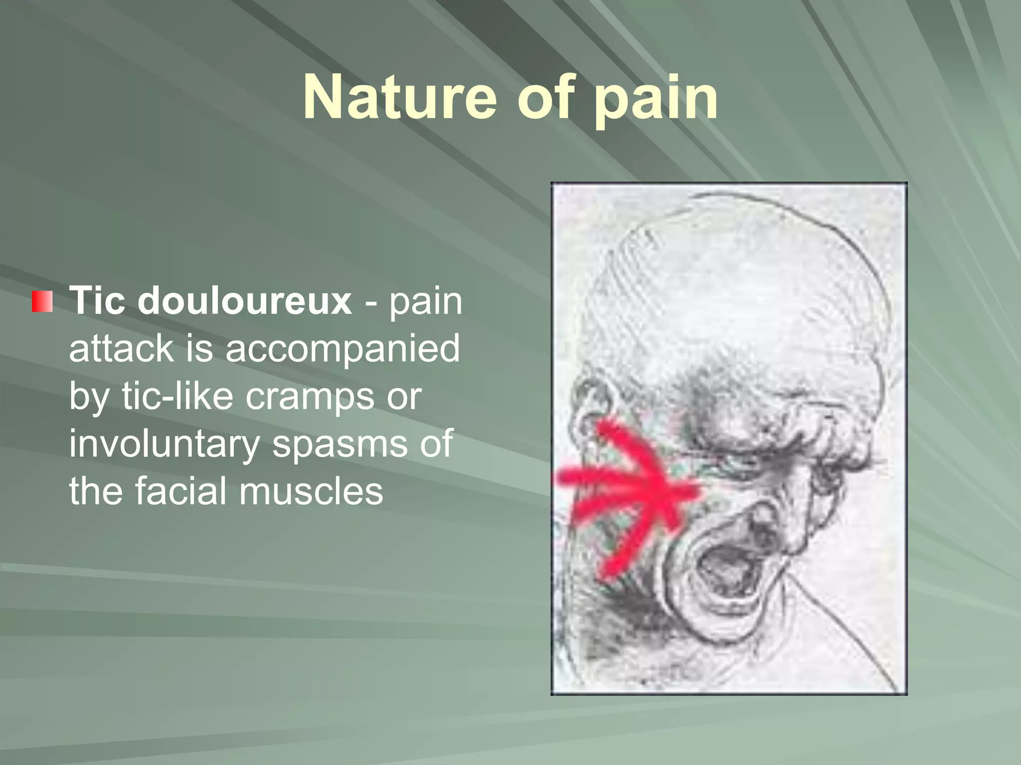

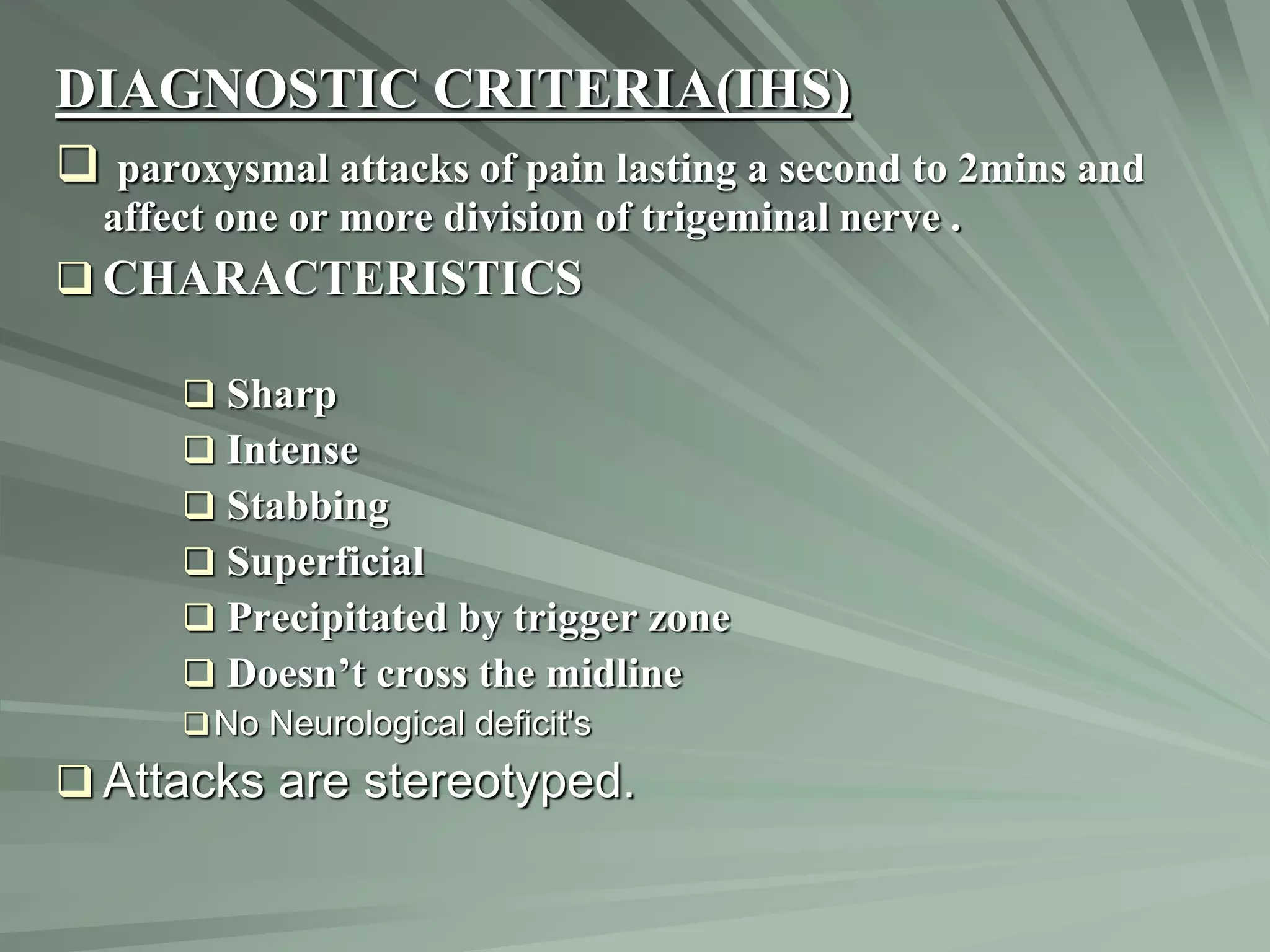


Trigeminal neuralgia, characterized by sudden, severe, recurring facial pain along the distribution of the trigeminal nerve, can arise from various causes including idiopathic, tumors, and vascular compression. The management of this condition involves a combination of medical therapies, primarily anticonvulsants, and surgical interventions if medical management is ineffective. Careful diagnosis and patient education are essential for effective treatment and management of trigeminal neuralgia.