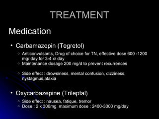
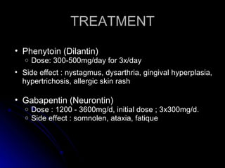
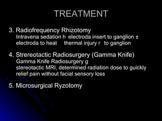
TRIGEMINAL NEURALGIA

            Presented by
            Wita I.Septina


           Supervised by
    Harmas Yazid Yusuf, drg. SpBM
INTRODUCTION
• Trigeminal Neuralgia (TN) is neuropathic facial pain
  arising from the trigeminal nerve.
• Incidence 4-5 cases : 100.000
• TN or Tic douloureux occur patients > 50 years.
• Male : Female ratio 2 : 3
• Unilateral (97%). Most affected V2 and V3.
• The pain is intense, usually sharp, electric shocklike
  pain in face, lasting periods of seconds to 2 minutes ,
ANATOMY TRIGEMINAL NERVE

• Cranial Nerve V
  o   Sensoric e Portio major
  o   Motoric Portio minor
  o   Sensoric + motoric n
      Gasseri
Fig 1. DistributionTrigeminal nerve
CLASSIFICATION
1.Typical Trigeminal Neuralgia
(Tic Douloureux)

• Most common form of TN
• Caused by blood vessels compressing the trigeminal nerve root
  enters the brain stem
• Irritation from repeated pulsations t caused hyperactivity of the
  trigeminal nerve nucleus p resulting TN pain
• Fig 2
(a)                       (b)                        (c)

Fig 2. Anatomy Trigeminal nerve and Trigeminal neuralgia
CLASSIFICATION
2. Atypical Trigeminal Neuralgia
• Unilateral
• Prominent constant
• Boring or burning pain
• Caused by vascular compression upon a specific part of
  trigeminal nerve (portio minor)
• A more severe from or progression of typical TN
CLASSIFICATION
3. Pre - Trigeminal Neuralgia
    Symptoms : odd sensations of pain or discomfort     before the first
    attack of TN pain


4. Multiple Sclerosis-Related Trigeminal
Neuralgia
•   The symptoms & characteristics identical o
•   2 - 4% patients with TN have multiple sclerosis (MS)
•   MS      formation of demyelinating plaques within the brain
•   First attack of pain y younger patients , bilateral
CLASSIFICATION
5. Secondary Trigeminal Neuralgia
• Caused by a lesion (tumor)
• A tumor y compresses or distorts the trigeminal nerve facial
  numbness, weakness of chewing muscles, constant pain
• Fig 3.


6. Post-Traumatic Trigeminal Neuralgia
• Develop following cranio-facial trauma, dental trauma, sinus trauma,
  destructive procedures (rhizotomies)
• Injury t
  cold, start immediately or days to years following injury
Fig. 3 MRI--- Tumor compressed trigeminal nerve
CLASSIFICATION

7. Failed Trigeminal Neuralgia
  Medications, microvascular decompression, and
  destructive rhizotomy procedure ineffective in controlling
  TN pain
ETIOLOGY

1.Blood vessels compression at the trigeminal
  nerve root
– Demyelination nerve
– A tumor compresses trigeminal nerve
– Injury to the trigeminal nerve
– Un known
Clinical Features
1. Severe paroxysmal pain
– The pain intense, stabbing, electrical shock- like, one
   side
– Frequently pain free between attacks.
– Lasting only seconds to two minutes
– Each attack spontaneously or be triggered by specific
   light stimulation
– Common triggers include touch, talking, eating, drinking,
   chewing, tooth brushing, hair combing and kissing.
Fig. 4 Progression of Trigeminal Neuralgia
DIAGNOSIS
•   Anamnesis
•   Clinical examination
•   CT scan and MRI
•   MRIA
Differential Diagnosis

1.Glossopharyngeal neuralgia
– Occipital neuralgia
– Paroxysmal hemicrania syndromes
– Migraine and cluster headaches
– Trigeminal neuropathy
TREATMENT

• Medication
• Surgical procedure
TREATMENT
Medication
• Carbamazepin (Tegretol)
  o Anticonvulsants, Drug of choice for TN, effective dose 600 -1200
    mg/ day for 3-4 x/ day
  o Maintenance dosage 200 mg/d to prevent recurrences

  o   Side effect : drowsiness, mental confusion, dizziness,
      nystagmus,ataxia


• Oxycarbazepine (Trileptal)
  o   Side effect : nausea, fatique, tremor
  o   Dose : 2 x 300mg, maximum dose : 2400-3000 mg/day
TREATMENT

• Phenytoin (Dilantin)
   o   Dose: 300-500mg/day for 3x/day
• Side effect : nystagmus, dysarthria, gingival hyperplasia,
  hypertrichosis, allergic skin rash

• Gabapentin (Neurontin)
   o   Dose : 1200 - 3600mg/d, initial dose ; 3x300mg/d.
   o   Side effect : somnolen, ataxia, fatique
TREATMENT

• Baclophen (Lioresal)
   Antispasmodic agents
   Initial dose : 2-3 x 5 mg/ day.
   Duration of action x
   Side effect : nausea, fatique
TREATMENT
Surgical Procedure
 For patients e medical therapy has failed   surgery is a viable and
 effective option

• Microvascular decompression
• Nerve Injury/ Destructive Procedure (Rhizotomy)
  1. Percutaneus Glycerol Rhizotomy
  • Percutaneus Balloon Compression Rhizotomy
  • Radiofrequency Rhizotomy
  • Stereotactic Radiosurgery (Gamma Knife)
  • Microsurgical Rhizotomy
TREATMENT
• Microvascular decompression
 o   non-destructive technique
 o   Under general anesthesia, incising the skin behind
     the ear (Craniotomy)
 o   Identify an arterial loop compressing the nerve n pad
     the vascular structure with Teflon felt
 o   Complication: CSF leaks, hearing loss, permanent
     anesthesia over the face
TREATMENT
• Nerve Injury/ DestructiveProcedure
  (Rhizotomy)

 1. Percutaneus Glycerol Rhizotomy
 The surgeon introduces a trocar or needle lateral to the
 corner of the mouth into foramen ovale l glycerol–ganglion
 Gasseri f nerve injury

 2. Percutaneus Ballon Compression Rhizotomy
 Under general anestesia – operator insert a balloon
 catheter through the the foramen ovale r the region of the
 ganglion
TREATMENT
3. Radiofrequency Rhizotomy
  Intravena sedation h electroda insert to ganglion
  electroda to heat thermal injury r to ganglion

4. Strereotactic Radiosurgery (Gamma Knife)
  Gamma Knife Radiosurgery g
  stereotactic MRI, determined radiation dose to guickly
  relief pain without facial sensory loss

5. Microsurgical Ryzotomy
CONCLUSION

• Trigeminal Neuralgia (TN) is neuropathic
  facial pain arising from the trigeminal
  nerve.


• Treatment for TN n
  initial therapy if pharmacologic treatment
  fails l surgical procedure.
Trigeminal neuralgia 2_

Trigeminal neuralgia 2_

  • 1.
    TRIGEMINAL NEURALGIA Presented by Wita I.Septina Supervised by Harmas Yazid Yusuf, drg. SpBM
  • 2.
    INTRODUCTION • Trigeminal Neuralgia(TN) is neuropathic facial pain arising from the trigeminal nerve. • Incidence 4-5 cases : 100.000 • TN or Tic douloureux occur patients > 50 years. • Male : Female ratio 2 : 3 • Unilateral (97%). Most affected V2 and V3. • The pain is intense, usually sharp, electric shocklike pain in face, lasting periods of seconds to 2 minutes ,
  • 3.
    ANATOMY TRIGEMINAL NERVE •Cranial Nerve V o Sensoric e Portio major o Motoric Portio minor o Sensoric + motoric n Gasseri
  • 4.
  • 5.
    CLASSIFICATION 1.Typical Trigeminal Neuralgia (TicDouloureux) • Most common form of TN • Caused by blood vessels compressing the trigeminal nerve root enters the brain stem • Irritation from repeated pulsations t caused hyperactivity of the trigeminal nerve nucleus p resulting TN pain • Fig 2
  • 6.
    (a) (b) (c) Fig 2. Anatomy Trigeminal nerve and Trigeminal neuralgia
  • 7.
    CLASSIFICATION 2. Atypical TrigeminalNeuralgia • Unilateral • Prominent constant • Boring or burning pain • Caused by vascular compression upon a specific part of trigeminal nerve (portio minor) • A more severe from or progression of typical TN
  • 8.
    CLASSIFICATION 3. Pre -Trigeminal Neuralgia Symptoms : odd sensations of pain or discomfort before the first attack of TN pain 4. Multiple Sclerosis-Related Trigeminal Neuralgia • The symptoms & characteristics identical o • 2 - 4% patients with TN have multiple sclerosis (MS) • MS formation of demyelinating plaques within the brain • First attack of pain y younger patients , bilateral
  • 9.
    CLASSIFICATION 5. Secondary TrigeminalNeuralgia • Caused by a lesion (tumor) • A tumor y compresses or distorts the trigeminal nerve facial numbness, weakness of chewing muscles, constant pain • Fig 3. 6. Post-Traumatic Trigeminal Neuralgia • Develop following cranio-facial trauma, dental trauma, sinus trauma, destructive procedures (rhizotomies) • Injury t cold, start immediately or days to years following injury
  • 10.
    Fig. 3 MRI---Tumor compressed trigeminal nerve
  • 11.
    CLASSIFICATION 7. Failed TrigeminalNeuralgia Medications, microvascular decompression, and destructive rhizotomy procedure ineffective in controlling TN pain
  • 12.
    ETIOLOGY 1.Blood vessels compressionat the trigeminal nerve root – Demyelination nerve – A tumor compresses trigeminal nerve – Injury to the trigeminal nerve – Un known
  • 13.
    Clinical Features 1. Severeparoxysmal pain – The pain intense, stabbing, electrical shock- like, one side – Frequently pain free between attacks. – Lasting only seconds to two minutes – Each attack spontaneously or be triggered by specific light stimulation – Common triggers include touch, talking, eating, drinking, chewing, tooth brushing, hair combing and kissing.
  • 14.
    Fig. 4 Progressionof Trigeminal Neuralgia
  • 15.
    DIAGNOSIS • Anamnesis • Clinical examination • CT scan and MRI • MRIA
  • 16.
    Differential Diagnosis 1.Glossopharyngeal neuralgia –Occipital neuralgia – Paroxysmal hemicrania syndromes – Migraine and cluster headaches – Trigeminal neuropathy
  • 17.
  • 18.
    TREATMENT Medication • Carbamazepin (Tegretol) o Anticonvulsants, Drug of choice for TN, effective dose 600 -1200 mg/ day for 3-4 x/ day o Maintenance dosage 200 mg/d to prevent recurrences o Side effect : drowsiness, mental confusion, dizziness, nystagmus,ataxia • Oxycarbazepine (Trileptal) o Side effect : nausea, fatique, tremor o Dose : 2 x 300mg, maximum dose : 2400-3000 mg/day
  • 19.
    TREATMENT • Phenytoin (Dilantin) o Dose: 300-500mg/day for 3x/day • Side effect : nystagmus, dysarthria, gingival hyperplasia, hypertrichosis, allergic skin rash • Gabapentin (Neurontin) o Dose : 1200 - 3600mg/d, initial dose ; 3x300mg/d. o Side effect : somnolen, ataxia, fatique
  • 20.
    TREATMENT • Baclophen (Lioresal) Antispasmodic agents Initial dose : 2-3 x 5 mg/ day. Duration of action x Side effect : nausea, fatique
  • 21.
    TREATMENT Surgical Procedure Forpatients e medical therapy has failed surgery is a viable and effective option • Microvascular decompression • Nerve Injury/ Destructive Procedure (Rhizotomy) 1. Percutaneus Glycerol Rhizotomy • Percutaneus Balloon Compression Rhizotomy • Radiofrequency Rhizotomy • Stereotactic Radiosurgery (Gamma Knife) • Microsurgical Rhizotomy
  • 22.
    TREATMENT • Microvascular decompression o non-destructive technique o Under general anesthesia, incising the skin behind the ear (Craniotomy) o Identify an arterial loop compressing the nerve n pad the vascular structure with Teflon felt o Complication: CSF leaks, hearing loss, permanent anesthesia over the face
  • 23.
    TREATMENT • Nerve Injury/DestructiveProcedure (Rhizotomy) 1. Percutaneus Glycerol Rhizotomy The surgeon introduces a trocar or needle lateral to the corner of the mouth into foramen ovale l glycerol–ganglion Gasseri f nerve injury 2. Percutaneus Ballon Compression Rhizotomy Under general anestesia – operator insert a balloon catheter through the the foramen ovale r the region of the ganglion
  • 24.
    TREATMENT 3. Radiofrequency Rhizotomy Intravena sedation h electroda insert to ganglion electroda to heat thermal injury r to ganglion 4. Strereotactic Radiosurgery (Gamma Knife) Gamma Knife Radiosurgery g stereotactic MRI, determined radiation dose to guickly relief pain without facial sensory loss 5. Microsurgical Ryzotomy
  • 25.
    CONCLUSION • Trigeminal Neuralgia(TN) is neuropathic facial pain arising from the trigeminal nerve. • Treatment for TN n initial therapy if pharmacologic treatment fails l surgical procedure.