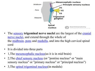
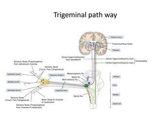
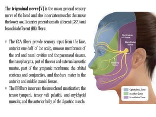
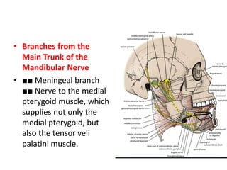
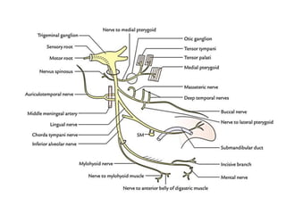
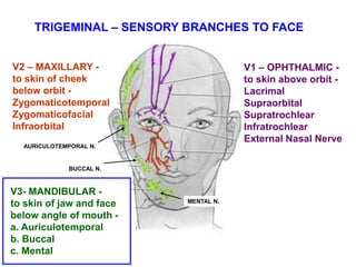
Trigeminal nerve
C N V
Functional components
C.N
• It has two roots
• Sensory root from mid pone on lateral side
• motor root come inferior to sensory root,and
pass through foramen ovale
Trigeminal divisions,ganglion,roots as we go towards
CNS.there in cns nuclei,then laminci
• The sensory trigeminal nerve nuclei are the largest of the cranial
nerve nuclei, and extend through the whole of
the midbrain, pons and medulla, and into the high cervical spinal
cord
• It is divided into three parts
• 1,The mesencephalic nucleus(as it is in mid brain)
• 2,The chief sensory nucleus (or "pontine nucleus" or "main
sensory nucleus" or "primary nucleus" or "principal nucleus")
• 3,The spinal trigeminal nucleus(in medula)
• Spinal tract of
trigeminal system.
• All fibers(trigeminal
laminesci) from the
nuclei go upwards
into
ventropostromedial
nucleus of
thallamus(trigemino-
thallamic tract)
• From here fibers
move up via posterior
limb of internal
capsule to the post
central
gyrus(somatosensory
cortex)
• Fibers of proprioception mainly from Muscles
Of Mastication from mandibular nerve goes to
mesencephalic nucleus
• Fine touch Fibers from face and mucosa of
nose and palate etc from Principle pontine
nucleus
• Pain and temp fibers are mainly connected to
spinal nucleus
Trigeminal path way
• Sensory nerve fibers for proprioception and fine touch
are heavly mylinated, while fibers for temp are less
mylinated and for pain are least mylinated.
• Fibers inside CNS are supported by schwan cell and
outside CNS are supported by oligodendrocytes,but for
7mm out side of pone same oligodendrocytes do
support the fibers out side CNS
• Those diseases which destroy central mylination
system can damage the sensory root of trigeminal
nerve as well e.g multiple sclerosis
It Moves from posterior cranial fossa into middle
cranial fossa
exits from anterolateral
surface of pons as large sensory root and small
motor root.
Ophthalmic
nerve
3. Nasociliary
1. Frontal N.
a) Supraorbital N.
b) Supratrochlear N.
Forehead, Upper Eyelid
2. Lacrimal N.
Upper eyelid
3. Nasociliary N.
a) Ant. and Post.
Ethmoidal N.
Nasal Cavity
Ethmoid Sinus , Tip of Nose
b) Long Ciliary N.
Sensory to
Cornea,sympathetic fiber
to dilator pupilae
c) Infratrochlear N.
Upper eyelid, Nose
V1 - OPHTHALMIC -
ALL GSA
1. Frontal
2. Lacrimal
V1
Supra-
trochlear
N.
Infra-
trochlear
N.
Supra-
orbital
N.
Long
Ciliary
Nn.
Ant. and
Post.
Ethmoidal
N.
The frontal nerve runs forward on the upper surface of the levator palpebrae
superioris muscle and divides into the supraorbital and supratrochlear nerves . These
nerves leave the orbital cavity and supply the frontal air sinus and the skin of the forehead
and the scalp.
The lacrimal nerve runs forward on the upper border of the lateral rectus muscle . It is
joined by the zygomaticotemporal branch of the maxillary nerve, which contains the
parasympathetic secretomotor fibers to the lacrimal gland. The lacrimal nerve then enters
the lacrimal gland and gives branches to the conjunctiva and the skin of the upper eyelid.
The nasociliary nerve crosses the optic nerve, runs forward on the upper border of
the medial rectus muscle and continues as the anterior ethmoid nerve through the
anterior ethmoidal foramen to enter the cranial cavity. It then descends through a slit at
the side of the crista galli to enter the nasal cavity. It gives off two internal nasal branches
and it then supplies the skin of the tip of the nose with the external nasal nerve. Its
branches include the following:
■■ Sensory fibers to the ciliary ganglion
■■ Long ciliary nerves that contain sympathetic fibers to the dilator pupillae muscle and
sensory fibers to the cornea
■■ Infratrochlear nerve that supplies the skin of the eyelids
■■ Posterior ethmoidal nerve that is sensory to the ethmoid and sphenoid sinuses
TRIGEMINAL – SENSORY BRANCHES TO FACE
V1 – OPHTHALMIC -
to skin above orbit -
a. Lacrimal
b. Supraorbital
c. Supratrochlear
d. Infratrochlear
e. External Nasal Nerve
(branch of Ant. Ethmoidal N.)
V2 – MAXILLARY -
to skin of cheek
below orbit -
Zygomaticotemporal
Zygomaticofacial
Infraorbital
V3- MANDIBULAR -
to skin of jaw and face
below angle of mouth -
Auriculotemporal
Buccal
Mental
LACRIMAL
N.
SUPRAORBITAL N.
SUPRATROCHLEAR N.
INFRATROCHLEAR N.
EXTERNAL
NASAL N.
Maxillary Nerve (V2) The maxillary nerve
arises from the trigeminal ganglion in the
middle cranial fossa. It passes forward in
the lateral wall of the cavernous sinus and
leaves the skull through the foramen
rotundum and crosses the pterygopalatine
fossa to enter the orbit through the inferior
orbital fissure . It then continues as the
infraorbital nerve in the infraorbital groove,
and it emerges on the face through the
infraorbital foramen. It gives sensory fibers
to the skin of the face and the side of the
nose
Branches
■■ Meningeal branches (supply middle cranial fossa
meninges)
■■ Zygomatic branch(via inferior orbital fissure) which
divides into the zygomaticotemporal and the
zygomaticofacial nerves that supply the skin of the face. The
zygomaticotemporal branch gives parasympathetic
secretomotor fibers to the lacrimal gland via the lacrimal
nerve.
■■ Ganglionic branches, which are two short nerves that
suspend the pterygopalatine ganglion in the pterygopalatine
fossa . They contain sensory fibers that have passed through
the ganglion from the nose, the palate, and the pharynx.
They also contain postganglionic parasympathetic fibers that
are going to the lacrimal gland.
■■ Posterior superior alveolar nerve
, which supplies the maxillary sinus
as well as the upper molar teeth and
adjoining parts of the gum and the
cheek
■■ Middle superior alveolar nerve,
which supplies the maxillary sinus as
well as the upper premolar teeth,
the gums, and the cheek
■■ Anterior superior alveolar nerve
, which supplies the maxillary sinus
as well as the upper canine and the
incisor teeth
Pterygomaxillary
Fissure
V2 - Maxillary enters
Pterygopalatine Fossa
V2 - MAXILLARY - GSA
note:
Pterygomaxillary
fissure is slit
between lateral
pterygoid plate
and maxillary bone;
Pterygopalatine
fossa is space
medial to slit
MAXILLARY BONE
LATERAL PTERYGOID PLATE
NOSE
Pterygopalatine Ganglion:The pterygopalatine
ganglion is a parasympathetic ganglion, which is
suspended from the maxillary nerve in the
pterygopalatine fossa . It is secretomotor to the
lacrimal and nasal glands .
Branches
■■ Orbital branches, which enter the orbit
through the inferior orbital fissure
■■ Greater and lesser palatine nerves , which
supply the palate, the tonsil, and the nasal
cavity
■■ Pharyngeal branch, which supplies the roof
of the nasopharynx.
1. Meningeal Branch
Dura of Middle
Cranial Fossa (not shown)
2. Ganglionic branches
a) Greater Palatine N.
Hard Palate
b) Lesser Palatine N.
Soft Palate
c) Nasopalatine
Nasal Cavity
Ant. part of hard Palate
d) Nasal Branches
Nasal Cavity
V2
Nasal
Branches
Naso-
Palatine
Greater
Palatine
Lesser
Palatine
V2 - MAXILLARY - GSA
Note: V2 branches from Pterygopalatine Fossa to Nasal Cavity are
deep and can only be seen on bisected head
Zygomatic
Infraorbital
3. Posterior
Superior Alveolar
Maxillary molar teeth
4. Infraorbital
Lower eyelid, nose,
upper lip
a) Ant. & Mid Sup
Alveolar
Other Maxillary teeth
5. Zygomatic - br.
a) Zygomaticofacial
b) Zygomaticotemporal
Skin of check, temporal
region
Post
Sup. Alveolar
V2 - MAXILLARY - GSA
Nose
V2
TRIGEMINAL – SENSORY BRANCHES TO FACE
V1 – OPHTHALMIC -
to skin above orbit -
Lacrimal
Supraorbital
Supratrochlear
Infratrochlear
External Nasal Nerve
V2 – MAXILLARY -
to skin of cheek
below orbit -
a. Zygomatico-
temporal
b. Zygomaticofacial
c. Infraorbital
V3- MANDIBULAR -
to skin of jaw and face
below angle of mouth -
Auriculotemporal
Buccal
Mental
INFRAORBITAL N.
ZYGOMATICOTEMPORAL N.
ZYGOMATICOFACIAL N.
V1 – OPHTHALMIC -
Sup. Orbital fissure –
GSA
V2 - MAXILLARY -
Foramen rotundum –
GSA
V3 – MANDIBULAR -
Foramen ovale – GSA
& SVE
TRIGEMINAL NERVE - V
V1
V2
V3
TRIGEMINAL NERVE SVE - FIRST BRANCHIAL ARCH MUSCLES
Mandibular Nerve (V3) The
mandibular nerve is both motor
and sensory . The sensory root
leaves the trigeminal ganglion and
passes out of the skull through the
foramen ovale to enter the
infratemporal fossa. The motor
root of the trigeminal nerve also
leaves the skull through the
foramen ovale and joins the
sensory root to form the trunk of
the mandibular nerve, and then
divides into a small anterior and a
large posterior division
• Branches from the
Main Trunk of the
Mandibular Nerve
• ■■ Meningeal branch
■■ Nerve to the medial
pterygoid muscle, which
supplies not only the
medial pterygoid, but
also the tensor veli
palatini muscle.
• Branches from the Anterior Division of the Mandibular Nerve
• ■■ Masseteric nerve to the masseter muscle
• ■■ Deep temporal nerves to the temporalis muscle
• ■■ Nerve to the lateral pterygoid muscle
• Buccal nerve to the skin and the mucous membrane of the
cheek . The buccal nerve does not supply the buccinator
muscle (which is supplied by the facial nerve), and it is the
only sensory branch of the anterior division of the mandibular
nerve.
Branches from the Posterior Division of the Mandibular
Nerve
■■ Auriculotemporal nerve, which supplies the skin of the
auricle , the external auditory meatus, the
temporomandibular joint, and the scalp. This nerve also
conveys postganglionic parasympathetic secretomotor fibers
from the otic ganglion to the parotid salivary gland.
■■ Lingual nerve, which descends in front of the inferior
alveolar nerve and enters the mouth . It then runs forward
on the side of the tongue and crosses the submandibular
duct. In its course, it is joined by the chorda tympani nerve ,
and it supplies the mucous membrane of the anterior two
thirds of the tongue and the floor of the mouth. It also gives
off preganglionic parasympathetic secretomotor fibers to the
submandibular ganglion.
• Inferior alveolar nerve , which
enters the mandibular canal to
supply the teeth of the lower jaw
and emerges through the mental
foramen (mental nerve) to supply
the skin of the chin. Before
entering the canal, it gives off the
mylohyoid nerve, which supplies
the mylohyoid muscle&anterior
belly of the digastric muscle.
• ■■ Communicating branch, which
frequently runs from the inferior
alveolar nerve to the lingual nerve
The branches of the posterior
division of the mandibular nerve
are sensory (except the nerve to
the mylohyoid muscle).
Deep
Temporal
1. Nervus Spinosus
Dura of Middle Cranial
Fossa (not shown)
2. Motor to
Medial Pterygoid
Tensor Tympani
Tensor Palati (not shown)
3. Ant Division
a. Nerve to Lat. Pterygoids
b. Masseteric N.
c. Deep Temporal
To Temporalis
d. Buccal Branch
Of Trigeminal
Sensory to Cheek
N. To Lat
Pterygoid
V3 - MANDIBULAR - GSA + SVE
Buccal
Branch
Of
Trigeminal
Auriculo-
Temporal
Post Division
a. Auriculotemporal
Skin of Temple, Scalp
External ear, TMJ
b. Lingual
Touch to Ant 2/3
Tongue
c. Inf. Alveolar N.
Sens to Lower Teeth
i) Mental N.
Skin Lower Jaw
ii) Nerve to Mylohyoid -
Motor to Mylohyoid
& Ant. Belly of Digastric
Inferior
Alveolar
Lingual
V3 - MANDIBULAR - GSA + SVE
MENTAL N.
N. TO
MYLOHYOID
TRIGEMINAL – SENSORY BRANCHES TO FACE
V1 – OPHTHALMIC -
to skin above orbit -
Lacrimal
Supraorbital
Supratrochlear
Infratrochlear
External Nasal Nerve
V2 – MAXILLARY -
to skin of cheek
below orbit -
Zygomaticotemporal
Zygomaticofacial
Infraorbital
V3- MANDIBULAR -
to skin of jaw and face
below angle of mouth -
a. Auriculotemporal
b. Buccal
c. Mental
AURICULOTEMPORAL N.
BUCCAL N.
MENTAL N.
The pterygoid canal (also vidian canal) is a passage in the skull leading from just
anterior to the foramen lacerum in the middle cranial fossa to the
pterygopalatine fossa. ... It transmits the nerve of pterygoid canal, artery of
the pterygoid canal and vein of the pterygoid canal.
cranial nerve 5.pptx

cranial nerve 5.pptx

  • 1.
  • 2.
  • 3.
  • 4.
    • It hastwo roots • Sensory root from mid pone on lateral side • motor root come inferior to sensory root,and pass through foramen ovale
  • 5.
    Trigeminal divisions,ganglion,roots aswe go towards CNS.there in cns nuclei,then laminci
  • 6.
    • The sensorytrigeminal nerve nuclei are the largest of the cranial nerve nuclei, and extend through the whole of the midbrain, pons and medulla, and into the high cervical spinal cord • It is divided into three parts • 1,The mesencephalic nucleus(as it is in mid brain) • 2,The chief sensory nucleus (or "pontine nucleus" or "main sensory nucleus" or "primary nucleus" or "principal nucleus") • 3,The spinal trigeminal nucleus(in medula)
  • 7.
    • Spinal tractof trigeminal system. • All fibers(trigeminal laminesci) from the nuclei go upwards into ventropostromedial nucleus of thallamus(trigemino- thallamic tract) • From here fibers move up via posterior limb of internal capsule to the post central gyrus(somatosensory cortex)
  • 11.
    • Fibers ofproprioception mainly from Muscles Of Mastication from mandibular nerve goes to mesencephalic nucleus • Fine touch Fibers from face and mucosa of nose and palate etc from Principle pontine nucleus • Pain and temp fibers are mainly connected to spinal nucleus
  • 13.
  • 15.
    • Sensory nervefibers for proprioception and fine touch are heavly mylinated, while fibers for temp are less mylinated and for pain are least mylinated. • Fibers inside CNS are supported by schwan cell and outside CNS are supported by oligodendrocytes,but for 7mm out side of pone same oligodendrocytes do support the fibers out side CNS • Those diseases which destroy central mylination system can damage the sensory root of trigeminal nerve as well e.g multiple sclerosis
  • 16.
    It Moves fromposterior cranial fossa into middle cranial fossa exits from anterolateral surface of pons as large sensory root and small motor root.
  • 20.
  • 21.
    3. Nasociliary 1. FrontalN. a) Supraorbital N. b) Supratrochlear N. Forehead, Upper Eyelid 2. Lacrimal N. Upper eyelid 3. Nasociliary N. a) Ant. and Post. Ethmoidal N. Nasal Cavity Ethmoid Sinus , Tip of Nose b) Long Ciliary N. Sensory to Cornea,sympathetic fiber to dilator pupilae c) Infratrochlear N. Upper eyelid, Nose V1 - OPHTHALMIC - ALL GSA 1. Frontal 2. Lacrimal V1 Supra- trochlear N. Infra- trochlear N. Supra- orbital N. Long Ciliary Nn. Ant. and Post. Ethmoidal N.
  • 23.
    The frontal nerveruns forward on the upper surface of the levator palpebrae superioris muscle and divides into the supraorbital and supratrochlear nerves . These nerves leave the orbital cavity and supply the frontal air sinus and the skin of the forehead and the scalp. The lacrimal nerve runs forward on the upper border of the lateral rectus muscle . It is joined by the zygomaticotemporal branch of the maxillary nerve, which contains the parasympathetic secretomotor fibers to the lacrimal gland. The lacrimal nerve then enters the lacrimal gland and gives branches to the conjunctiva and the skin of the upper eyelid. The nasociliary nerve crosses the optic nerve, runs forward on the upper border of the medial rectus muscle and continues as the anterior ethmoid nerve through the anterior ethmoidal foramen to enter the cranial cavity. It then descends through a slit at the side of the crista galli to enter the nasal cavity. It gives off two internal nasal branches and it then supplies the skin of the tip of the nose with the external nasal nerve. Its branches include the following: ■■ Sensory fibers to the ciliary ganglion ■■ Long ciliary nerves that contain sympathetic fibers to the dilator pupillae muscle and sensory fibers to the cornea ■■ Infratrochlear nerve that supplies the skin of the eyelids ■■ Posterior ethmoidal nerve that is sensory to the ethmoid and sphenoid sinuses
  • 25.
    TRIGEMINAL – SENSORYBRANCHES TO FACE V1 – OPHTHALMIC - to skin above orbit - a. Lacrimal b. Supraorbital c. Supratrochlear d. Infratrochlear e. External Nasal Nerve (branch of Ant. Ethmoidal N.) V2 – MAXILLARY - to skin of cheek below orbit - Zygomaticotemporal Zygomaticofacial Infraorbital V3- MANDIBULAR - to skin of jaw and face below angle of mouth - Auriculotemporal Buccal Mental LACRIMAL N. SUPRAORBITAL N. SUPRATROCHLEAR N. INFRATROCHLEAR N. EXTERNAL NASAL N.
  • 26.
    Maxillary Nerve (V2)The maxillary nerve arises from the trigeminal ganglion in the middle cranial fossa. It passes forward in the lateral wall of the cavernous sinus and leaves the skull through the foramen rotundum and crosses the pterygopalatine fossa to enter the orbit through the inferior orbital fissure . It then continues as the infraorbital nerve in the infraorbital groove, and it emerges on the face through the infraorbital foramen. It gives sensory fibers to the skin of the face and the side of the nose
  • 28.
    Branches ■■ Meningeal branches(supply middle cranial fossa meninges) ■■ Zygomatic branch(via inferior orbital fissure) which divides into the zygomaticotemporal and the zygomaticofacial nerves that supply the skin of the face. The zygomaticotemporal branch gives parasympathetic secretomotor fibers to the lacrimal gland via the lacrimal nerve. ■■ Ganglionic branches, which are two short nerves that suspend the pterygopalatine ganglion in the pterygopalatine fossa . They contain sensory fibers that have passed through the ganglion from the nose, the palate, and the pharynx. They also contain postganglionic parasympathetic fibers that are going to the lacrimal gland.
  • 29.
    ■■ Posterior superioralveolar nerve , which supplies the maxillary sinus as well as the upper molar teeth and adjoining parts of the gum and the cheek ■■ Middle superior alveolar nerve, which supplies the maxillary sinus as well as the upper premolar teeth, the gums, and the cheek ■■ Anterior superior alveolar nerve , which supplies the maxillary sinus as well as the upper canine and the incisor teeth
  • 30.
    Pterygomaxillary Fissure V2 - Maxillaryenters Pterygopalatine Fossa V2 - MAXILLARY - GSA note: Pterygomaxillary fissure is slit between lateral pterygoid plate and maxillary bone; Pterygopalatine fossa is space medial to slit MAXILLARY BONE LATERAL PTERYGOID PLATE NOSE
  • 32.
    Pterygopalatine Ganglion:The pterygopalatine ganglionis a parasympathetic ganglion, which is suspended from the maxillary nerve in the pterygopalatine fossa . It is secretomotor to the lacrimal and nasal glands . Branches ■■ Orbital branches, which enter the orbit through the inferior orbital fissure ■■ Greater and lesser palatine nerves , which supply the palate, the tonsil, and the nasal cavity ■■ Pharyngeal branch, which supplies the roof of the nasopharynx.
  • 33.
    1. Meningeal Branch Duraof Middle Cranial Fossa (not shown) 2. Ganglionic branches a) Greater Palatine N. Hard Palate b) Lesser Palatine N. Soft Palate c) Nasopalatine Nasal Cavity Ant. part of hard Palate d) Nasal Branches Nasal Cavity V2 Nasal Branches Naso- Palatine Greater Palatine Lesser Palatine V2 - MAXILLARY - GSA Note: V2 branches from Pterygopalatine Fossa to Nasal Cavity are deep and can only be seen on bisected head
  • 34.
    Zygomatic Infraorbital 3. Posterior Superior Alveolar Maxillarymolar teeth 4. Infraorbital Lower eyelid, nose, upper lip a) Ant. & Mid Sup Alveolar Other Maxillary teeth 5. Zygomatic - br. a) Zygomaticofacial b) Zygomaticotemporal Skin of check, temporal region Post Sup. Alveolar V2 - MAXILLARY - GSA Nose V2
  • 35.
    TRIGEMINAL – SENSORYBRANCHES TO FACE V1 – OPHTHALMIC - to skin above orbit - Lacrimal Supraorbital Supratrochlear Infratrochlear External Nasal Nerve V2 – MAXILLARY - to skin of cheek below orbit - a. Zygomatico- temporal b. Zygomaticofacial c. Infraorbital V3- MANDIBULAR - to skin of jaw and face below angle of mouth - Auriculotemporal Buccal Mental INFRAORBITAL N. ZYGOMATICOTEMPORAL N. ZYGOMATICOFACIAL N.
  • 36.
    V1 – OPHTHALMIC- Sup. Orbital fissure – GSA V2 - MAXILLARY - Foramen rotundum – GSA V3 – MANDIBULAR - Foramen ovale – GSA & SVE TRIGEMINAL NERVE - V V1 V2 V3
  • 38.
    TRIGEMINAL NERVE SVE- FIRST BRANCHIAL ARCH MUSCLES
  • 39.
    Mandibular Nerve (V3)The mandibular nerve is both motor and sensory . The sensory root leaves the trigeminal ganglion and passes out of the skull through the foramen ovale to enter the infratemporal fossa. The motor root of the trigeminal nerve also leaves the skull through the foramen ovale and joins the sensory root to form the trunk of the mandibular nerve, and then divides into a small anterior and a large posterior division
  • 40.
    • Branches fromthe Main Trunk of the Mandibular Nerve • ■■ Meningeal branch ■■ Nerve to the medial pterygoid muscle, which supplies not only the medial pterygoid, but also the tensor veli palatini muscle.
  • 44.
    • Branches fromthe Anterior Division of the Mandibular Nerve • ■■ Masseteric nerve to the masseter muscle • ■■ Deep temporal nerves to the temporalis muscle • ■■ Nerve to the lateral pterygoid muscle • Buccal nerve to the skin and the mucous membrane of the cheek . The buccal nerve does not supply the buccinator muscle (which is supplied by the facial nerve), and it is the only sensory branch of the anterior division of the mandibular nerve.
  • 47.
    Branches from thePosterior Division of the Mandibular Nerve ■■ Auriculotemporal nerve, which supplies the skin of the auricle , the external auditory meatus, the temporomandibular joint, and the scalp. This nerve also conveys postganglionic parasympathetic secretomotor fibers from the otic ganglion to the parotid salivary gland. ■■ Lingual nerve, which descends in front of the inferior alveolar nerve and enters the mouth . It then runs forward on the side of the tongue and crosses the submandibular duct. In its course, it is joined by the chorda tympani nerve , and it supplies the mucous membrane of the anterior two thirds of the tongue and the floor of the mouth. It also gives off preganglionic parasympathetic secretomotor fibers to the submandibular ganglion.
  • 48.
    • Inferior alveolarnerve , which enters the mandibular canal to supply the teeth of the lower jaw and emerges through the mental foramen (mental nerve) to supply the skin of the chin. Before entering the canal, it gives off the mylohyoid nerve, which supplies the mylohyoid muscle&anterior belly of the digastric muscle. • ■■ Communicating branch, which frequently runs from the inferior alveolar nerve to the lingual nerve The branches of the posterior division of the mandibular nerve are sensory (except the nerve to the mylohyoid muscle).
  • 49.
    Deep Temporal 1. Nervus Spinosus Duraof Middle Cranial Fossa (not shown) 2. Motor to Medial Pterygoid Tensor Tympani Tensor Palati (not shown) 3. Ant Division a. Nerve to Lat. Pterygoids b. Masseteric N. c. Deep Temporal To Temporalis d. Buccal Branch Of Trigeminal Sensory to Cheek N. To Lat Pterygoid V3 - MANDIBULAR - GSA + SVE Buccal Branch Of Trigeminal
  • 50.
    Auriculo- Temporal Post Division a. Auriculotemporal Skinof Temple, Scalp External ear, TMJ b. Lingual Touch to Ant 2/3 Tongue c. Inf. Alveolar N. Sens to Lower Teeth i) Mental N. Skin Lower Jaw ii) Nerve to Mylohyoid - Motor to Mylohyoid & Ant. Belly of Digastric Inferior Alveolar Lingual V3 - MANDIBULAR - GSA + SVE MENTAL N. N. TO MYLOHYOID
  • 51.
    TRIGEMINAL – SENSORYBRANCHES TO FACE V1 – OPHTHALMIC - to skin above orbit - Lacrimal Supraorbital Supratrochlear Infratrochlear External Nasal Nerve V2 – MAXILLARY - to skin of cheek below orbit - Zygomaticotemporal Zygomaticofacial Infraorbital V3- MANDIBULAR - to skin of jaw and face below angle of mouth - a. Auriculotemporal b. Buccal c. Mental AURICULOTEMPORAL N. BUCCAL N. MENTAL N.
  • 53.
    The pterygoid canal(also vidian canal) is a passage in the skull leading from just anterior to the foramen lacerum in the middle cranial fossa to the pterygopalatine fossa. ... It transmits the nerve of pterygoid canal, artery of the pterygoid canal and vein of the pterygoid canal.