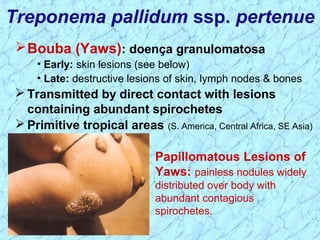
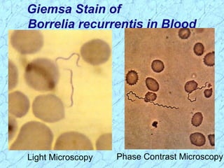
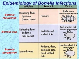
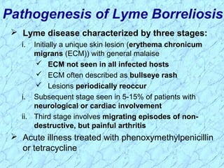
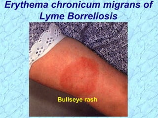
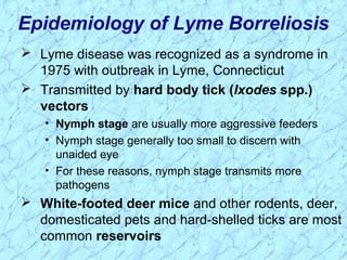
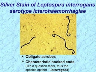
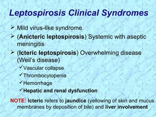
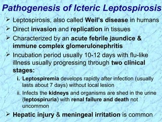
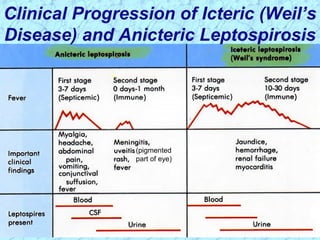
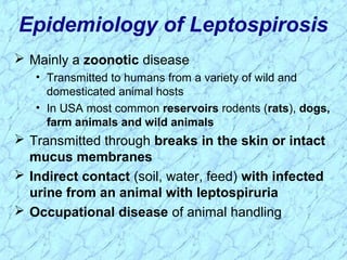
Treponema pallidum causes syphilis in humans. It has a long incubation period and can progress through primary, secondary, latent, and tertiary stages if left untreated. Borrelia recurrentis causes epidemic relapsing fever which is transmitted between humans by body lice. Borrelia burgdorferi causes Lyme disease transmitted by certain ticks and causes a characteristic bullseye rash. Leptospira interrogans causes leptospirosis, a zoonotic disease transmitted by contact with infected animal urine, which can range from mild to severe with jaundice and organ failure.
3. Características Gerais das Espiroquetas
Gram-negativas
• Espiroqueta vem do grego “cabelo encaracolado”
Células extremamente finas e podem ser longas
Células helicoidais com extremidades cônicas
Móveis através de flagelos periplásmicos
• Diferente número de flagelos e diferentes inserções em
Treponema, Borrelia e Leptospira
11. Treponema pallidum ssp. endemicum
Bejel (sífilis endêmica)
• Initial lesions: nondescript oral lesions
• Secondary lesions: oral papules and mucosal patches
• Late: gummas (granulomas) of skin, bones &
nasopharynx
Transmitted person-to-person by contaminated
eating utensils
Tropical/subtropical areas (Africa, Asia & Australia)
12. Treponema pallidum ssp. pertenue
Papillomatous Lesions of
Yaws: painless nodules widely
distributed over body with
abundant contagious
spirochetes.
Bouba (Yaws): doença granulomatosa
• Early: skin lesions (see below)
• Late: destructive lesions of skin, lymph nodes & bones
Transmitted by direct contact with lesions
containing abundant spirochetes
Primitive tropical areas (S. America, Central Africa, SE Asia)
13. Treponema carateum
Pinta: Primariamente restrita à pele
• 1-3 week incubation period
• Initial lesions: small pruritic papules
• Secondary: enlarged plaques persist for
months to years
• Late: disseminated, recurrent
hypopigmentation or depigmentation of
skin lesions; scarring & disfigurement
Transmitted by direct contact with skin
lesions
Primitive tropical areas
(Mexico, Central & South America)
Hypopigmented Skin Lesions
of Pinta: depigmentation is
commonly seen as a late sequel with
all treponemal diseases
18. Muito finos para serem vistos pela microscopia
óptica, apesar de serem corados pelo Gram
• Espiroquetas móveis pela microscopia de campo
escuro
• Imunofluorescência direta ou impregnação pela prata
Patógeno intra-celular
Não crescem in vitro
Não sobrevivem fora do hospedeiro
Características do Treponema
pallidum
19. Epidemiologia do T. pallidum
Transmitidos por contato sexual direto ou
transmissão materno fetal
Não são altamente contagiosos (~30% de chance
de adquirir doença após exposição única a parceiro
infectado). Taxa de transmissão depende do estágio
da doença
Longo período de incubação durante o qual o
hospedeiro não transmite a doença
• Epidemiologia útil para rastrear contatos e
administrar tratamento preventivo
Prostituição permanece aspecto epidemiológico
central na transmissão
20. Patogênese do T. pallidum
Destruição de tecidos e lesões são primariamente
uma consequência da resposta imune dos
pacientes
Sífilis é uma doença dos vasos sanguíneos e das
áreas perivasculares
Apesar de uma vigorosa resposta imune do
hospedeiro o microorganismo é capaz de persistir
por décadas
• Infecção não é completamente controlada ou
erradicada
• Nos estágios iniciais, há inibição da imunidade celular
• Nos estágios tardios da doença, as lesões tendem a ser
localizadas
21. Fatores de Virulência -T. pallidum
Outer membrane proteins promote adherence
Hyaluronidase may facilitate perivascular
infiltration
Antiphagocytic coating of fibronectin
Tissue destruction and lesions are primarily
result of host’s immune response
(immunopathology)
22. Doença primária envolve a invasão das membranas
mucosas, multiplicação rápida e ampla
disseminação através dos vasos linfáticos e
circulação sistêmica
Ocorre antes do desenvolvimento da lesão primária
10-90 dias (usualmente 3-4 semanas) após contato
inicial o hospedeiro apresenta uma resposta
inflamatória no sítio da inoculação resultando na
lesão sifilítica, chamada de cancro (não dolorosa)
• Cancro muda de duro a ulcerativo com abundante
disseminação de espiroquetas
• Edema das paredes capilares e linfonodos regionais
• Lesão Primária cicatriza espontaneamente em dois meses,
levando a falsa sensação de alívio
Patogênese do T. pallidum (cont.)
Sífilis Primária
23. Doença secundária aparece 2-10 semanas após
lesão primária
Rash mucocutâneo amplamente disseminado
Lesões secundárias da pele e membranas
mucosas são altamente contagiosas
Resposta imunológica generalizada
Patogênese do T. pallidum (cont.)
Sífilis secundária
25. Após estágio secundário da doença, hospedeiro
entra em período latente
•Primeiros 4 anos = latente precoce
•Período subsequente = latente tardio
Cerca de 40% dos pacientes em estágio latente
progridem para doença sifilítica tardia latente
terciária (estágio terciário)
Patogênese do T. pallidum (cont.)
Estágio Latente da Sífilis
26. Sífilis Terciária caracterizada por lesões cutâneas
granulomatosas localizadas (gomas) nas quais
poucos organismos estão presentes
Neurosífilis tardia se desenvolve em cerca de 1/6
dos casos não tratados, usualmente mais de 5 anos
após infecção inicial
• Demência, convulsões, etc.
Involvimento cardiovascular aparece 10-40 anos
após infecção inicial resultando em insuficiência
cardíaca e morte
Patogênese do T. pallidum (cont.)
Sífilis Terciária
27. Progressão da Sífilis Não Tratada
Tertiary Stage
Late benign Gomas na pele e partes moles
28. Congenital syphilis results from transplacental
infection
T. pallidum septicemia in the developing fetus and
widespread dissemination
Abortion, neonatal mortality, and late mental or
physical problems resulting from scars from the
active disease and progression of the active disease
state
Pathogenesis of T. pallidum (cont.)
Congenital Syphilis
30. Prevention & Treatment of Syphilis
Penicillin remains drug of choice
• WHO monitors treatment recommendations
• 7-10 days continuously for early stage
• At least 21 days continuously beyond the early stage
Prevention with barrier methods (e.g., condoms)
Prophylactic treatment of contacts identified
through epidemiological tracing
31. Diagnostic Tests for Syphilis
NOTE: Treponemal antigen tests indicate experience with a treponemal
infection, but cross-react with antigens other than T. pallidum ssp.
pallidum. Since pinta and yaws are rare in USA, positive treponemal
antigen tests are usually indicative of syphilitic infection.
(Original Wasserman Test)
41. Associated with poverty, crowding, and warfare
Arthropod vectors
• Louse-borne borreliosis = Epidemic Relapsing Fever
Transmitted person-to-person by human body lice
(vectors) from infected human reservoir
Infect host only when louse is injured, e.g., during
scratching
Therefore, a single louse can only infect a single person
Lice leave host that develops a fever and seek normal
temperature host
• Tick-borne borreliosis = Endemic Relapsing Fever
Sporadic cases
Transmitted by soft body ticks (vectors) from small
mammal reservoir
Ticks can multiply and infect new human hosts
Epidemiology of Relapsing Fever
42. Pathogenesis of Relapsing Fever
Relapsing fever (a.k.a., tick fever, borreliosis, famine
fever)
• Acute infection with 2-14 day (~ 6 day) incubation period
• Followed by recurring febrile episodes
• Constant spirochaetemia that worsens during febrile
stages
Epidemic Relapsing Fever = Louse-borne borreliosis
• Borrelia recurrentis
Endemic Relapsing Fever = Tick-borne borreliosis
• Borrelia spp.
45. Pathogenesis of Lyme Borreliosis
Lyme disease characterized by three stages:
i. Initially a unique skin lesion (erythema chronicum
migrans (ECM)) with general malaise
ECM not seen in all infected hosts
ECM often described as bullseye rash
Lesions periodically reoccur
i. Subsequent stage seen in 5-15% of patients with
neurological or cardiac involvement
ii. Third stage involves migrating episodes of non-
destructive, but painful arthritis
Acute illness treated with phenoxymethylpenicillin
or tetracycline
48. Bacteria and Syndromes that Cause
Cross-Reactions with Lyme
Borreliosis Serological Tests
49. Lyme disease was recognized as a syndrome in
1975 with outbreak in Lyme, Connecticut
Transmitted by hard body tick (Ixodes spp.)
vectors
• Nymph stage are usually more aggressive feeders
• Nymph stage generally too small to discern with
unaided eye
• For these reasons, nymph stage transmits more
pathogens
White-footed deer mice and other rodents, deer,
domesticated pets and hard-shelled ticks are most
common reservoirs
Epidemiology of Lyme Borreliosis
53. Silver Stain of Leptospira interrogans
serotype icterohaemorrhagiae
Obligate aerobes
Characteristic hooked ends
(like a question mark, thus the
species epithet – interrogans)
54. Leptospirosis Clinical Syndromes
Mild virus-like syndrome
(Anicteric leptospirosis) Systemic with aseptic
meningitis
(Icteric leptospirosis) Overwhelming disease
(Weil’s disease)
Vascular collapse
Thrombocytopenia
Hemorrhage
Hepatic and renal dysfunction
NOTE: Icteric refers to jaundice (yellowing of skin and mucus
membranes by deposition of bile) and liver involvement
55. Leptospirosis, also called Weil’s disease in humans
Direct invasion and replication in tissues
Characterized by an acute febrile jaundice &
immune complex glomerulonephritis
Incubation period usually 10-12 days with flu-like
illness usually progressing through two clinical
stages:
i. Leptospiremia develops rapidly after infection (usually
lasts about 7 days) without local lesion
ii. Infects the kidneys and organisms are shed in the urine
(leptospiruria) with renal failure and death not
uncommon
Hepatic injury & meningeal irritation is common
Pathogenesis of Icteric Leptospirosis
56. Clinical Progression of Icteric (Weil’s
Disease) and Anicteric Leptospirosis
(pigmented
part of eye)
57. Epidemiology of Leptospirosis
Mainly a zoonotic disease
• Transmitted to humans from a variety of wild and
domesticated animal hosts
• In USA most common reservoirs rodents (rats), dogs,
farm animals and wild animals
Transmitted through breaks in the skin or intact
mucus membranes
Indirect contact (soil, water, feed) with infected
urine from an animal with leptospiruria
Occupational disease of animal handling