Tips for using and customizing a medical PPT on thoracic duct anatomy
•Download as PPTX, PDF•
0 likes•130 views

This document provides tips for using a PowerPoint presentation on the thoracic duct: 1. The presentation can be freely downloaded, edited, and modified. Many of the slides are blank except for the title to facilitate active learning. 2. The instructor should first show blank slides related to a topic and ask students what they know, then show the next slide with information. 3. This process of blank slide then information slide should be repeated for revisions and self study. 4. The presentation covers the introduction, formation, course, relations, tributaries and applied anatomy of the thoracic duct.

Recommended
More Related Content
What's hot
What's hot (20)
Similar to Tips for using and customizing a medical PPT on thoracic duct anatomy
Similar to Tips for using and customizing a medical PPT on thoracic duct anatomy (20)
More from Pradeep Pande
More from Pradeep Pande (20)
Recently uploaded
Recently uploaded (20)
Tips for using and customizing a medical PPT on thoracic duct anatomy
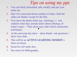
- 1. Tips on using my ppt. 1. You can freely download, edit, modify and put your name etc. 2. Don’t be concerned about number of slides. Half the slides are blanks except for the title. 3. First show the blank slides (eg. Aetiology ) > Ask students what they already know about ethology of today's topic. > Then show next slide which enumerates aetiologies. 4. At the end rerun the show – show blank> ask questions > show next slide. 5. This will be an ACTIVE LEARNING SESSION x three revisions. 6. Good for self study also. 7. See notes for bibliography.
- 4. Introduction Formation Course Relations Tributaries Applied anatomy Learning objectives 4
- 6. Introduction - Is the largest lymphatic trunk which drains chyle(product of fat digestion) & most lymph of body. - Extent- Upper abdomen at lower border of T12 to lower part of neck, crossing post & sup mediastinum - 45cms long & 0.5cms wide - Appears Beaded due to presence of many valves in its lumen 6
- 7. Drains lymph from whole of body except • Rt side of head & neck • Rt upper limb • Rt lung & thoracic wall • Rt side of heart and rt surface of liver Area of drainage 7
- 8. 8
- 9. Course of Thoracic Duct 9
- 10. Course: 10
- 11. Course: Begins in abdomen at lower border of T12 as a continuation of cisterna chyli Enters post mediastinum through aortic opening of diaphragm(T12) At T5 shifts to left & runs in superior mediastinum At C7 (root of neck) arches laterally, then downwards Ends at angle formed by union of left int jugular vein & lt subclavian vein, (regurge of blood prevented by a pair of valves) 11
- 12. Tributaries: Receives lymph from both halves below diaphragm through Cisternae chyli & Lt half above diaphragm 1. From post IC lymph nodes of lower 6 spaces 2. From upper lumbar nodes(paraaortic LN) 3. From Post Mediastinal lymph nodes & post IC LNs of upper 6 IC spaces 4. From axilla through Lt Subclavian trunk 5. From nodes in Lt ½ of H & N thru Lt Jugular trunk 6. From Lt half of thorax (Lt lung & Lt side of heart) through Lt Bronchomediastinal trunk 12
- 13. Applied anatomy Obstruction of Thoracic duct – Due to mature filarial parasites lymph vessels get burst chylothorax, chyloperitoneum, chyluria. Cervical part of thoracic duct is damaged in block dissection of neck Thoracic duct is very thin walled and colourless so more prone for injury during surgery in post mediastinum. 13
- 14. Get this ppt in mobile 1. Download Microsoft PowerPoint from play store. 2. Open Google assistant 3. Open Google lens. 4. Scan qr code from next slide.
- 15. Get this ppt in mobile
- 16. Get my ppt collection • https://www.slideshare.net/drpradeeppande/ edit_my_uploads • https://www.dropbox.com/sh/x600md3cvj8 5woy/AACVMHuQtvHvl_K8ehc3ltkEa?dl =0 • https://www.facebook.com/doctorpradeeppa nde/?ref=pages_you_manage