SlideShare a Scribd company logo
1 of 18
Download to read offline
Anatomy Of The Nose
The nose is divided into two parts;
1. External nose
2. Internal nose
1.External Nose
The external nose is pyramidal in shape present anteriorly on the face. It upper
narrow part forms Root of Nose while broad lower part with openings is called
Base of Nose. Base in directed downward. The pyramidal nose consists of
osteocartilaginous framework covered by muscles and skin.
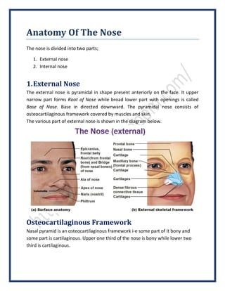
The various part of external nose is shown in the diagram below.
Osteocartilaginous Framework
Nasal pyramid is an osteocartilaginous framework i-e some part of it bony and
some part is cartilaginous. Upper one third of the nose is bony while lower two
third is cartilaginous.
Bony Part
Upper one third of the external framework of nose is contributed by hard bone.
The bony part is formed by two nasal bones which meet in midline and rest on
upper part of nasal process of the frontal bones and are also themselves held
between the frontal processes of the maxillae.
Cartilaginous Part
The cartilaginous part of nose is formed by three type of cartilages;
1) Upper lateral cartilages
2) Lower lateral cartilages (alar cartilages)
3) Lesser alar cartilages (sesamoid cartilages)
4) Septal cartilage
Upper Lateral Cartilages
These cartilages extend from the undersurface of the nasal bones above, to the
alar cartilages below. Anteriorly, these cartilages fuse with each other and with
the upper border of the septal cartilage in the midline. The lower edge of upper
lateral cartilages is free and can be seen intranasally as limen vestibuli or nasal
valve on each side separately.
Lower Lateral Cartilages (Alar Cartilages)
Each of the alar cartilages is U-shaped which has two crus i-e lateral crus and
medial crus. The lateral crus form the ala of nose and medial crus becomes the
part of columella. The lateral crus of alar cartilage overlap the lower edge of
upper lateral cartilage on each side (covers the free edge of upper lateral
cartilages which form limen vestibuli or nasal valve on each side).
Lesser Alar Cartilages (Sesamoid Cartilages)
These are two or more in numbers which lie above and lateral to the alar
cartilages. Perichondrium and periosteum connects these cartilages with one
another and with the adjoining bones. The free edge or margin of nostril is
formed by the fibrofatty tissue in most of the part (posterior of the margin) and
not by the alar cartilages (alar cartilages form only some portion of anterior of the
margin of nostril).
Septal Cartilage
Septal cartilage is the midline structure with its anterosuperior border running
from undersurface of nasal bone to the nasal tip. It provides support to the
dorsum of cartilaginous part of the nose.
Clinical: - After removal of septal cartilage (completely or most of its part) as
submucosal resection (SMR) operation, support of dorsum of nose is lost and
supratip depression results. In order to correct it, we have to provide artificial
support by surgery.
Musculature Of Nose
Nasal musculature covers the osteocartilaginous framework of the nose and is
responsible for the movement of the nasal tip, ala and the overlying skin. These
are the procerus, nasalis (transverse and alar parts), levator labii superioris
alaeque nasi, anterior and posterior dilator nares and depressor septi.
Nasal Skin
The skin over the nasal bones and the upper lateral cartilages is thin and freely
mobile, while that covering the alar cartilages is thick and closely adherent and
rich of sebaceous gland.
Clinical: - The hypertrophy of sebaceous gland in nasal skin gives rise to a
lobulated tumor which is called rhinophyma.
2.Internal Nose
Nasal septum divides the internal nose into right and left nasal cavities. Each nasal
cavity has communication with the exterior through naris or nostril and with the
Nasopharynx through posterior nasal aperture or choana. Nasal cavity is
partitioned into two areas i-e skin lined portion, the vestibule and a mucosa lined
portion, the nasal cavity proper.
a) Vestibule of nose
b) Nasal cavity proper
a) Vestibule Of Nose
Vestibule is anterior and inferior part of the nasal cavity. It is lined by skin
(keratinized stratified squamous epithelium) and contains sebaceous glands, hair
follicles and hair called vibrissae.
Note: - The hairs in the vestibule of nose are called vibrissae.
The upper limit of the nasal vestibule is demarcated by nasal valve (limen nasi) a
structure visible on the lateral wall while seen from inside. The limen vestibuli is
actually the lower free edge of upper lateral cartilage seen intranasally and is
same structure named as limen nasi.
Nasal Valve
Nasal valve is bounded laterally by the lower border of upper lateral cartilage
(limen vestibuli) and fibrofatty tissue and anterior end of inferior turbinate.
Medially the structure present is nasal septum, and caudally the floor of pyriform
aperture.
Note: - The angle between the nasal septum and lower border of upper lateral
cartilage is about 300
.
Nasal Valve Area
Nasal valve area is the least cross sectional area of the nose that functions to
regulate airflow and resistance on inspiration. It is the cross sectional area
bounded by the structures forming the nasal valve.
b) Nasal Cavity Proper
The nasal cavity proper is the rest portion of internal nose other than the
vestibule. Each nasal cavity has a lateral wall, a medial wall, a roof and a floor.
i. Lateral nasal wall
ii. Medial nasal wall
iii. Roof
iv. Floor
i. Lateral Nasal Wall
On the lateral wall of the nose, three and occasionally four turbinates or conchae
are well demarcated. Conchae or turbinates are scroll like bony projections
covered by the mucous membrane. The spaces between the turbinates are called
meatuses.
Note: - Inferior turbinate is the most prominent bony projection and the superior
turbinate is the least prominent bony projection.
Inferior Turbinate
Inferior turbinate is the separate bone while the other turbinates are the bony
projections of various bones. Below it, into the inferior meatus, opens the
nasolacrimal duct. At the terminal end of the nasolacrimal duct, a mucosal valve
called Hasner’s valve is present that guard it.
Middle Turbinate
Middle turbinate is ethmoturbinal i-e middle turbinate is the part of ethmoid
bone. It’s attachment to the lateral wall is by a bony lamella called ground or
basal lamella which is in S-shaped manner, not in straight line. In the anterior
third, it lies in sagittal plane and is attached to lateral edge of cribriform plate. In
the middle third, it lies in frontal plane and is attached to lamina papyracea while
in its posterior third, it runs horizontally and forms roof of the middle meatus and
is attached to lamina papyracea and medial wall of maxillary sinus.
The anterior group of paranasal sinuses is the ostia of various sinuses draining
anterior to basal lamina while the posterior group of paranasal sinuses is those
opening into posterior and superior to basal lamina.
Middle Meatus
Middle meatus is related with several important structures which are important
in endoscopic surgery of the sinuses.
Uncinate process is a hook like structure running in from anterosuperior to
posteroinferior direction. It posterior border is sharp and runs parallel to anterior
border of bulla ethmoidalis. The gap between the two is called is hiatus
semilunaris (inferior), a two dimensional space 1-2 mm in width.
The anteroinferior border of uncinate process is attached to the lateral wall and
the posteroinferior end of uncinate process is attached to inferior turbinate
dividing the membranous part of lower middle meatus into anterior and posterior
fontanelle.
Clinical: - The fontanel area is devoid of bone and consists of membrane only and
leads into maxillary sinus when perforated.
Upper attachment of uncinate process shows great variations and may be
inserted into the lateral nasal wall, upward into the base of skull or medially into
the middle turbinate which accounts for variations in drainage of frontal sinus.
The space limited medially by the uncinate process and frontal process of maxilla
and sometimes lacrimal bone and laterally by the lamina papyracea is called
infundibulum.
The natural ostium of the maxillary sinus is situated in the lower part of
infundibulum. The accessory ostium or ostia of maxillary sinus are sometimes
seen in the anterior or posterior fontanel.
Bulla Ethmoidalis
Bulla ethmoidalis is an ethmoidal cell situated behind the uncinate process. The
anterior surface of the bulla forms the posterior boundary of hiatus semilunaris.
Bulla ethmoidalis may be a pnematized cell or a solid bony prominence depending
upon the degree of pnematization. It may be extended superiorly to the base of
skull and posteriorly to fuse with the ground lamella. Sometimes, space is present
above or behind the bulla, it is called suprabullar or retrobullar recesses
respectively.
The suprabullar and retrobullar recesses together form the lateral sinus (sinus
lateralis of Grunwald). The lateral sinus is thus bounded superiorly by the skull
base, laterally by lamina papyracea, medially by middle turbinate and inferiorly by
the bulla ethmoidalis. Posteriorly, the sinus lateralis may extend up to basal
lamella of meddle turbinate.
The cleft like communication between the bulla and skull base and opening into
middle meatus in also called hiatus semilunaris superior in contrast to hiatus
semilunaris inferior referred to before.
Atrium Of The Middle Meatus
Atrium of the middle meatus is a shallow depression lying in front of middle
turbinate and above the nasal vestibule.
Agger Nasi
Agger nasi is an elevation just anterior to the attachment of middle turbinate.
When pnematized it contains air cells, the agger nasi cells, which communicate
with frontal recess. An enlarged agger nasi cell may encroach on frontal recess
area, constricting it and causing mechanical obstruction to frontal sinus drainage.
The pnematization of middle turbinate leads to an enlarged ballooned out middle
turbinate called concha bullosa. It drains into frontal recess directly or through
agger nasi cells. Haller cells are air cells situated in the roof of maxillary sinus.
They are peneumatized from anterior or posterior ethmoid cells. Enlargement of
Haller cells encroaches on ethmoid infundibulum, impeding draining of maxillary
sinus.
Superior Turbinate
Superior turbinate is also an ethmoturbinal i-e a part of ethmoidal bone. It is
situated posterior and superior to middle turbinate. It may also get pnematized
by one or more cells. It is an important landmark to identify ostium of sphenoid
sinus which lies medial to it.
Superior Meatus
Likewise other meatus, superior meatus is the space present below the superior
turbinate respectively. Posterior ethmoidal cells have opening into it. Posterior
ethmoidal cells vary from 1-5 in numbers. Onodi cell is a posterior ethmoidal cell
which may grow posteriorly by the side of sphenoid sinus or superior to it for as
much distance as 1.5 cm from the anterior surface of sphenoid.
Clinical: - Onodi cell have surgical importance as the optic nerve may be related to
its lateral wall.
Sphenoethmoidal Recess
Sphenoethmoidal recess is situated above the superior turbinate. Sphenoid sinus
opens into it.
Supreme Turbinate
Sometimes, a fourth turbinate called supreme turbinate is also present above the
superior turbinate and has a narrow meatus beneath it.
The ostium of sphenoid sinus is situated in the sphenoehtmoidal recess medial to
the superior or supreme turbinate. It can be located endosceopically about 1 cm
above the upper margin of posterior choana close to the posterior border of the
septum.
ii. Medial Nasal Wall
Nasal septum forms the medial wall of the nose. The detail anatomy is described
separately as it is a structure of utmost importance and has surgical value in
clinical cases.
iii. Roof
The roof the nose has three parts i-e anterior part, middle part and posterior part
of roof. The anterior sloping part of the roof is formed by the nasal bones,
posterior sloping part is formed by the body of sphenoid bone and the middle
horizontal part is formed by cribriform plate of ethmoid through which the
olfactory nerves enter the nasal cavity.
iv. Floor
The floor of the nose is formed by palatine process of the maxilla in its anterior
three-fourths and horizontal part of the palatine bone in its posterior one-fourth.
Lining Membrane Of Internal Nose
Lining membrane of the internal nose is different is it various part i-e vestibule
and other areas of internal nose have different lining membrane related with
their functions.
a) Vestibule
Vestibule is lined by normal skin containing hair, hair follicles and sebaceous
glands.
b) Olfactory Region
Upper one third of the lateral wall (up to superior concha), corresponding part of
nasal septum and the roof of nasal cavity form the olfactory region. Here mucous
membrane in paler in color.
c) Respiratory Region
Lower two thirds of the nasal cavity form the respiratory region. Here mucous
membrane shows variable thickness being thickest over nasal conchae especially
at their ends, quite thick over the nasal septum but very thin in the meatuses and
floor of the nose. This part of the membrane is highly vascular and also contains
erectile tissue. The lining epithelium is pseudostratified ciliated columnar
epithelium which contains plenty of goblet cells. In the submucous layer of
mucous membrane are situated serous, mucous, both serous and mucous
secreting glands, the ducts of which open on the surface of mucosa.
Nerve Supply Of The Nose
Three types of nerves supply the nose;
1. Olfactory nerves
2. The nerves of common sensation
3. Autonomic nerves
1. Olfactory Nerves
These nerves carry the sense of smell and supply the olfactory region of the nose.
These are the central filaments of the olfactory cells and are arranged into 12-20
nerves which pass through the cribriform plate and end in the olfactory bulb.
Clinical: - These nerves can carry the sheaths of dura, arachnoid and pia with
them into the nose. So the injury to these nerves may open CSF space leading to
CSF rhinorrhea or meningitis.
2. The Nerves Of Common Sensation
The following nerves carry common sensations from the nose;
 Anterior ethmoidal nerve
 The braches of sphenopalatine ganglion
 The braches of infraorbital nerve (These supply vestibules of the nose both
on its medial and lateral side)
The most of the posterior two-third of nasal cavity (both septum and nasal wall)
are supplied by braches of sphenopalatine ganglion.
Clinical 1: - The sensation from this area can be blocked by placing a pledge of
cotton soaked in anaesthetic solution near the sphenopalatine foramen situated
at the posterior extremity of middle turbinate.
Clinical 2: -Anterior ethmoidal nerve which supplies anterior and superior part of
the nasal cavity (lateral wall and septum) cam be blocked by placing the pledge
high up on the inside of nasal bones where the nerve enters.
3. Autonomic Nerves
The parasympathetic nerve fibers supply the nasal glands and control the nasal
secretions. These come from greater superficial petrosal nerve, travel in the nerve
of pterygoid canal (vidian nerve) and reach the sphenopalatine ganglion where
these relay before reaching the nasal cavity. These nerves also supply the blood
vessels of the nose and cause vasodilation.
Sympathetic nerve fibers come from upper two thoracic segments of spinal cord,
pass through superior cervical ganglion, travel in deep petrosal nerve and join the
parasympathetic fibers of greater petrosal nerve to form the nerve of pterygoid
canal (vidian nerve). These nerves reach the nasal cavity without relay in the
sphenopalatine ganglion. The stimulation of these nerves causes vasoconstriction.
Clinical: - The excessive rhinorrhea in cases of vasomotor and allergic rhinitis can
be controlled by secretion of the vidian nerve.
Blood Supply Of The Nose
Branches from both the internal and external carotid systems supply the nose.
The detail of these arteries will be in anatomy section.
Lymphatic Drainage
Lymphatics from the external nose and anterior part of nasal cavity drain into
submandibular lymph nodes while those from the rest of nasal cavity drain into
upper jugular nodes either directly or through the retropharyngeal nodes.
Lymphatics of the upper part of the nasal cavity communicate with subarachnoid
space along the olfactory nerves.
Feedback us @ http://mukhdoom.com for more data & info.
Like us @ https://www.facebook.com/StairsOfSuccess

More Related Content

What's hot

Anatomy of nose and paranasal sinus
Anatomy of nose and paranasal sinusAnatomy of nose and paranasal sinus
Anatomy of nose and paranasal sinusMBBS IMS MSU
 
palatine tonsil, its anatomy, diseases and their management
palatine tonsil, its anatomy, diseases and their managementpalatine tonsil, its anatomy, diseases and their management
palatine tonsil, its anatomy, diseases and their managementVaibhav Lahane
 
Anatomy of middle ear
Anatomy of middle earAnatomy of middle ear
Anatomy of middle earRazal M
 
Anatomy of nose and para nasal sinuses . by DR. MD. KHURSHID PERVEJ. GMC PATIALA
Anatomy of nose and para nasal sinuses . by DR. MD. KHURSHID PERVEJ. GMC PATIALAAnatomy of nose and para nasal sinuses . by DR. MD. KHURSHID PERVEJ. GMC PATIALA
Anatomy of nose and para nasal sinuses . by DR. MD. KHURSHID PERVEJ. GMC PATIALAkbristi12
 
Pterygopalatine fossa and approaches by Dr.Ashwin Menon
Pterygopalatine fossa and approaches by Dr.Ashwin MenonPterygopalatine fossa and approaches by Dr.Ashwin Menon
Pterygopalatine fossa and approaches by Dr.Ashwin MenonDr.Ashwin Menon
 
Anatomy of lateral wall of nose & pns ajay m
Anatomy of lateral wall of nose & pns ajay mAnatomy of lateral wall of nose & pns ajay m
Anatomy of lateral wall of nose & pns ajay mAjay Manickam
 
External ear,tympanic membrane and auditory tube Dr.N.Mugunthan.M.S.,
External ear,tympanic membrane and auditory tube  Dr.N.Mugunthan.M.S.,External ear,tympanic membrane and auditory tube  Dr.N.Mugunthan.M.S.,
External ear,tympanic membrane and auditory tube Dr.N.Mugunthan.M.S.,mgmcri1234
 
Cavity of larynx
Cavity of larynxCavity of larynx
Cavity of larynxJinijazz93
 
Pharynx Dr.N.Mugunthan.M.S.,
Pharynx  Dr.N.Mugunthan.M.S.,Pharynx  Dr.N.Mugunthan.M.S.,
Pharynx Dr.N.Mugunthan.M.S.,mgmcri1234
 
Embryology & anatomy of external ear
Embryology &  anatomy of external earEmbryology &  anatomy of external ear
Embryology & anatomy of external earDr. Pruthvi Raj S
 

What's hot (20)

Anatomy of nose and paranasal sinus
Anatomy of nose and paranasal sinusAnatomy of nose and paranasal sinus
Anatomy of nose and paranasal sinus
 
Anatomy of pharynx
Anatomy of pharynxAnatomy of pharynx
Anatomy of pharynx
 
palatine tonsil, its anatomy, diseases and their management
palatine tonsil, its anatomy, diseases and their managementpalatine tonsil, its anatomy, diseases and their management
palatine tonsil, its anatomy, diseases and their management
 
Anatomy of middle ear
Anatomy of middle earAnatomy of middle ear
Anatomy of middle ear
 
Anatomy of nose and para nasal sinuses . by DR. MD. KHURSHID PERVEJ. GMC PATIALA
Anatomy of nose and para nasal sinuses . by DR. MD. KHURSHID PERVEJ. GMC PATIALAAnatomy of nose and para nasal sinuses . by DR. MD. KHURSHID PERVEJ. GMC PATIALA
Anatomy of nose and para nasal sinuses . by DR. MD. KHURSHID PERVEJ. GMC PATIALA
 
Physiology of Nose
Physiology of Nose Physiology of Nose
Physiology of Nose
 
Middle ear
Middle earMiddle ear
Middle ear
 
Pterygopalatine fossa and approaches by Dr.Ashwin Menon
Pterygopalatine fossa and approaches by Dr.Ashwin MenonPterygopalatine fossa and approaches by Dr.Ashwin Menon
Pterygopalatine fossa and approaches by Dr.Ashwin Menon
 
Anatomy of nose
Anatomy of noseAnatomy of nose
Anatomy of nose
 
Pterygopalatine fossa
Pterygopalatine  fossaPterygopalatine  fossa
Pterygopalatine fossa
 
Lateral wall of nose
Lateral wall of noseLateral wall of nose
Lateral wall of nose
 
Anatomy of The Nose
Anatomy of The NoseAnatomy of The Nose
Anatomy of The Nose
 
Anatomy of lateral wall of nose & pns ajay m
Anatomy of lateral wall of nose & pns ajay mAnatomy of lateral wall of nose & pns ajay m
Anatomy of lateral wall of nose & pns ajay m
 
Lateral Wall of Nose
Lateral Wall of NoseLateral Wall of Nose
Lateral Wall of Nose
 
External ear,tympanic membrane and auditory tube Dr.N.Mugunthan.M.S.,
External ear,tympanic membrane and auditory tube  Dr.N.Mugunthan.M.S.,External ear,tympanic membrane and auditory tube  Dr.N.Mugunthan.M.S.,
External ear,tympanic membrane and auditory tube Dr.N.Mugunthan.M.S.,
 
Cavity of larynx
Cavity of larynxCavity of larynx
Cavity of larynx
 
Pharynx Dr.N.Mugunthan.M.S.,
Pharynx  Dr.N.Mugunthan.M.S.,Pharynx  Dr.N.Mugunthan.M.S.,
Pharynx Dr.N.Mugunthan.M.S.,
 
Embryology & anatomy of external ear
Embryology &  anatomy of external earEmbryology &  anatomy of external ear
Embryology & anatomy of external ear
 
Embryology nose and paranasal sinuses
Embryology nose and paranasal sinusesEmbryology nose and paranasal sinuses
Embryology nose and paranasal sinuses
 
Muscles of Pharynx
Muscles of PharynxMuscles of Pharynx
Muscles of Pharynx
 

Viewers also liked (18)

Nasal mucosa
Nasal mucosa Nasal mucosa
Nasal mucosa
 
Nose
NoseNose
Nose
 
Histo Respiratory1
Histo Respiratory1Histo Respiratory1
Histo Respiratory1
 
Anatomi hidung dan tenggorokan
Anatomi hidung dan tenggorokanAnatomi hidung dan tenggorokan
Anatomi hidung dan tenggorokan
 
Grafts in Nasal Surgery v1
Grafts in Nasal Surgery v1Grafts in Nasal Surgery v1
Grafts in Nasal Surgery v1
 
Nose And Paranasal Sinusefinal
Nose And Paranasal SinusefinalNose And Paranasal Sinusefinal
Nose And Paranasal Sinusefinal
 
Diseases of ocular motility
Diseases of ocular motilityDiseases of ocular motility
Diseases of ocular motility
 
Axial skeleton
Axial skeletonAxial skeleton
Axial skeleton
 
Epistaxis management
Epistaxis managementEpistaxis management
Epistaxis management
 
First aid & emergency care of the injured
First aid & emergency care of the injured First aid & emergency care of the injured
First aid & emergency care of the injured
 
Anatomi hidung
Anatomi hidungAnatomi hidung
Anatomi hidung
 
Surgical anatomy of nose
Surgical anatomy of noseSurgical anatomy of nose
Surgical anatomy of nose
 
Common ocular emergencies
Common ocular emergenciesCommon ocular emergencies
Common ocular emergencies
 
MEKANISME PERNAPASAN BURUNG
MEKANISME PERNAPASAN BURUNGMEKANISME PERNAPASAN BURUNG
MEKANISME PERNAPASAN BURUNG
 
Muscles Of Facial Expression
Muscles Of Facial ExpressionMuscles Of Facial Expression
Muscles Of Facial Expression
 
Endoscopic anatomy of nose and PNS
Endoscopic anatomy of nose and PNSEndoscopic anatomy of nose and PNS
Endoscopic anatomy of nose and PNS
 
Epistaxis
EpistaxisEpistaxis
Epistaxis
 
Avian respiration
Avian respirationAvian respiration
Avian respiration
 

Similar to Anatomy of the Nose | SurgicoMed.com

EXTERNAL ANATOMY OF NOS by dr gangaE .pptx
EXTERNAL ANATOMY OF NOS by dr gangaE .pptxEXTERNAL ANATOMY OF NOS by dr gangaE .pptx
EXTERNAL ANATOMY OF NOS by dr gangaE .pptxabhishektengli2
 
ANATOMY OF NOSE AND PNS.pptx
ANATOMY OF NOSE AND PNS.pptxANATOMY OF NOSE AND PNS.pptx
ANATOMY OF NOSE AND PNS.pptxSatishray9
 
nasal polyp ,ayurvedic view
nasal polyp ,ayurvedic viewnasal polyp ,ayurvedic view
nasal polyp ,ayurvedic viewSwathi Anand
 
Anatomy of nose and paranasal sinuses
Anatomy of nose and paranasal sinusesAnatomy of nose and paranasal sinuses
Anatomy of nose and paranasal sinusesRajesh Kundu
 
Anatomy of lateral wall of nose
Anatomy of lateral wall of noseAnatomy of lateral wall of nose
Anatomy of lateral wall of noseMohammed Nishad N
 
Anatomy of the nose ppt-1.pptx
Anatomy of the nose ppt-1.pptxAnatomy of the nose ppt-1.pptx
Anatomy of the nose ppt-1.pptxBaksantino123
 
Anatomy and physiology of ENT organs
Anatomy and physiology of ENT organsAnatomy and physiology of ENT organs
Anatomy and physiology of ENT organsKapil Dhital
 
Anatomy and physiology of ENT organs
Anatomy and physiology of ENT organsAnatomy and physiology of ENT organs
Anatomy and physiology of ENT organsKapil Dhital
 
Anatomy of nose (Applied)
Anatomy of nose (Applied)Anatomy of nose (Applied)
Anatomy of nose (Applied)BASIT ALI KHAN
 
Anatomy of Internal Nose
Anatomy of Internal Nose  Anatomy of Internal Nose
Anatomy of Internal Nose Arjun Graison
 
Cross Sectional Anatomy of Paranasal sinus
Cross Sectional Anatomy of Paranasal sinus Cross Sectional Anatomy of Paranasal sinus
Cross Sectional Anatomy of Paranasal sinus Sarbesh Tiwari
 
2- Nasal cavity & Pharynx xxxxxxxxxxxx
2-  Nasal cavity  & Pharynx xxxxxxxxxxxx2-  Nasal cavity  & Pharynx xxxxxxxxxxxx
2- Nasal cavity & Pharynx xxxxxxxxxxxxegodoc222
 
Pharynx and palate
Pharynx and palatePharynx and palate
Pharynx and palateSankaraNYKAM
 
Surgical anatomy of nose
Surgical anatomy of noseSurgical anatomy of nose
Surgical anatomy of noseAugustine raj
 
FUNCTIONAL ANATOMY OF THE NOSE.pptx
FUNCTIONAL ANATOMY OF THE NOSE.pptxFUNCTIONAL ANATOMY OF THE NOSE.pptx
FUNCTIONAL ANATOMY OF THE NOSE.pptxBaksantino123
 
2.FUNCTIONAL ANATOMY OF THE NOSE.pptx
2.FUNCTIONAL ANATOMY OF THE NOSE.pptx2.FUNCTIONAL ANATOMY OF THE NOSE.pptx
2.FUNCTIONAL ANATOMY OF THE NOSE.pptxBaksantino123
 

Similar to Anatomy of the Nose | SurgicoMed.com (20)

EXTERNAL ANATOMY OF NOS by dr gangaE .pptx
EXTERNAL ANATOMY OF NOS by dr gangaE .pptxEXTERNAL ANATOMY OF NOS by dr gangaE .pptx
EXTERNAL ANATOMY OF NOS by dr gangaE .pptx
 
Anatomy of nose
Anatomy of noseAnatomy of nose
Anatomy of nose
 
ANATOMY OF NOSE AND PNS.pptx
ANATOMY OF NOSE AND PNS.pptxANATOMY OF NOSE AND PNS.pptx
ANATOMY OF NOSE AND PNS.pptx
 
nasal polyp ,ayurvedic view
nasal polyp ,ayurvedic viewnasal polyp ,ayurvedic view
nasal polyp ,ayurvedic view
 
Anatomy of nose and paranasal sinuses
Anatomy of nose and paranasal sinusesAnatomy of nose and paranasal sinuses
Anatomy of nose and paranasal sinuses
 
Anatomy of lateral wall of nose
Anatomy of lateral wall of noseAnatomy of lateral wall of nose
Anatomy of lateral wall of nose
 
Anatomy of the nose ppt-1.pptx
Anatomy of the nose ppt-1.pptxAnatomy of the nose ppt-1.pptx
Anatomy of the nose ppt-1.pptx
 
Anatomy and physiology of ENT organs
Anatomy and physiology of ENT organsAnatomy and physiology of ENT organs
Anatomy and physiology of ENT organs
 
Anatomy and physiology of ENT organs
Anatomy and physiology of ENT organsAnatomy and physiology of ENT organs
Anatomy and physiology of ENT organs
 
Anatomy of nose (Applied)
Anatomy of nose (Applied)Anatomy of nose (Applied)
Anatomy of nose (Applied)
 
Anatomy of Internal Nose
Anatomy of Internal Nose  Anatomy of Internal Nose
Anatomy of Internal Nose
 
Cross Sectional Anatomy of Paranasal sinus
Cross Sectional Anatomy of Paranasal sinus Cross Sectional Anatomy of Paranasal sinus
Cross Sectional Anatomy of Paranasal sinus
 
2- Nasal cavity & Pharynx xxxxxxxxxxxx
2-  Nasal cavity  & Pharynx xxxxxxxxxxxx2-  Nasal cavity  & Pharynx xxxxxxxxxxxx
2- Nasal cavity & Pharynx xxxxxxxxxxxx
 
Pharynx and palate
Pharynx and palatePharynx and palate
Pharynx and palate
 
Surgical anatomy of nose
Surgical anatomy of noseSurgical anatomy of nose
Surgical anatomy of nose
 
osteology.pptx
osteology.pptxosteology.pptx
osteology.pptx
 
FUNCTIONAL ANATOMY OF THE NOSE.pptx
FUNCTIONAL ANATOMY OF THE NOSE.pptxFUNCTIONAL ANATOMY OF THE NOSE.pptx
FUNCTIONAL ANATOMY OF THE NOSE.pptx
 
2.FUNCTIONAL ANATOMY OF THE NOSE.pptx
2.FUNCTIONAL ANATOMY OF THE NOSE.pptx2.FUNCTIONAL ANATOMY OF THE NOSE.pptx
2.FUNCTIONAL ANATOMY OF THE NOSE.pptx
 
Anatomy of human ear
Anatomy of human earAnatomy of human ear
Anatomy of human ear
 
Nose
NoseNose
Nose
 

More from Mukhdoom BaharAli

25 Important Cases In Ear Nose Throat | SurgicoMed.com
25 Important Cases In Ear Nose Throat | SurgicoMed.com25 Important Cases In Ear Nose Throat | SurgicoMed.com
25 Important Cases In Ear Nose Throat | SurgicoMed.comMukhdoom BaharAli
 
Dns (Deviated Nasal Septum) | SurgicoMed.com
Dns (Deviated Nasal Septum) | SurgicoMed.comDns (Deviated Nasal Septum) | SurgicoMed.com
Dns (Deviated Nasal Septum) | SurgicoMed.comMukhdoom BaharAli
 
Anatomy of Ear | SurgicoMed.com
Anatomy of Ear | SurgicoMed.comAnatomy of Ear | SurgicoMed.com
Anatomy of Ear | SurgicoMed.comMukhdoom BaharAli
 
Housing & its Basic Requirements | SurgicoMed.com
Housing & its Basic Requirements | SurgicoMed.comHousing & its Basic Requirements | SurgicoMed.com
Housing & its Basic Requirements | SurgicoMed.comMukhdoom BaharAli
 
Concept of Disease Control | SurgicoMed.com
Concept of Disease Control | SurgicoMed.comConcept of Disease Control | SurgicoMed.com
Concept of Disease Control | SurgicoMed.comMukhdoom BaharAli
 
Biostatistics (Basic Definitions & Concepts) | SurgicoMed.com
Biostatistics (Basic Definitions & Concepts) | SurgicoMed.comBiostatistics (Basic Definitions & Concepts) | SurgicoMed.com
Biostatistics (Basic Definitions & Concepts) | SurgicoMed.comMukhdoom BaharAli
 
An Introduction To Community Medicine (Basic Definitions) | SurgicoMed.com
An Introduction To Community Medicine (Basic Definitions) | SurgicoMed.comAn Introduction To Community Medicine (Basic Definitions) | SurgicoMed.com
An Introduction To Community Medicine (Basic Definitions) | SurgicoMed.comMukhdoom BaharAli
 
Typhoid Fever | SurgicoMed.com
Typhoid Fever | SurgicoMed.comTyphoid Fever | SurgicoMed.com
Typhoid Fever | SurgicoMed.comMukhdoom BaharAli
 
Tuberculosis | SurgicoMed.com
Tuberculosis | SurgicoMed.comTuberculosis | SurgicoMed.com
Tuberculosis | SurgicoMed.comMukhdoom BaharAli
 
Hydronephrosis | SurgicoMed.com
Hydronephrosis | SurgicoMed.comHydronephrosis | SurgicoMed.com
Hydronephrosis | SurgicoMed.comMukhdoom BaharAli
 

More from Mukhdoom BaharAli (11)

Ent ospe | SurgicoMed.com
Ent ospe | SurgicoMed.comEnt ospe | SurgicoMed.com
Ent ospe | SurgicoMed.com
 
25 Important Cases In Ear Nose Throat | SurgicoMed.com
25 Important Cases In Ear Nose Throat | SurgicoMed.com25 Important Cases In Ear Nose Throat | SurgicoMed.com
25 Important Cases In Ear Nose Throat | SurgicoMed.com
 
Dns (Deviated Nasal Septum) | SurgicoMed.com
Dns (Deviated Nasal Septum) | SurgicoMed.comDns (Deviated Nasal Septum) | SurgicoMed.com
Dns (Deviated Nasal Septum) | SurgicoMed.com
 
Anatomy of Ear | SurgicoMed.com
Anatomy of Ear | SurgicoMed.comAnatomy of Ear | SurgicoMed.com
Anatomy of Ear | SurgicoMed.com
 
Housing & its Basic Requirements | SurgicoMed.com
Housing & its Basic Requirements | SurgicoMed.comHousing & its Basic Requirements | SurgicoMed.com
Housing & its Basic Requirements | SurgicoMed.com
 
Concept of Disease Control | SurgicoMed.com
Concept of Disease Control | SurgicoMed.comConcept of Disease Control | SurgicoMed.com
Concept of Disease Control | SurgicoMed.com
 
Biostatistics (Basic Definitions & Concepts) | SurgicoMed.com
Biostatistics (Basic Definitions & Concepts) | SurgicoMed.comBiostatistics (Basic Definitions & Concepts) | SurgicoMed.com
Biostatistics (Basic Definitions & Concepts) | SurgicoMed.com
 
An Introduction To Community Medicine (Basic Definitions) | SurgicoMed.com
An Introduction To Community Medicine (Basic Definitions) | SurgicoMed.comAn Introduction To Community Medicine (Basic Definitions) | SurgicoMed.com
An Introduction To Community Medicine (Basic Definitions) | SurgicoMed.com
 
Typhoid Fever | SurgicoMed.com
Typhoid Fever | SurgicoMed.comTyphoid Fever | SurgicoMed.com
Typhoid Fever | SurgicoMed.com
 
Tuberculosis | SurgicoMed.com
Tuberculosis | SurgicoMed.comTuberculosis | SurgicoMed.com
Tuberculosis | SurgicoMed.com
 
Hydronephrosis | SurgicoMed.com
Hydronephrosis | SurgicoMed.comHydronephrosis | SurgicoMed.com
Hydronephrosis | SurgicoMed.com
 

Recently uploaded

URLs and Routing in the Odoo 17 Website App
URLs and Routing in the Odoo 17 Website AppURLs and Routing in the Odoo 17 Website App
URLs and Routing in the Odoo 17 Website AppCeline George
 
Introduction to ArtificiaI Intelligence in Higher Education
Introduction to ArtificiaI Intelligence in Higher EducationIntroduction to ArtificiaI Intelligence in Higher Education
Introduction to ArtificiaI Intelligence in Higher Educationpboyjonauth
 
MENTAL STATUS EXAMINATION format.docx
MENTAL     STATUS EXAMINATION format.docxMENTAL     STATUS EXAMINATION format.docx
MENTAL STATUS EXAMINATION format.docxPoojaSen20
 
The Most Excellent Way | 1 Corinthians 13
The Most Excellent Way | 1 Corinthians 13The Most Excellent Way | 1 Corinthians 13
The Most Excellent Way | 1 Corinthians 13Steve Thomason
 
mini mental status format.docx
mini    mental       status     format.docxmini    mental       status     format.docx
mini mental status format.docxPoojaSen20
 
Paris 2024 Olympic Geographies - an activity
Paris 2024 Olympic Geographies - an activityParis 2024 Olympic Geographies - an activity
Paris 2024 Olympic Geographies - an activityGeoBlogs
 
Contemporary philippine arts from the regions_PPT_Module_12 [Autosaved] (1).pptx
Contemporary philippine arts from the regions_PPT_Module_12 [Autosaved] (1).pptxContemporary philippine arts from the regions_PPT_Module_12 [Autosaved] (1).pptx
Contemporary philippine arts from the regions_PPT_Module_12 [Autosaved] (1).pptxRoyAbrique
 
Mastering the Unannounced Regulatory Inspection
Mastering the Unannounced Regulatory InspectionMastering the Unannounced Regulatory Inspection
Mastering the Unannounced Regulatory InspectionSafetyChain Software
 
Presentation by Andreas Schleicher Tackling the School Absenteeism Crisis 30 ...
Presentation by Andreas Schleicher Tackling the School Absenteeism Crisis 30 ...Presentation by Andreas Schleicher Tackling the School Absenteeism Crisis 30 ...
Presentation by Andreas Schleicher Tackling the School Absenteeism Crisis 30 ...EduSkills OECD
 
Employee wellbeing at the workplace.pptx
Employee wellbeing at the workplace.pptxEmployee wellbeing at the workplace.pptx
Employee wellbeing at the workplace.pptxNirmalaLoungPoorunde1
 
Crayon Activity Handout For the Crayon A
Crayon Activity Handout For the Crayon ACrayon Activity Handout For the Crayon A
Crayon Activity Handout For the Crayon AUnboundStockton
 
Hybridoma Technology ( Production , Purification , and Application )
Hybridoma Technology  ( Production , Purification , and Application  ) Hybridoma Technology  ( Production , Purification , and Application  )
Hybridoma Technology ( Production , Purification , and Application ) Sakshi Ghasle
 
How to Make a Pirate ship Primary Education.pptx
How to Make a Pirate ship Primary Education.pptxHow to Make a Pirate ship Primary Education.pptx
How to Make a Pirate ship Primary Education.pptxmanuelaromero2013
 
Sanyam Choudhary Chemistry practical.pdf
Sanyam Choudhary Chemistry practical.pdfSanyam Choudhary Chemistry practical.pdf
Sanyam Choudhary Chemistry practical.pdfsanyamsingh5019
 
microwave assisted reaction. General introduction
microwave assisted reaction. General introductionmicrowave assisted reaction. General introduction
microwave assisted reaction. General introductionMaksud Ahmed
 

Recently uploaded (20)

URLs and Routing in the Odoo 17 Website App
URLs and Routing in the Odoo 17 Website AppURLs and Routing in the Odoo 17 Website App
URLs and Routing in the Odoo 17 Website App
 
Introduction to ArtificiaI Intelligence in Higher Education
Introduction to ArtificiaI Intelligence in Higher EducationIntroduction to ArtificiaI Intelligence in Higher Education
Introduction to ArtificiaI Intelligence in Higher Education
 
MENTAL STATUS EXAMINATION format.docx
MENTAL     STATUS EXAMINATION format.docxMENTAL     STATUS EXAMINATION format.docx
MENTAL STATUS EXAMINATION format.docx
 
The Most Excellent Way | 1 Corinthians 13
The Most Excellent Way | 1 Corinthians 13The Most Excellent Way | 1 Corinthians 13
The Most Excellent Way | 1 Corinthians 13
 
mini mental status format.docx
mini    mental       status     format.docxmini    mental       status     format.docx
mini mental status format.docx
 
Paris 2024 Olympic Geographies - an activity
Paris 2024 Olympic Geographies - an activityParis 2024 Olympic Geographies - an activity
Paris 2024 Olympic Geographies - an activity
 
Contemporary philippine arts from the regions_PPT_Module_12 [Autosaved] (1).pptx
Contemporary philippine arts from the regions_PPT_Module_12 [Autosaved] (1).pptxContemporary philippine arts from the regions_PPT_Module_12 [Autosaved] (1).pptx
Contemporary philippine arts from the regions_PPT_Module_12 [Autosaved] (1).pptx
 
Mastering the Unannounced Regulatory Inspection
Mastering the Unannounced Regulatory InspectionMastering the Unannounced Regulatory Inspection
Mastering the Unannounced Regulatory Inspection
 
TataKelola dan KamSiber Kecerdasan Buatan v022.pdf
TataKelola dan KamSiber Kecerdasan Buatan v022.pdfTataKelola dan KamSiber Kecerdasan Buatan v022.pdf
TataKelola dan KamSiber Kecerdasan Buatan v022.pdf
 
Presentation by Andreas Schleicher Tackling the School Absenteeism Crisis 30 ...
Presentation by Andreas Schleicher Tackling the School Absenteeism Crisis 30 ...Presentation by Andreas Schleicher Tackling the School Absenteeism Crisis 30 ...
Presentation by Andreas Schleicher Tackling the School Absenteeism Crisis 30 ...
 
Employee wellbeing at the workplace.pptx
Employee wellbeing at the workplace.pptxEmployee wellbeing at the workplace.pptx
Employee wellbeing at the workplace.pptx
 
Crayon Activity Handout For the Crayon A
Crayon Activity Handout For the Crayon ACrayon Activity Handout For the Crayon A
Crayon Activity Handout For the Crayon A
 
Staff of Color (SOC) Retention Efforts DDSD
Staff of Color (SOC) Retention Efforts DDSDStaff of Color (SOC) Retention Efforts DDSD
Staff of Color (SOC) Retention Efforts DDSD
 
Hybridoma Technology ( Production , Purification , and Application )
Hybridoma Technology  ( Production , Purification , and Application  ) Hybridoma Technology  ( Production , Purification , and Application  )
Hybridoma Technology ( Production , Purification , and Application )
 
How to Make a Pirate ship Primary Education.pptx
How to Make a Pirate ship Primary Education.pptxHow to Make a Pirate ship Primary Education.pptx
How to Make a Pirate ship Primary Education.pptx
 
Código Creativo y Arte de Software | Unidad 1
Código Creativo y Arte de Software | Unidad 1Código Creativo y Arte de Software | Unidad 1
Código Creativo y Arte de Software | Unidad 1
 
Sanyam Choudhary Chemistry practical.pdf
Sanyam Choudhary Chemistry practical.pdfSanyam Choudhary Chemistry practical.pdf
Sanyam Choudhary Chemistry practical.pdf
 
Model Call Girl in Tilak Nagar Delhi reach out to us at 🔝9953056974🔝
Model Call Girl in Tilak Nagar Delhi reach out to us at 🔝9953056974🔝Model Call Girl in Tilak Nagar Delhi reach out to us at 🔝9953056974🔝
Model Call Girl in Tilak Nagar Delhi reach out to us at 🔝9953056974🔝
 
microwave assisted reaction. General introduction
microwave assisted reaction. General introductionmicrowave assisted reaction. General introduction
microwave assisted reaction. General introduction
 
Model Call Girl in Bikash Puri Delhi reach out to us at 🔝9953056974🔝
Model Call Girl in Bikash Puri  Delhi reach out to us at 🔝9953056974🔝Model Call Girl in Bikash Puri  Delhi reach out to us at 🔝9953056974🔝
Model Call Girl in Bikash Puri Delhi reach out to us at 🔝9953056974🔝
 

Anatomy of the Nose | SurgicoMed.com

  • 1. Anatomy Of The Nose The nose is divided into two parts; 1. External nose 2. Internal nose 1.External Nose The external nose is pyramidal in shape present anteriorly on the face. It upper narrow part forms Root of Nose while broad lower part with openings is called Base of Nose. Base in directed downward. The pyramidal nose consists of osteocartilaginous framework covered by muscles and skin. The various part of external nose is shown in the diagram below. Osteocartilaginous Framework Nasal pyramid is an osteocartilaginous framework i-e some part of it bony and some part is cartilaginous. Upper one third of the nose is bony while lower two third is cartilaginous.
  • 2. Bony Part Upper one third of the external framework of nose is contributed by hard bone. The bony part is formed by two nasal bones which meet in midline and rest on upper part of nasal process of the frontal bones and are also themselves held between the frontal processes of the maxillae.
  • 3. Cartilaginous Part The cartilaginous part of nose is formed by three type of cartilages; 1) Upper lateral cartilages 2) Lower lateral cartilages (alar cartilages) 3) Lesser alar cartilages (sesamoid cartilages) 4) Septal cartilage Upper Lateral Cartilages These cartilages extend from the undersurface of the nasal bones above, to the alar cartilages below. Anteriorly, these cartilages fuse with each other and with the upper border of the septal cartilage in the midline. The lower edge of upper
  • 4. lateral cartilages is free and can be seen intranasally as limen vestibuli or nasal valve on each side separately. Lower Lateral Cartilages (Alar Cartilages) Each of the alar cartilages is U-shaped which has two crus i-e lateral crus and medial crus. The lateral crus form the ala of nose and medial crus becomes the part of columella. The lateral crus of alar cartilage overlap the lower edge of upper lateral cartilage on each side (covers the free edge of upper lateral cartilages which form limen vestibuli or nasal valve on each side). Lesser Alar Cartilages (Sesamoid Cartilages) These are two or more in numbers which lie above and lateral to the alar cartilages. Perichondrium and periosteum connects these cartilages with one another and with the adjoining bones. The free edge or margin of nostril is formed by the fibrofatty tissue in most of the part (posterior of the margin) and not by the alar cartilages (alar cartilages form only some portion of anterior of the margin of nostril).
  • 5. Septal Cartilage Septal cartilage is the midline structure with its anterosuperior border running from undersurface of nasal bone to the nasal tip. It provides support to the dorsum of cartilaginous part of the nose. Clinical: - After removal of septal cartilage (completely or most of its part) as submucosal resection (SMR) operation, support of dorsum of nose is lost and supratip depression results. In order to correct it, we have to provide artificial support by surgery. Musculature Of Nose Nasal musculature covers the osteocartilaginous framework of the nose and is responsible for the movement of the nasal tip, ala and the overlying skin. These are the procerus, nasalis (transverse and alar parts), levator labii superioris alaeque nasi, anterior and posterior dilator nares and depressor septi. Nasal Skin The skin over the nasal bones and the upper lateral cartilages is thin and freely mobile, while that covering the alar cartilages is thick and closely adherent and rich of sebaceous gland. Clinical: - The hypertrophy of sebaceous gland in nasal skin gives rise to a lobulated tumor which is called rhinophyma. 2.Internal Nose Nasal septum divides the internal nose into right and left nasal cavities. Each nasal cavity has communication with the exterior through naris or nostril and with the Nasopharynx through posterior nasal aperture or choana. Nasal cavity is partitioned into two areas i-e skin lined portion, the vestibule and a mucosa lined portion, the nasal cavity proper. a) Vestibule of nose b) Nasal cavity proper
  • 6. a) Vestibule Of Nose Vestibule is anterior and inferior part of the nasal cavity. It is lined by skin (keratinized stratified squamous epithelium) and contains sebaceous glands, hair follicles and hair called vibrissae. Note: - The hairs in the vestibule of nose are called vibrissae. The upper limit of the nasal vestibule is demarcated by nasal valve (limen nasi) a structure visible on the lateral wall while seen from inside. The limen vestibuli is actually the lower free edge of upper lateral cartilage seen intranasally and is same structure named as limen nasi. Nasal Valve Nasal valve is bounded laterally by the lower border of upper lateral cartilage (limen vestibuli) and fibrofatty tissue and anterior end of inferior turbinate. Medially the structure present is nasal septum, and caudally the floor of pyriform aperture. Note: - The angle between the nasal septum and lower border of upper lateral cartilage is about 300 . Nasal Valve Area Nasal valve area is the least cross sectional area of the nose that functions to regulate airflow and resistance on inspiration. It is the cross sectional area bounded by the structures forming the nasal valve. b) Nasal Cavity Proper The nasal cavity proper is the rest portion of internal nose other than the vestibule. Each nasal cavity has a lateral wall, a medial wall, a roof and a floor. i. Lateral nasal wall ii. Medial nasal wall iii. Roof iv. Floor
  • 7. i. Lateral Nasal Wall On the lateral wall of the nose, three and occasionally four turbinates or conchae are well demarcated. Conchae or turbinates are scroll like bony projections covered by the mucous membrane. The spaces between the turbinates are called meatuses. Note: - Inferior turbinate is the most prominent bony projection and the superior turbinate is the least prominent bony projection. Inferior Turbinate Inferior turbinate is the separate bone while the other turbinates are the bony projections of various bones. Below it, into the inferior meatus, opens the nasolacrimal duct. At the terminal end of the nasolacrimal duct, a mucosal valve called Hasner’s valve is present that guard it.
  • 8. Middle Turbinate Middle turbinate is ethmoturbinal i-e middle turbinate is the part of ethmoid bone. It’s attachment to the lateral wall is by a bony lamella called ground or basal lamella which is in S-shaped manner, not in straight line. In the anterior third, it lies in sagittal plane and is attached to lateral edge of cribriform plate. In the middle third, it lies in frontal plane and is attached to lamina papyracea while in its posterior third, it runs horizontally and forms roof of the middle meatus and is attached to lamina papyracea and medial wall of maxillary sinus. The anterior group of paranasal sinuses is the ostia of various sinuses draining anterior to basal lamina while the posterior group of paranasal sinuses is those opening into posterior and superior to basal lamina. Middle Meatus Middle meatus is related with several important structures which are important in endoscopic surgery of the sinuses. Uncinate process is a hook like structure running in from anterosuperior to posteroinferior direction. It posterior border is sharp and runs parallel to anterior border of bulla ethmoidalis. The gap between the two is called is hiatus semilunaris (inferior), a two dimensional space 1-2 mm in width.
  • 9. The anteroinferior border of uncinate process is attached to the lateral wall and the posteroinferior end of uncinate process is attached to inferior turbinate dividing the membranous part of lower middle meatus into anterior and posterior fontanelle. Clinical: - The fontanel area is devoid of bone and consists of membrane only and leads into maxillary sinus when perforated. Upper attachment of uncinate process shows great variations and may be inserted into the lateral nasal wall, upward into the base of skull or medially into the middle turbinate which accounts for variations in drainage of frontal sinus. The space limited medially by the uncinate process and frontal process of maxilla and sometimes lacrimal bone and laterally by the lamina papyracea is called infundibulum. The natural ostium of the maxillary sinus is situated in the lower part of infundibulum. The accessory ostium or ostia of maxillary sinus are sometimes seen in the anterior or posterior fontanel.
  • 10. Bulla Ethmoidalis Bulla ethmoidalis is an ethmoidal cell situated behind the uncinate process. The anterior surface of the bulla forms the posterior boundary of hiatus semilunaris. Bulla ethmoidalis may be a pnematized cell or a solid bony prominence depending upon the degree of pnematization. It may be extended superiorly to the base of skull and posteriorly to fuse with the ground lamella. Sometimes, space is present above or behind the bulla, it is called suprabullar or retrobullar recesses respectively. The suprabullar and retrobullar recesses together form the lateral sinus (sinus lateralis of Grunwald). The lateral sinus is thus bounded superiorly by the skull base, laterally by lamina papyracea, medially by middle turbinate and inferiorly by the bulla ethmoidalis. Posteriorly, the sinus lateralis may extend up to basal lamella of meddle turbinate. The cleft like communication between the bulla and skull base and opening into middle meatus in also called hiatus semilunaris superior in contrast to hiatus semilunaris inferior referred to before. Atrium Of The Middle Meatus Atrium of the middle meatus is a shallow depression lying in front of middle turbinate and above the nasal vestibule.
  • 11. Agger Nasi Agger nasi is an elevation just anterior to the attachment of middle turbinate. When pnematized it contains air cells, the agger nasi cells, which communicate with frontal recess. An enlarged agger nasi cell may encroach on frontal recess area, constricting it and causing mechanical obstruction to frontal sinus drainage. The pnematization of middle turbinate leads to an enlarged ballooned out middle turbinate called concha bullosa. It drains into frontal recess directly or through agger nasi cells. Haller cells are air cells situated in the roof of maxillary sinus. They are peneumatized from anterior or posterior ethmoid cells. Enlargement of Haller cells encroaches on ethmoid infundibulum, impeding draining of maxillary sinus. Superior Turbinate Superior turbinate is also an ethmoturbinal i-e a part of ethmoidal bone. It is situated posterior and superior to middle turbinate. It may also get pnematized by one or more cells. It is an important landmark to identify ostium of sphenoid sinus which lies medial to it. Superior Meatus Likewise other meatus, superior meatus is the space present below the superior turbinate respectively. Posterior ethmoidal cells have opening into it. Posterior ethmoidal cells vary from 1-5 in numbers. Onodi cell is a posterior ethmoidal cell which may grow posteriorly by the side of sphenoid sinus or superior to it for as much distance as 1.5 cm from the anterior surface of sphenoid. Clinical: - Onodi cell have surgical importance as the optic nerve may be related to its lateral wall. Sphenoethmoidal Recess Sphenoethmoidal recess is situated above the superior turbinate. Sphenoid sinus opens into it. Supreme Turbinate Sometimes, a fourth turbinate called supreme turbinate is also present above the superior turbinate and has a narrow meatus beneath it.
  • 12. The ostium of sphenoid sinus is situated in the sphenoehtmoidal recess medial to the superior or supreme turbinate. It can be located endosceopically about 1 cm above the upper margin of posterior choana close to the posterior border of the septum. ii. Medial Nasal Wall Nasal septum forms the medial wall of the nose. The detail anatomy is described separately as it is a structure of utmost importance and has surgical value in clinical cases. iii. Roof The roof the nose has three parts i-e anterior part, middle part and posterior part of roof. The anterior sloping part of the roof is formed by the nasal bones, posterior sloping part is formed by the body of sphenoid bone and the middle horizontal part is formed by cribriform plate of ethmoid through which the olfactory nerves enter the nasal cavity. iv. Floor The floor of the nose is formed by palatine process of the maxilla in its anterior three-fourths and horizontal part of the palatine bone in its posterior one-fourth. Lining Membrane Of Internal Nose Lining membrane of the internal nose is different is it various part i-e vestibule and other areas of internal nose have different lining membrane related with their functions. a) Vestibule Vestibule is lined by normal skin containing hair, hair follicles and sebaceous glands. b) Olfactory Region Upper one third of the lateral wall (up to superior concha), corresponding part of nasal septum and the roof of nasal cavity form the olfactory region. Here mucous membrane in paler in color.
  • 13. c) Respiratory Region Lower two thirds of the nasal cavity form the respiratory region. Here mucous membrane shows variable thickness being thickest over nasal conchae especially at their ends, quite thick over the nasal septum but very thin in the meatuses and floor of the nose. This part of the membrane is highly vascular and also contains erectile tissue. The lining epithelium is pseudostratified ciliated columnar epithelium which contains plenty of goblet cells. In the submucous layer of mucous membrane are situated serous, mucous, both serous and mucous secreting glands, the ducts of which open on the surface of mucosa. Nerve Supply Of The Nose Three types of nerves supply the nose; 1. Olfactory nerves 2. The nerves of common sensation 3. Autonomic nerves 1. Olfactory Nerves These nerves carry the sense of smell and supply the olfactory region of the nose. These are the central filaments of the olfactory cells and are arranged into 12-20 nerves which pass through the cribriform plate and end in the olfactory bulb. Clinical: - These nerves can carry the sheaths of dura, arachnoid and pia with them into the nose. So the injury to these nerves may open CSF space leading to CSF rhinorrhea or meningitis.
  • 14. 2. The Nerves Of Common Sensation The following nerves carry common sensations from the nose;  Anterior ethmoidal nerve  The braches of sphenopalatine ganglion  The braches of infraorbital nerve (These supply vestibules of the nose both on its medial and lateral side) The most of the posterior two-third of nasal cavity (both septum and nasal wall) are supplied by braches of sphenopalatine ganglion.
  • 15. Clinical 1: - The sensation from this area can be blocked by placing a pledge of cotton soaked in anaesthetic solution near the sphenopalatine foramen situated at the posterior extremity of middle turbinate. Clinical 2: -Anterior ethmoidal nerve which supplies anterior and superior part of the nasal cavity (lateral wall and septum) cam be blocked by placing the pledge high up on the inside of nasal bones where the nerve enters. 3. Autonomic Nerves The parasympathetic nerve fibers supply the nasal glands and control the nasal secretions. These come from greater superficial petrosal nerve, travel in the nerve of pterygoid canal (vidian nerve) and reach the sphenopalatine ganglion where
  • 16. these relay before reaching the nasal cavity. These nerves also supply the blood vessels of the nose and cause vasodilation. Sympathetic nerve fibers come from upper two thoracic segments of spinal cord, pass through superior cervical ganglion, travel in deep petrosal nerve and join the parasympathetic fibers of greater petrosal nerve to form the nerve of pterygoid canal (vidian nerve). These nerves reach the nasal cavity without relay in the sphenopalatine ganglion. The stimulation of these nerves causes vasoconstriction. Clinical: - The excessive rhinorrhea in cases of vasomotor and allergic rhinitis can be controlled by secretion of the vidian nerve. Blood Supply Of The Nose Branches from both the internal and external carotid systems supply the nose. The detail of these arteries will be in anatomy section.
  • 17. Lymphatic Drainage Lymphatics from the external nose and anterior part of nasal cavity drain into submandibular lymph nodes while those from the rest of nasal cavity drain into upper jugular nodes either directly or through the retropharyngeal nodes. Lymphatics of the upper part of the nasal cavity communicate with subarachnoid space along the olfactory nerves.
  • 18. Feedback us @ http://mukhdoom.com for more data & info. Like us @ https://www.facebook.com/StairsOfSuccess