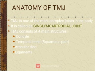
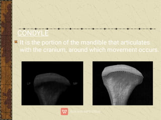
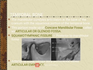
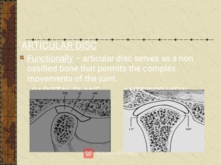
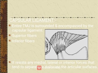
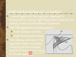
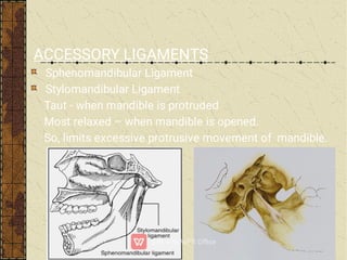
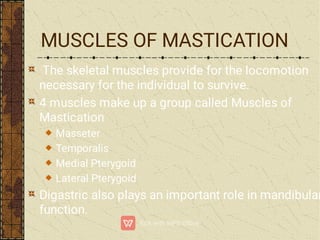
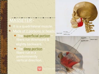
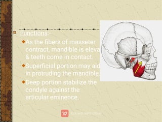
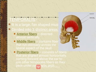
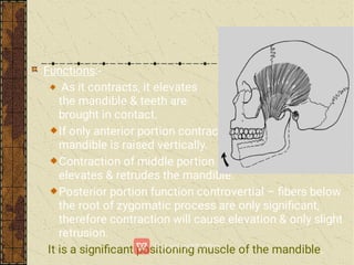
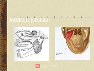
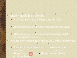
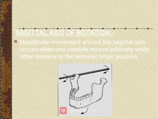
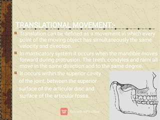
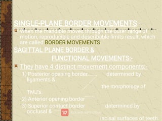
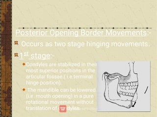
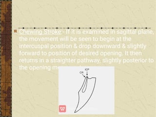
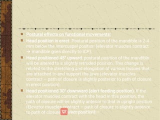
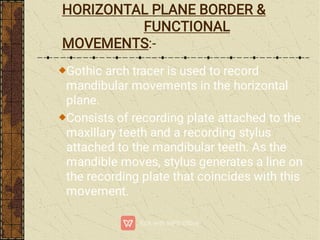
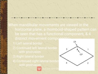
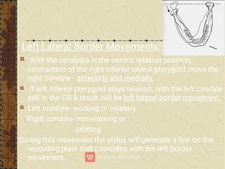
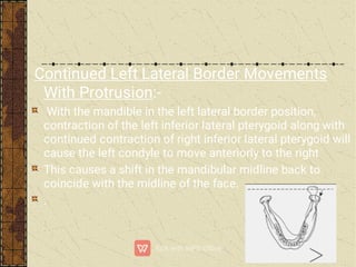
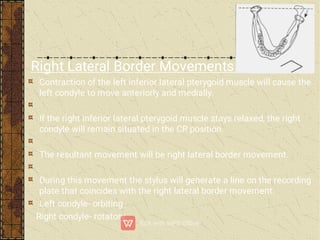
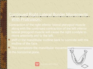
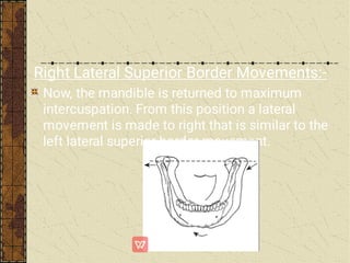
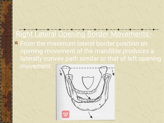
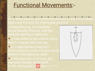
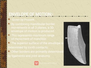
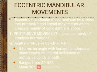
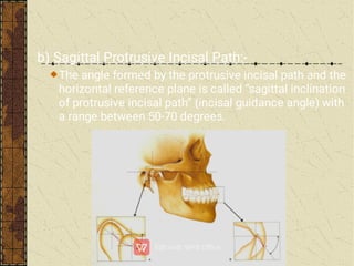
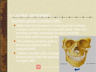
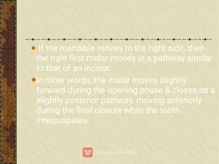
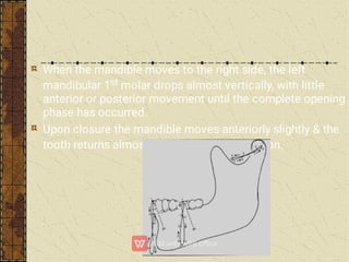
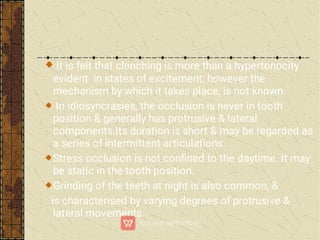
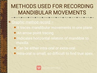
MANDIBULAR
MOVEMENTS
MANDIBULAR
MOVEMENTS
CONTENTS
Introduction
Review of Literature
Anatomy of TMJ
Muscles of Mastication
Neurologic structures & neuromuscular functions
Mandibular Movements
Eccentric Mandibular Movements
Clinical Signicance of Mandibular Movements
Major Functions of Masticatory System
Methods Used For Recording Mandibular Movements
Conclusion
References
INTRODUCTION
The masticatory system is extremely complex. It
is made up of three linked chains. First link is
maxilla & mandible, second link is TMJ & third is
muscles & ligaments.
First two, are considered to be passive & the third
one is an active link.
During performance of various functions there is a
delicate balance between various components.
It is important to study mandibular movements as
it enables us to plan arrangement of teeth &
selection of articulators so that articial
prosthesis is in harmony with stomatognathic
system.
Woelfel J.B., Hickey J.C., Stacy R.W. & Rinear L. (1960) –
conducted a study on electromyographic analysis of jaw
movements. The objective of the study were-
1)To determine the range of variability of muscular activity in
jaw
movements.
2)To determine the range of variability in a series of
electromyograms.
3)To provide an analysis of the role played by the external
pterygoid muscles in trained (learned) jaw movements.
REVIEW OF LITERATURE
They concluded that:-
1) The temperal muscle is capable of unilateral and
fractional
response but does not show increased activity in any part
during
protrusion or uncontrolled openings.
2)The right and left digastric muscles did not function
individually.Their greatest activity was during uncotrolled
openings and retrusion of the mandible.
3)The masseter muscle had the greatest activity during
clenching
into centric occlusion.
4)The external pterygoid muscle was very active during
contralateral excursions, uncontrolled openings, and
Lundeen T.F. & Mendoza F.(1984) – conducted a study on
comparison of Bennett shift measured at the hinge axis &
arbitrary hinge axis position. In their study they used flag
system & a facebow system. They modied the facebow
by substitution of a Hinge Axis Locator. The end of Hinge
Axis Locator was modied with a plastic tting to
facilitate the attachment of measuring pointer. They
concluded that there was no signicant difference in the
Bennett shift measurements made at the hinge axis & the
arbitrary hinge axis positions when measured in 0.25mm
increments. The average immediate Bennett shift was
1.05mm on the left side & 1.12mm on the right side when
rm guidance was used.
Colaizzi F.A., Micheal C.G., Javid N.S.& Gibbs C.H. (1988) –
conducted a comparative study of the condylar & incisal
border movements on complete denture wearers & natural
dentition subjects.
5 edentulous patients having previous denture experience
of 10-21yrs were selected. 2 different occlusal schemes
tested were teeth without cuspal inclines(0Âş teeth) & teeth
with cuspal inclines(30Âş teeth). Patients were asked to
wear dentures with 0Âş teeth for 2wks which were replaced
with 30Âş teeth for next 2wks. 10 natural dentition subjects
with good occlusion were recorded. Movements were
recorded by replicator system. Jaw motion was measured
with photo-optical transducers.
They concluded that,
Denture teeth of both 0 degree & 30 degree designs
produced a rounded, poorly dened intercuspal-like
position in contrast to a sharp well-dened intercuspal
position in the natural dentition subjects.
Denture wearers had a shallow angle of disooclusion
in the frontal view compared with the natural dentition
subjects.
Side shift(Bennett movement) was more than twice as
great in the long-term denture wearers as in the natural
dentition subjects.
Non-working side side shift was greater on the left than
on the right side for the denture wearers.
In the denture wearers, the working-side condylar
movements were greater with a more lateral protrusive
component than in the natural dentition subjects.
ANATOMY OF TMJ
TMJ is one of the most complex joints in the body.
It is called as GINGLYMOARTRODIAL JOINT.
TMJ consists of 4 main structures:-
Condyle
Temporal bone (Squamous part)
Articular disc
Ligaments
CONDYLE
It is the portion of the mandible that articulates
with the cranium, around which movement occurs.
TEMPORAL BONE
The mandibular condyles articulates at the base of the
cranium with the squamous portion of the temporal bone.
This portion made up of Concave Mandibular Fossa called
as ARTICULAR OR GLENOID FOSSA.
SQUAMOTYMPANIC FISSURE – Posterior to mandibular
fossa.
Anterior to fossa convex bony prominence called
ARTICULAR EMINENCE.
ARTICULAR DISC
Functionally – articular disc serves as a non
ossied bone that permits the complex
movements of the joint.
SAGITTAL PLANE ANTERIOR VIEW
Attachment of Articular Disc:-
e
LIGAMENTS:-
Play an important role in protecting the structures.
Ligaments do not enter actively into joint function,
rather they act as passive restraining devices to
limit & restrict border movements.
3 functional ligaments support the TMJ are:-
Collateral ligament
Capsular ligament
Temporomandibular ligament
2 accessory ligaments are:-
Sphenomandibular ligament
Stylomandibular ligament
COLLATERAL(DISCAL) LIGAMENTS:-
They attach the medial & lateral borders of
articular disc to the poles of the condyle.
Commonly called as DISCAL LIGAMENTS.
2 TYPES:-
Medial discal ligament
Lateral discal ligament
These ligaments are responsible for dividing joint
mediolaterally into superior & inferior cavities &
are collagenous connective tissue bers.
Function – Restricts movement of the disc away
from the condyle.
Attachments of the discal ligament permit the
disc to be rotated anteriorly & posteriorly on the
articular surface of the condyle.
These ligaments are responsible for the hinging
movement of the TMJ.
CAPSULAR LIGAMENT:-
Entire TMJ is surrounded & encompassed by the
capsular ligament.
Superior bers
Inferior bers
It resists any medial, lateral or inferior forces that
tend to separate or dislocate the articular surfaces.
TEMPOROMANDIBULAR LIGAMENT:-
Lateral aspect of the capsular ligament is
reinforced by strong, tight bers that make up the
lateral or temporomandibular ligament.
The TM ligament is composed of :-
Outer oblique portion
Inner horizontal portion
The oblique portion of TM
ligament resist excessive
dropping of the condyle;
therefore limiting the extent
of mouth opening.
ACCESSORY LIGAMENTS
Sphenomandibular Ligament
Stylomandibular Ligament
Taut - when mandible is protruded
Most relaxed – when mandible is opened.
So, limits excessive protrusive movement of mandible.
MUSCLES OF MASTICATION
The skeletal muscles provide for the locomotion
necessary for the individual to survive.
4 muscles make up a group called Muscles of
Mastication
Masseter
Temporalis
Medial Pterygoid
Lateral Pterygoid
Digastric also plays an important role in mandibular
function.
MASSETER:-
It is a quadrilateral muscle.
Made of 2 portions or heads:-
The superficial portion –
bers run downward &
slightly backward.
The deep portion –
bers run in a
predominently
vertical direction.
Functions:-
As the bers of masseter
contract, mandible is elevated
& teeth come in contact.
Supercial portion may aid
in protruding the mandible.
Deep portion stabilize the
condyle against the
articular eminence.
TEMPORALIS:-
It is a large, fan shaped muscle.
Divided into 3 distinct areas:-
Anterior fibers – directed
almost vertically.
Middle fibers – contains fibers
that run obliquely across the
lateral aspect of the skull.
Posterior fibers – consists of fibers
that are aligned almost horizontally,
coming forward above the ear to
join other temporalis bers as they
pass under zygomatic arch.
Functions:-
As it contracts, it elevates
the mandible & teeth are
brought in contact.
If only anterior portion contracts-
mandible is raised vertically.
Contraction of middle portion –
elevates & retrudes the mandible.
Posterior portion function controvertial – fibers below
the root of zygomatic process are only signicant,
therefore contraction will cause elevation & only slight
retrusion.
It is a signicant positioning muscle of the mandible.
MEDIAL PTERYGOID:-
Functions:-
When bers contracts, mandible is elevated.
Helps in protruding the mandible.
Unilateral contraction will bring about a
mediotrusive movement of the mandible.
LATERAL PTERYGOID:-
2 different portions or bellies:-
Inferior
Superior
Inferior Lateral Pterygoid:-
Function:-
When contracts simultaneously,
the condyles are pulled down
the articular eminences &
the mandible is protruded.
Unilateral contraction creates a mediotrusive
movement of the condyle & causes a lateral movement
of the mandible to the opposite side.
When it functions with mandibular depressors – the
mandible is lowered & the condyles glide forward &
downward on the articular eminences.
Superior Lateral Pterygoid:-
It is smaller than inferior
lateral pterygoid.
Function:-
During opening the
superior lateral pterygoid
remains inactive, becoming
active only in conjunction
with elevator muscles.
It is active during power
stroke & when teeth are
held together.
DIGASTRICS:-
Not considered a muscle of mastication, but it
does have an important influence on the function
of the mandible.
Divided into 2 portions:-
Posterior belly
Anterior belly
Function:-
When right & left digastrics
contract & the suprahyoid &
infrahyoid muscles x the
hyoid bone, the mandible is
depressed & pulled backward &
the teeth are bought out of contact.
When mandible is stabilized,
the digastric muscles with the
suprahyoid & infrahyoid muscles
elevate the hyoid bone, which
is necessary function for swallowing.
NEUROLOGIC STRUCTURE &
NEUROMUSCULAR FUNCTION
Function of masticatory system is complex. A
highly rened neurologic control system regulates
& coordinates the activities of entire masticatory
system.
NEUROLOGIC STRUCTURES:-
The masticatory system consists of following
receptors to monitor the status of its components:
-
1) MUSCLE SPINDLE-
Skeletal muscle consists of two types of muscle bres
–
1)
2)

A bundle of intrafusal bres bound by a
connective tissue sheath is called muscle
spindle.
Within each spindle the nuclei of the
intrafuscal bers are arranged in 2 distinct
fashions:-
Chainlike(nuclear chain type)
Clumped(nuclear bag type)
There are two types of afferent nerves that
supply the intrafusal bres. They are:
1) Primary endings or annulospiral endings
2) Secondary endings or flower spray endings
Efferent supply of intrafusal bers is by
When muscle is stretched:-
Intrafusal & extrafusal bers are stretched
Annulospiral & flower spray endings are activated
Afferent neurons carry information to trigeminal
mesencephalic nucleus
The CNS then sends back impulse via 2 efferent pathways:
-
Fusimotor nerve bers or ι efferent motor
neurons gamma
efferent(for (for extrafusal bers)
intrafusal bers)
2) GOLGI TENDON ORGANS-
Located in muscle tendon between muscle bres and
their attachment to bone.
They primarily monitor tension, whereas the muscle
spindles primarily monitor muscle length.
3) PACINIAN CORPUSCLES-
The pacinian corpuscles are large oval organs made up
of concentric lamellae of connective tissue. They are
widely distributed.
They serve principally for the perception of the
movements and rm pressure.
4) NOCICEPTORS-
They are sensory receptors that are stimulated
by injury & transmit injury information to CNS by
way of afferent nerve bers.
REFLEX ACTION:-
2 general reflex actions are important:-
1) MYOTACTIC REFLEX or stretch reflex-
Is the only monosynaptic reflex.
Sudden stretching of skeletal muscle

Afferant nerve activity from the spindle

Trigeminal mesencephalic nucleus

Afferent ber synapse in trigeminal motor nucleus with
Îą- efferent motor neurons

Efferent bres carry information to extrafusal bres

Muscle contraction
Myotactic reflex is an important determinant of
rest position of the jaw.
It is a principal determinant of muscle tonus in
elevator muscles.
2) NOCICEPTIVE REFLEX or flexor reflex-
Polysynaptic reflex to noxious stimuli & hence, considered to be
protective.
Sudden biting on hard object

Noxious stimuli

Afferent nerves carry impulse to trigeminal spinal
tract nucleus where they synapse with interneurons

Excitatory and inhibitory interneurons

Synapse with efferent neurons in the trigeminal
motor nucleus ( called antagonistic inhibition)
Influence of Higher Centers:-
Although the cortex is the main determinant of
function of brainstem.
Within brainstem, is a pool of neurons that control
rhythmic muscle activity such as breathing,
walking & chewing.
This pool of neurons is called ‘central pattern
generator.’
It is responsible for precise timing of activity
between antagonistic muscles so that specic
functions can be carried out.
CLASSIFICATION:-
I) According to Sharry:-
a) According to direction - Opening and closing
movements
Protrusion and retraction
Lateral gliding movements
b) According to tooth contact - Movements with tooth
contact
Movements without tooth contact
c) Limitation by joint structure - Border movements
Intra border movements
d) Functions of masticatory system - Mastication
Deglutition
Speech
Respiration
II) According to the type of movement occurs in
TMJ:-
a) Rotational
b) Translation
III) According to the planes of border movements:-
a) Sagittal plane border movement
b) Horizontal plane border movements
c) Frontal plane border movements
MANDIBULAR MOVEMENTS
TYPES:-
2 types of movement occur in TMJ:-
Rotational
Translational
ROTATIONAL MOVEMENT:-
Rotation – “movement of
a body about its axis.”
In masticatory system,
rotation occurs when the
mouth opens and closes
around a xed point or axis
within the condyles.
HORIZONTAL AXIS OF ROTATION:-
Mandibular movement around the horizontal axis is an
opening and closing motion, referred to as Hinge
Movement and horizontal axis around which it occurs is
therefore referred to as “HINGE AXIS”.
The Hinge movement is the only
example of mandibular activity
in which a “pure” rotational
movement occurs.
TERMINAL HINGE AXIS
When the condyles are in their most superior position in
the articular fossae and the mouth is purely rotated open,
the axis around which movement occurs is called the
‘Terminal Hinge Axis’.
FRONTAL (VERTICAL) AXIS OF ROTATION:-
Mandibular movement around the frontal axis
occurs when one condyle moves anteriorly out of
terminal hinge position with the vertical axis of
opposite condyle remaining in the terminal hinge
position.
SAGITTAL AXIS OF ROTATION:-
Mandibular movement around the sagittal axis
occurs when one condyle moves inferiorly while
other remains in the terminal hinge position.
TRANSLATIONAL MOVEMENT:-
Translation can be dened as a movement in which every
point of the moving object has simultaneously the same
velocity and direction.
In masticatory system it occurs when the mandible moves
forward during protrusion. The teeth, condyles and rami all
move in the same direction and to the same degree.
It occurs within the superior cavity
of the joint, between the superior
surface of the articular disc and the inferior
surface of the articular fossa.
SINGLE-PLANE BORDER MOVEMENTS:-
When the mandible moves through the outer range of
motion, reproducible and describable limits result, which
are called BORDER MOVEMENTS.
SAGITTAL PLANE BORDER &
FUNCTIONAL MOVEMENTS:-
They have 4 distinct movement components:-
1) Posterior opening border determined by
ligaments &
the morphology of
TMJ’s.
2) Anterior opening border
3) Superior contact border determined by
occlusal &
incisal surfaces of teeth.
Posterior Opening Border Movements:-
Occurs as two stage hinging movements.
1st stage:-
Condyles are stabilized in their
most superior positions in the
articular fossae.( i.e.terminal
hinge position).
The mandible can be lowered
(i.e. mouth opening) in a pure
rotational movement without
translation of condyles.
In CR, the mandible can be rotated around the
horizontal axis to a distance of 20-25mm as
measured between the incisal edges of
maxillary and mandibular incisors.
At this point of opening, the T.M. ligaments
tighten, after which continued opening results in
an anterior & inferior translation of condyles.
2nd Stage:-
As the condyle translates the axis of rotation of the
mandible shifts into the bodies of rami likely to be the
area of attachment of sphenomandibular ligament,
resulting in the second stage of the posterior opening
border movement.
During this stage, the condyles move anteriorly
and inferiorly and the mandible moves
posteriorly and inferiorly.
Maximum opening is reached when capsular
ligaments prevent further movement of the
condyles.
Maximum opening range is 40-60mm.
Anterior Opening Border Movements:-
With the mandible maximally opened, closure
accompanied by contraction of inferior lateral
pterygoids(which keep the condyles positioned
anteriorly) will generate the anterior border
movement.
Because the maximum protrusive position is
determined in part by stylomandibular ligaments,
when closure occurs, tightening of ligaments
produces a posterior movement of the condyles.
The posterior movement of the condyle from the
maximally open position to maximally protruded
position produces eccentricity in the anterior
border movement. Therefore, it is not a pure hinge
movement.
Superior Contact Border Movements:-
Throughout this entire border movement tooth
contact is present.
It depends on:-
Amount of variation between centric relation and
maximum intercuspation.
The steepness of the cuspal inclines of the posterior
teeth.
Amount of vertical and horizontal overlap of anterior
teeth
Lingual morphology of maxillary anterior teeth.
General interarch relationships of the teeth.
The initial tooth contact in
terminal hinge axis or centric
relation occurs between the
mesial inclines of maxillary
tooth & distal inclines of
mandibular tooth.
When muscular force is
applied to the mandible,
a superoanterior movement
or shift will occur until the
intercuspal position is reached.
The slide from CR to maximum intercuspation,
may have a lateral component.
Average distance is 1.25Âą1mm.
In the intercuspal position, the opposing anterior
teeth usually contact.
When the mandible is protruded,
from maximum intercuspation,
contact between the incisal
edges of the mandibular anterior
teeth & lingual inclines of
maxillary anterior teeth result
in an anteroinferior movement
of the mandible.
This continues until the
maxillary and mandibular
anterior teeth are in edge
to edge relationship, at which
a horizontal movement
continues until incisal edges
of mandibular teeth pass
beyond the edges of maxillary teeth.
At this point mandible moves
in a superior direction until
the posterior teeth contact.
The occlusal surfaces of
posterior teeth then dictate
the remaining pathway to
the maximum protrusive
movement, which joins
with the most superior portion
of the anterior opening border movement.
Functional Movements:-
Functional movement occurs during functional
activity of the mandible. They usually take place
within the border movements & therefore,
considered as free movements.
Most functional movements require maximum
intercuspation & therefore typically begin at &
below the intercuspal position.
When mandible is at rest, it is found to be located
approximately 2 to 4mm below the intercuspal
position. This is called the Clinical Rest Position.
Postural position – Since,
clinical rest position is not a
true resting position, the
position in which mandible
is maintained is termed as
‘postural position.’
Chewing Stroke:- If it is examined in sagittal plane,
the movement will be seen to begin at the
intercuspal position & drop downward & slightly
forward to position of desired opening. It then
returns in a straighter pathway, slightly posterior to
the opening movement.
Postural effects on functional movements:
Head position is erect: Postural position of the mandible is 2-4
mm below the intercuspal position (elevator muscles contract
 mandible goes directly to ICP).
Head positioned 45° upward: postural position of the mandible
will be altered to a slightly retruded position. This change is
related to the stretching and elongation of various tissues that
are attached to and support the jaws (elevator muscles
contract – path of closure is slightly posterior to path of closure
in erect position).
Head positioned 30° downward (alert feeding position). If the
elevator muscles contract with the head in this position, the
path of closure will be slightly anterior to that in upright position.
(Elevator muscles contract – path of closure is slightly anterior
to path of closure in erect position).
HORIZONTAL PLANE BORDER &
FUNCTIONAL
MOVEMENTS:-
Gothic arch tracer is used to record
mandibular movements in the horizontal
plane.
Consists of recording plate attached to the
maxillary teeth and a recording stylus
attached to the mandibular teeth. As the
mandible moves, stylus generates a line on
the recording plate that coincides with this
movement.
When mandibular movements are viewed in the
horizontal plane, a rhomboid-shaped pattern can
be seen that has a functional component, & 4
distinct movement components:-
1) Left lateral border
2) Continued left lateral border
with protrusion
3) Right lateral border
4) Continued right lateral border
with protrusion
Left Lateral Border Movements:-
With the condyles in the centric relation position,
contraction of the right inferior lateral pterygoid move the
right condyle - anteriorly and medially.
If left inferior pterygoid stays relaxed, with the left condyle
still in the CR & result will be left lateral border movement.
Left condyle- working or rotatory
Right condyle- non-working or
orbiting
During this movement the stylus will generate a line on the
recording plate that coincides with the left border
movement.
Continued Left Lateral Border Movements
With Protrusion:-
With the mandible in the left lateral border position,
contraction of the left inferior lateral pterygoid along with
continued contraction of right inferior lateral pterygoid will
cause the left condyle to move anteriorly to the right
This causes a shift in the mandibular midline back to
coincide with the midline of the face.
.
Right Lateral Border Movements:-
Contraction of the left inferior lateral pterygoid muscle will cause the
left condyle to move anteriorly and medially.
If the right inferior lateral pterygoid muscle stays relaxed, the right
condyle will remain situated in the CR position.
The resultant movement will be right lateral border movement.
During this movement the stylus will generate a line on the recording
plate that coincides with the right lateral border movement.
Left condyle- orbiting
Right condyle- rotatory
Continued Right Lateral Border Movements
With Protrusion:-
Contraction of the right inferior lateral pterygoid muscle
along with the continued contraction of the left inferior
lateral pterygoid muscle will cause the right condyle to
move anteriorly and to the left.
shift in the mandibular midline back to coincide with the
midline of the face.
This completes the mandibular movements
in the horizontal plane.
Functional Movements:-
As in the sagittal plane, functional movement in the
horizontal plane most often occur near the intercuspal
position.
During chewing the range of jaw movements begins some
distance from maximum intercuspal position; but as the
food is broken down into smaller particles, jaw action
moves closer and closer to intercuspal position.
FRONTAL(VERTICAL) BORDER &
FUNCTIONAL MOVEMENTS:-
A shield-shaped pattern can be seen that has a
functional component, & four distinct movement
components:-
1. Left lateral superior border.
2. Left lateral opening border.
3. Right lateral superior border.
4. Right lateral opening border.
With the mandible in maximum intercuspation
a lateral movement is made to the left.
A recording device will disclose an inferiorly
concave path being generated.
The precise mixture of this path is primarily determined by the
morphology and interarch relationships of the maxillary and
mandibular teeth that are in contact during this movement.
Of secondary influence are the condyle-disc-fossa relationships and
morphology of the working or rotating side TMJ.
The maximum lateral extent of this movement is determined by the
ligaments of the rotating joint.
Left Lateral Superior Border Movements:-
Left Lateral Opening Border Movements:-
From the maximum left lateral superior border position,
an opening movement of the mandible produces a
laterally convex path. As maximum opening is approached,
ligaments tighten and produce a medially directed
movement that causes a shift back in the mandibular
midline to coincide with the midline of the face.
Right Lateral Superior Border Movements:-
Now, the mandible is returned to maximum
intercuspation. From this position a lateral
movement is made to right that is similar to the
left lateral superior border movement.
Right Lateral Opening Border Movements:-
From the maximum lateral border position an
opening movement of the mandible produces a
laterally convex path similar to that of left opening
movement.
Functional Movements:-
As in other planes, functional movements in the frontal plane
begin and end and the intercuspal position.
During chewing, the mandible
drops directly inferiorly until the
desired opening is achieved.
It then shifts to the side on which
bolus is placed and rises up.
As it approaches maximum
intercuspation, bolus is broken down
between the opposing teeth.
In the nal mm of closure, the
mandible quickly shifts back
to the intercuspal position.
ENVELOPE OF MOTION:-
Given by Posselt.
By combining mandibular border
movements in all 3 planes, a 3D
envelope of motion is produced.
This represents maximum range
of movement of mandible.
The superior surface of the envelope is
determined by tooth contacts.
Other borders are primarily determined
by ligaments and joint anatomy.
ECCENTRIC MANDIBULAR
MOVEMENTS
Eccentric mandibular movement can be divided
into protrusive and lateral movements which
consists mainly of condylar translations:-
1) PROTRUSIVE MOVEMENT:- consists mainly of
condylar translations
a)Sagittal Protrusive Condylar Path:-
It forms an angle with horizontal reference
plane known as sagittal inclination of
protrusive condylar path.
Ranges from 5Âş- 55Âş.
Mean 30Âş.
b) Sagittal Protrusive Incisal Path:-
The angle formed by the protrusive incisal path and the
horizontal reference plane is called “sagittal inclination
of protrusive incisal path” (incisal guidance angle) with
a range between 50-70 degrees.
2) LATERAL MOVEMENT:-
Sagittal Lateral Condylar Path:-
When lateral movement is executed the working
condyle rotates & moves outward while, other non
working condyle translates forward, medially
downward orbiting around the rotating working condyle.
The orbiting condylar path is
known as sagittal lateral
condylar path.
Lateral condylar path is longer
& more steep than the protrusive
condylar path.
Fischer Angle:- The angle formed between the
sagittal protrusive condylar path & sagittal
lateral condylar path(approx 5Âş).
The angle formed by the sagittal lateral
condylar path & horizontal reference plane is
known as “sagittal inclination of lateral condylar
path” with a range 11º - 61º & mean 35º.
Bennett in 1908, studied working condylar path &
called as BENNETT MOVEMENT, reffered as
laterotrusion.ment
Bennett movement refers to condylar movement
on the working side &,
Bennett Shift is the bodily side shift of the
mandible on working side in horizontal direction.
Bennett movement is consequent to the medial
movement of orbiting condyle & is regulated by:-
Morphology of medial wall of mandibular fossa.
Inner horizontal part of TM ligament which
attaches to the lateral pole of rotating condyle.
Bennett movement has 3 components:-
Amount
Timing
Direction
1) AMOUNT:-
More medial the wall from the medial pole of the
orbiting condyle, closes the T.M. ligament attachment
of the rotating condyle the greater will be the Bennett
movement.
When the Bennett movement occurs early, a shift is
seen before the condyle begins to translate from the
fossa. This is called as ‘IMMEDIATE SIDE SHIFT.’
Beyond this the condyle moves forward, downward &
inward, this is known as ‘PROGRESSIVE SIDE SHIFT.’
2) TIMING:-
Amount of immediate side shift and progressive side
shift. The rate or amount of descent of contralateral
condyle & the rotation & lateral shift of working condyle.
Immediate side shift – is the 1st movement the
mandible makes when initiating lateral excursions. The
non-working condyle moves from centric position
medially against the medial & superior wall of the
articular fossa to a distance of approx. 1mm.(range
0-2.6mm).
This is not an exact 90Âş or a right angled medial move
in horizontal plane.
Progressive side shift:- Beyond the immediate side
shift the condyles move forward, downward and inward.
This movement component is called progressive side
shift. Its value is 7.5 degrees.
The angle formed by lateral
horizontal condylar path &
sagittal plane is called
‘BENNETT ANGLE’
(varies 2Âş -44Âş mean 16Âş).
3) DIRECTION:-
The direction of Bennett movement depends primarily
on the direction taken by the rotating condyle during
the bodily movement. The direction of the shift of the
rotating condyle during Bennett movement is
determined by the TM joint undergoing rotation.
CLINICAL SIGNIFICANCE
A prosthodontist designs a prosthesis to replace
the lost teeth, for replacement of missing teeth &
restoring function. Knowledge of the mandibular
movements essential, it helps the dentist in:
- Selecting and programming of articulators
- Treating TMJ disturbances.
- Arranging articial teeth.
- Development of occlusal scheme.
PROTRUSIVE MOVEMENT:-
1) Condylar guidance and anterior guidance:
These are the two end controlling factors of the
mandibular movement when the movement of a solid
body is governed by contacting surfaces at either ends of
that body, the direction of movement of any point within
the body is determined by its location in rotation to two
guiding surfaces. Then,
a) Since a second molar is closer to the condylar
guidance than is a lateral incisor, the condylar guidance
has a greater effects on the direction of movement of
lower second molar than it does on lower lateral incisor.
b) Anterior guidance has a greater effect on the
direction of movements of the lower canine
than it does on the lower molar of the two,
anterior guidance has the greater effect of
direction of movement of tooth movement
during mandibular movement, as all of the
teeth are closer to anterior guidance than the
condylar guidance.
Effects of condylar guidance and anterior guidance on
cusp height and fossa depth:
a) The lesser the condylar guidance angle, the shorter the
cusps
must be.
b) The greater the condylar guidance angle, the longer the
cusps
may be
c) The greater the horizontal overlap of the maxillary anterior
teeth, the shorter the cusps of the posterior teeth must be.
d)The lesser the horizontal overlap the longer the cusps of
the
posterior teeth may be.
e) The lesser the vertical overlap, the shorter the cusps of
the
posterior teeth must be.
2) Bennet’s Movement:-
a)The bodily lateral movement or lateral shift of the
mandible during lateral jaw movement.
- Movement responsible for lateral chewing stroke.
- Movement during which the greater lateral force is
exerted.
- It is extremely important that articulating surfaces are is
strict harmony with this side shift.
b) Effect on cusp height:
- Greater the side shift of the mandible shorter the
cusps must be.
- The lesser the side shift of the mandible longer
the
cusps may be.
3) Value of True Hinge Axis:-
a) It is a starting point of lateral movements.
b) Allows the transfer of opening axis of the articulation so
that occlusion would be on the same arc of closure as the
lower jaw.
c) The interocclusal centric relation record is made in
terminal hinge position and is used to orient the
mandibular cast to the maxillary cast.
d) Hinge axis method of orienting casts in an articulator
permits control of the vertical dimension on the
articulation.
4) Centric Relation and Centric Occlusion :-
Centric Relation:-
a) It is a posterior border position and posterior limit of the
envelope of motion.
b) It is repeatable position recordable position and a
physiologically acceptable position for mastication and
speech.
c) It is an optimum position of jaws for the health comfort
and
function of TMJ.
Centric occlusion – It is the relationship of opposition
of occlusal surfaces of teeth which provides the
MAJOR FUNCTIONS OF
MASTICATORY SYSTEM
MASTICATION:-
It is the act of chewing food.
Mastication is made up of rhythmic & well controlled
separation & closure of the maxillary & mandibular
teeth.
In frontal plane, it has a ‘tear shaped’ pattern.
It has:- a) Opening Phase
b) Closing Phase –
i) Crushing Phase
ii) Grinding Phase.
a) Opening phase:-
The mandible drops downward from the intercuspal
position to a point where the incisal edges are approx.
16-18mm apart.
It then moves laterally 5-6mm from the midline as the
closing movement begins.
b) Closing phase:-
Here, 1st phase is the crushing phase in which the food
is trapped between the teeth.
When teeth approach each other, the lateral
displacement is lessened so that when the teeth are
only 3mm apart, the jaw occupies a position only
As the mandible continues to close, the grinding phase
begins.
During the grinding phase, the mandible is guided by
the occlusal surfaces of the teeth back to the
intercuspal position, which causes the cuspal inclines
of the teeth to pass over each other permitting chewing
& grinding of the bolus of food.
During the opening phase, the mandible moves slightly
anteriorly.
During the closing phase, it follows a slightly posterior
pathway, ending in an anterior movement back to the
maximum intercuspal position.
If the movement of a mandibular incisor is followed in
the sagittal plane during a typical chewing stroke, it will
be seen that during the opening phase the mandible
moves slightly anteriorly.
During the closing phase it follows a slightly posterior
pathway, ending in an anterior movement back to the
maximum intercuspal position.
The amount of anterior movement depends on the
contact pattern of the anterior teeth & the stage of
If the mandible moves to the right side, then
the right rst molar moves in a pathway similar
to that of an incisor.
In other words, the molar moves slightly
forward during the opening phase & closes on a
slightly posterior pathway, moving anteriorly
during the nal closure while the tooth
intercuspates.
When the mandible moves to the right side, the left
mandibular 1st molar drops almost vertically, with little
anterior or posterior movement until the complete opening
phase has occurred.
Upon closure the mandible moves anteriorly slightly & the
tooth returns almost directly to intercuspation.
SWALLOWING(DEGLUTITION):-
It is a series of co-ordinated muscular contractions
that moves a bolus of food from the oral cavity through
the oesophagus to the stomach.
Stabilization of the mandible is an important part of
swallowing.
The mandible must be xed so contraction of
suprahyoid & infrahyoid muscles can control proper
movement of the hyoid bone needed for swallowing.
a) Somatic swallow – teeth used for stabilizing
mandible
b) Visceral swallow – mandible is braced by placing the
tongue forward between the dental
arches & gum pads.
EMPTY MOVEMENTS:-
Dened in a negative way, the comprise all occlusions
other than those taking place in mastication or
swallowing.
They may be observed in some conditions of mental
stress & in periodontal disease.
The occlusions developed in stress situation may be
either intermittent or continuous.
Stress occlusions – day time – clenching
idiosyncrasies
night time – clenching
grinding
It is felt that clenching is more than a hypertonocity
evident in states of excitement; however the
mechanism by which it takes place, is not known.
In idiosyncrasies, the occlusion is never in tooth
position & generally has protrusive & lateral
components.Its duration is short & may be regarded as
a series of intermittent articulations.
Stress occlusion is not conned to the daytime. It may
be static in the tooth position.
Grinding of the teeth at night is also common, &
is characterised by varying degrees of protrusive &
lateral movements.
METHODS USED FOR RECORDING
MANDIBULAR MOVEMENTS
Graphic method record –
It traces mandibular movements in one plane.
An arrow point tracing.
Indicates horizontal relation of mandible to
maxilla.
Can be either intra-oral or extra-oral.
Intra-oral is small, so dicult to nd true apex.
Computer-monitored radionucleide tracking of
three-dimensional mandibular movement.(By
Salomon 1979)
Materials & Methods:-
Recording equipment is divided into two parts, the
Gamma camera, has a 30cm diameter detecting head
and is equipped with a 4mm pinhole collimeter to
provide an enlarged image of the moving source.
Computer is linked to camera and runs under RTII Real
time operating system with “gamma 11” software
monitoring the data collection.
Various peripherals, including disc drives & a video
color television monitor, are connected to the
Emitting source: The source which is tracked during the
experiment has to be placed on the patient and is
prepared from Technetium portable generator.
- A source is prepared by depositing a few microdrops of a
high concentrated solution of Tc99m in saline on to a piece
of non-absorbing cardboard each drop is the evaporated
in a oven.
- It is possible to obtain an activity of 100 to 150ÂľCi on a
drop as small as 0.5mm diameter paper is then cutout to
form a square of 1cm, sealed and taped to the point of
interest at the time of experiment.
Patient Recordings: After the radioactive source is
placed on his chin, the patient sits in front of the
collimator with his head in a resting position.
Datas are collected in a list mode for several
seconds (e.g. few chewing cycles), results are
processed by the computer are movements and
displayd on the color television monitor.
A control oscilloscope on camera is used for
positioning & immediate viewing.
STEREOGRAPHICS:-
The TMJ stereographic system (Swanson. 1966; Mensor.
1973)
Swanson, (1979) requires the development of intra-oral
clutches with an adjustable central bearing screw
incorporated to provide smooth jaw movements. Four
studs embedded in the upper clutch allow intra-oral
moulding of border movements in soft acrylic added to
the lower clutch. These intra-oral engravings provide a
permanent three-dimensional record of guided jaw
movements, and are then employed to generate the
equivalent condylar characteristics on the TMJ articulator
guided by the intra-oral engravings.
In this way permanent condylar mouldings are made, that
incorporate condylar inclination, and progressive and
PANTOGRAPHS:-
Pantograph is used clinically to measure
mandibular movements.
2 Types – Mechanical
Electronic.
Graphical methods of tracking jaw movement,
both intra-oral and extra-oral have been used since
the turn of the century for complete denture
construction; the extra-oral technique was
pioneered by (Gysi 1913) and was the forerunner
of the pantograph as it is known today.
It includes six tracing platforms and styli to graph
gothic arch as well as jaw and condylar
movements.
A vertical and a horizontal tracing table are located
on each side of the patient's face overlying the TM
joints, and a pair of horizontal tables,
approximately at the level of the plane of
occlusion, is located below the eyes.
A stylus is used on each table to record border
movements on a pressure sensitive material.
The tracing procedure is carried out to record
terminal hinge axis as the reference point and
lateral border paths are traced whilst the jaw is
The tracings obtained may then be carefully
removed from the patient by locking the upper
and lower face bows together and detaching from
the clutches.
The clutches are then removed from the patient
and reassembled and placed on the matched
articulator.
The articulator condylar settings are adjusted to
allow border tracings captured on the pantograph
to be duplicated by articulator.
REPLICATOR:-
By Messerman(1964).
It is a system for studying human jaw motion. All jaw
motions are measured & recorded on magnetic tape. Jaw
motion is measured with six incremental photo-optical
transducers mounted between maxillary & mandibular
reference facebows. Border hinge movements are
recorded with the replicator.
There are 3 important advances:-
No drift – Reference positions such as intercuspal
positions must remain highly accurate.
Ease & accuracy of recording data on magnetic tape
with digital system.
Incremental data its suitably for ecient entry into
computer.
KINESIOGRAPH:-
It was developed by Jankelson et al(1975).
A magnetometer jaw motion recording system, is
limited to incisal point measurements & is
susceptible to reference drift from head
movement. Although molar & condylar
movements cannot be shown on kinesiograph, it
is easy to attach to patient.
Incisal movement & velocities can be displayed
on oscilloscope screen for visual inspection.
CONCLUSION

MANDIBULAR MOVEMENTS-2.pdf complete denture ***********

  • 1.
  • 2.
    CONTENTS Introduction Review of Literature Anatomyof TMJ Muscles of Mastication Neurologic structures & neuromuscular functions Mandibular Movements Eccentric Mandibular Movements Clinical Signicance of Mandibular Movements Major Functions of Masticatory System Methods Used For Recording Mandibular Movements Conclusion References
  • 3.
    INTRODUCTION The masticatory systemis extremely complex. It is made up of three linked chains. First link is maxilla & mandible, second link is TMJ & third is muscles & ligaments. First two, are considered to be passive & the third one is an active link. During performance of various functions there is a delicate balance between various components. It is important to study mandibular movements as it enables us to plan arrangement of teeth & selection of articulators so that articial prosthesis is in harmony with stomatognathic system.
  • 4.
    Woelfel J.B., HickeyJ.C., Stacy R.W. & Rinear L. (1960) – conducted a study on electromyographic analysis of jaw movements. The objective of the study were- 1)To determine the range of variability of muscular activity in jaw movements. 2)To determine the range of variability in a series of electromyograms. 3)To provide an analysis of the role played by the external pterygoid muscles in trained (learned) jaw movements. REVIEW OF LITERATURE
  • 5.
    They concluded that:- 1)The temperal muscle is capable of unilateral and fractional response but does not show increased activity in any part during protrusion or uncontrolled openings. 2)The right and left digastric muscles did not function individually.Their greatest activity was during uncotrolled openings and retrusion of the mandible. 3)The masseter muscle had the greatest activity during clenching into centric occlusion. 4)The external pterygoid muscle was very active during contralateral excursions, uncontrolled openings, and
  • 6.
    Lundeen T.F. &Mendoza F.(1984) – conducted a study on comparison of Bennett shift measured at the hinge axis & arbitrary hinge axis position. In their study they used flag system & a facebow system. They modified the facebow by substitution of a Hinge Axis Locator. The end of Hinge Axis Locator was modified with a plastic fitting to facilitate the attachment of measuring pointer. They concluded that there was no significant difference in the Bennett shift measurements made at the hinge axis & the arbitrary hinge axis positions when measured in 0.25mm increments. The average immediate Bennett shift was 1.05mm on the left side & 1.12mm on the right side when firm guidance was used.
  • 7.
    Colaizzi F.A., MichealC.G., Javid N.S.& Gibbs C.H. (1988) – conducted a comparative study of the condylar & incisal border movements on complete denture wearers & natural dentition subjects. 5 edentulous patients having previous denture experience of 10-21yrs were selected. 2 different occlusal schemes tested were teeth without cuspal inclines(0º teeth) & teeth with cuspal inclines(30º teeth). Patients were asked to wear dentures with 0º teeth for 2wks which were replaced with 30º teeth for next 2wks. 10 natural dentition subjects with good occlusion were recorded. Movements were recorded by replicator system. Jaw motion was measured with photo-optical transducers.
  • 8.
    They concluded that, Dentureteeth of both 0 degree & 30 degree designs produced a rounded, poorly dened intercuspal-like position in contrast to a sharp well-dened intercuspal position in the natural dentition subjects. Denture wearers had a shallow angle of disooclusion in the frontal view compared with the natural dentition subjects. Side shift(Bennett movement) was more than twice as great in the long-term denture wearers as in the natural dentition subjects. Non-working side side shift was greater on the left than on the right side for the denture wearers. In the denture wearers, the working-side condylar movements were greater with a more lateral protrusive component than in the natural dentition subjects.
  • 9.
    ANATOMY OF TMJ TMJis one of the most complex joints in the body. It is called as GINGLYMOARTRODIAL JOINT. TMJ consists of 4 main structures:- Condyle Temporal bone (Squamous part) Articular disc Ligaments
  • 10.
    CONDYLE It is theportion of the mandible that articulates with the cranium, around which movement occurs.
  • 11.
    TEMPORAL BONE The mandibularcondyles articulates at the base of the cranium with the squamous portion of the temporal bone. This portion made up of Concave Mandibular Fossa called as ARTICULAR OR GLENOID FOSSA. SQUAMOTYMPANIC FISSURE – Posterior to mandibular fossa. Anterior to fossa convex bony prominence called ARTICULAR EMINENCE.
  • 12.
    ARTICULAR DISC Functionally –articular disc serves as a non ossified bone that permits the complex movements of the joint. SAGITTAL PLANE ANTERIOR VIEW
  • 13.
  • 14.
    e LIGAMENTS:- Play an importantrole in protecting the structures. Ligaments do not enter actively into joint function, rather they act as passive restraining devices to limit & restrict border movements. 3 functional ligaments support the TMJ are:- Collateral ligament Capsular ligament Temporomandibular ligament 2 accessory ligaments are:- Sphenomandibular ligament Stylomandibular ligament
  • 15.
    COLLATERAL(DISCAL) LIGAMENTS:- They attachthe medial & lateral borders of articular disc to the poles of the condyle. Commonly called as DISCAL LIGAMENTS. 2 TYPES:- Medial discal ligament Lateral discal ligament
  • 16.
    These ligaments areresponsible for dividing joint mediolaterally into superior & inferior cavities & are collagenous connective tissue fibers. Function – Restricts movement of the disc away from the condyle. Attachments of the discal ligament permit the disc to be rotated anteriorly & posteriorly on the articular surface of the condyle. These ligaments are responsible for the hinging movement of the TMJ.
  • 17.
    CAPSULAR LIGAMENT:- Entire TMJis surrounded & encompassed by the capsular ligament. Superior bers Inferior bers It resists any medial, lateral or inferior forces that tend to separate or dislocate the articular surfaces.
  • 18.
    TEMPOROMANDIBULAR LIGAMENT:- Lateral aspectof the capsular ligament is reinforced by strong, tight bers that make up the lateral or temporomandibular ligament. The TM ligament is composed of :- Outer oblique portion Inner horizontal portion The oblique portion of TM ligament resist excessive dropping of the condyle; therefore limiting the extent of mouth opening.
  • 19.
    ACCESSORY LIGAMENTS Sphenomandibular Ligament StylomandibularLigament Taut - when mandible is protruded Most relaxed – when mandible is opened. So, limits excessive protrusive movement of mandible.
  • 20.
    MUSCLES OF MASTICATION Theskeletal muscles provide for the locomotion necessary for the individual to survive. 4 muscles make up a group called Muscles of Mastication Masseter Temporalis Medial Pterygoid Lateral Pterygoid Digastric also plays an important role in mandibular function.
  • 21.
    MASSETER:- It is aquadrilateral muscle. Made of 2 portions or heads:- The superficial portion – fibers run downward & slightly backward. The deep portion – fibers run in a predominently vertical direction.
  • 22.
    Functions:- As the bersof masseter contract, mandible is elevated & teeth come in contact. Supercial portion may aid in protruding the mandible. Deep portion stabilize the condyle against the articular eminence.
  • 23.
    TEMPORALIS:- It is alarge, fan shaped muscle. Divided into 3 distinct areas:- Anterior fibers – directed almost vertically. Middle fibers – contains fibers that run obliquely across the lateral aspect of the skull. Posterior fibers – consists of fibers that are aligned almost horizontally, coming forward above the ear to join other temporalis fibers as they pass under zygomatic arch.
  • 24.
    Functions:- As it contracts,it elevates the mandible & teeth are brought in contact. If only anterior portion contracts- mandible is raised vertically. Contraction of middle portion – elevates & retrudes the mandible. Posterior portion function controvertial – fibers below the root of zygomatic process are only significant, therefore contraction will cause elevation & only slight retrusion. It is a significant positioning muscle of the mandible.
  • 25.
  • 26.
    Functions:- When bers contracts,mandible is elevated. Helps in protruding the mandible. Unilateral contraction will bring about a mediotrusive movement of the mandible.
  • 27.
    LATERAL PTERYGOID:- 2 differentportions or bellies:- Inferior Superior
  • 28.
    Inferior Lateral Pterygoid:- Function:- Whencontracts simultaneously, the condyles are pulled down the articular eminences & the mandible is protruded. Unilateral contraction creates a mediotrusive movement of the condyle & causes a lateral movement of the mandible to the opposite side. When it functions with mandibular depressors – the mandible is lowered & the condyles glide forward & downward on the articular eminences.
  • 29.
    Superior Lateral Pterygoid:- Itis smaller than inferior lateral pterygoid. Function:- During opening the superior lateral pterygoid remains inactive, becoming active only in conjunction with elevator muscles. It is active during power stroke & when teeth are held together.
  • 30.
    DIGASTRICS:- Not considered amuscle of mastication, but it does have an important influence on the function of the mandible. Divided into 2 portions:- Posterior belly Anterior belly
  • 31.
    Function:- When right &left digastrics contract & the suprahyoid & infrahyoid muscles x the hyoid bone, the mandible is depressed & pulled backward & the teeth are bought out of contact. When mandible is stabilized, the digastric muscles with the suprahyoid & infrahyoid muscles elevate the hyoid bone, which is necessary function for swallowing.
  • 32.
    NEUROLOGIC STRUCTURE & NEUROMUSCULARFUNCTION Function of masticatory system is complex. A highly refined neurologic control system regulates & coordinates the activities of entire masticatory system. NEUROLOGIC STRUCTURES:- The masticatory system consists of following receptors to monitor the status of its components: - 1) MUSCLE SPINDLE- Skeletal muscle consists of two types of muscle fibres –
  • 33.
    1) 2)  A bundle ofintrafusal fibres bound by a connective tissue sheath is called muscle spindle. Within each spindle the nuclei of the intrafuscal fibers are arranged in 2 distinct fashions:- Chainlike(nuclear chain type) Clumped(nuclear bag type) There are two types of afferent nerves that supply the intrafusal fibres. They are: 1) Primary endings or annulospiral endings 2) Secondary endings or flower spray endings Efferent supply of intrafusal fibers is by
  • 34.
    When muscle isstretched:- Intrafusal & extrafusal fibers are stretched Annulospiral & flower spray endings are activated Afferent neurons carry information to trigeminal mesencephalic nucleus The CNS then sends back impulse via 2 efferent pathways: - Fusimotor nerve fibers or α efferent motor neurons gamma efferent(for (for extrafusal fibers) intrafusal fibers)
  • 35.
    2) GOLGI TENDONORGANS- Located in muscle tendon between muscle bres and their attachment to bone. They primarily monitor tension, whereas the muscle spindles primarily monitor muscle length. 3) PACINIAN CORPUSCLES- The pacinian corpuscles are large oval organs made up of concentric lamellae of connective tissue. They are widely distributed. They serve principally for the perception of the movements and rm pressure.
  • 36.
    4) NOCICEPTORS- They aresensory receptors that are stimulated by injury & transmit injury information to CNS by way of afferent nerve bers.
  • 37.
    REFLEX ACTION:- 2 generalreflex actions are important:- 1) MYOTACTIC REFLEX or stretch reflex- Is the only monosynaptic reflex. Sudden stretching of skeletal muscle  Afferant nerve activity from the spindle  Trigeminal mesencephalic nucleus  Afferent fiber synapse in trigeminal motor nucleus with α- efferent motor neurons  Efferent fibres carry information to extrafusal fibres  Muscle contraction
  • 38.
    Myotactic reflex isan important determinant of rest position of the jaw. It is a principal determinant of muscle tonus in elevator muscles.
  • 39.
    2) NOCICEPTIVE REFLEXor flexor reflex- Polysynaptic reflex to noxious stimuli & hence, considered to be protective. Sudden biting on hard object  Noxious stimuli  Afferent nerves carry impulse to trigeminal spinal tract nucleus where they synapse with interneurons  Excitatory and inhibitory interneurons  Synapse with efferent neurons in the trigeminal motor nucleus ( called antagonistic inhibition)
  • 40.
    Influence of HigherCenters:- Although the cortex is the main determinant of function of brainstem. Within brainstem, is a pool of neurons that control rhythmic muscle activity such as breathing, walking & chewing. This pool of neurons is called ‘central pattern generator.’ It is responsible for precise timing of activity between antagonistic muscles so that specific functions can be carried out.
  • 41.
    CLASSIFICATION:- I) According toSharry:- a) According to direction - Opening and closing movements Protrusion and retraction Lateral gliding movements b) According to tooth contact - Movements with tooth contact Movements without tooth contact c) Limitation by joint structure - Border movements Intra border movements d) Functions of masticatory system - Mastication Deglutition Speech Respiration
  • 42.
    II) According tothe type of movement occurs in TMJ:- a) Rotational b) Translation III) According to the planes of border movements:- a) Sagittal plane border movement b) Horizontal plane border movements c) Frontal plane border movements
  • 43.
    MANDIBULAR MOVEMENTS TYPES:- 2 typesof movement occur in TMJ:- Rotational Translational ROTATIONAL MOVEMENT:- Rotation – “movement of a body about its axis.” In masticatory system, rotation occurs when the mouth opens and closes around a fixed point or axis within the condyles.
  • 44.
    HORIZONTAL AXIS OFROTATION:- Mandibular movement around the horizontal axis is an opening and closing motion, referred to as Hinge Movement and horizontal axis around which it occurs is therefore referred to as “HINGE AXIS”. The Hinge movement is the only example of mandibular activity in which a “pure” rotational movement occurs. TERMINAL HINGE AXIS When the condyles are in their most superior position in the articular fossae and the mouth is purely rotated open, the axis around which movement occurs is called the ‘Terminal Hinge Axis’.
  • 45.
    FRONTAL (VERTICAL) AXISOF ROTATION:- Mandibular movement around the frontal axis occurs when one condyle moves anteriorly out of terminal hinge position with the vertical axis of opposite condyle remaining in the terminal hinge position.
  • 46.
    SAGITTAL AXIS OFROTATION:- Mandibular movement around the sagittal axis occurs when one condyle moves inferiorly while other remains in the terminal hinge position.
  • 47.
    TRANSLATIONAL MOVEMENT:- Translation canbe dened as a movement in which every point of the moving object has simultaneously the same velocity and direction. In masticatory system it occurs when the mandible moves forward during protrusion. The teeth, condyles and rami all move in the same direction and to the same degree. It occurs within the superior cavity of the joint, between the superior surface of the articular disc and the inferior surface of the articular fossa.
  • 48.
    SINGLE-PLANE BORDER MOVEMENTS:- Whenthe mandible moves through the outer range of motion, reproducible and describable limits result, which are called BORDER MOVEMENTS. SAGITTAL PLANE BORDER & FUNCTIONAL MOVEMENTS:- They have 4 distinct movement components:- 1) Posterior opening border determined by ligaments & the morphology of TMJ’s. 2) Anterior opening border 3) Superior contact border determined by occlusal & incisal surfaces of teeth.
  • 49.
    Posterior Opening BorderMovements:- Occurs as two stage hinging movements. 1st stage:- Condyles are stabilized in their most superior positions in the articular fossae.( i.e.terminal hinge position). The mandible can be lowered (i.e. mouth opening) in a pure rotational movement without translation of condyles.
  • 50.
    In CR, themandible can be rotated around the horizontal axis to a distance of 20-25mm as measured between the incisal edges of maxillary and mandibular incisors. At this point of opening, the T.M. ligaments tighten, after which continued opening results in an anterior & inferior translation of condyles.
  • 51.
    2nd Stage:- As thecondyle translates the axis of rotation of the mandible shifts into the bodies of rami likely to be the area of attachment of sphenomandibular ligament, resulting in the second stage of the posterior opening border movement.
  • 52.
    During this stage,the condyles move anteriorly and inferiorly and the mandible moves posteriorly and inferiorly. Maximum opening is reached when capsular ligaments prevent further movement of the condyles. Maximum opening range is 40-60mm.
  • 53.
    Anterior Opening BorderMovements:- With the mandible maximally opened, closure accompanied by contraction of inferior lateral pterygoids(which keep the condyles positioned anteriorly) will generate the anterior border movement.
  • 54.
    Because the maximumprotrusive position is determined in part by stylomandibular ligaments, when closure occurs, tightening of ligaments produces a posterior movement of the condyles. The posterior movement of the condyle from the maximally open position to maximally protruded position produces eccentricity in the anterior border movement. Therefore, it is not a pure hinge movement.
  • 55.
    Superior Contact BorderMovements:- Throughout this entire border movement tooth contact is present. It depends on:- Amount of variation between centric relation and maximum intercuspation. The steepness of the cuspal inclines of the posterior teeth. Amount of vertical and horizontal overlap of anterior teeth Lingual morphology of maxillary anterior teeth. General interarch relationships of the teeth.
  • 56.
    The initial toothcontact in terminal hinge axis or centric relation occurs between the mesial inclines of maxillary tooth & distal inclines of mandibular tooth. When muscular force is applied to the mandible, a superoanterior movement or shift will occur until the intercuspal position is reached.
  • 57.
    The slide fromCR to maximum intercuspation, may have a lateral component. Average distance is 1.25Âą1mm. In the intercuspal position, the opposing anterior teeth usually contact. When the mandible is protruded, from maximum intercuspation, contact between the incisal edges of the mandibular anterior teeth & lingual inclines of maxillary anterior teeth result in an anteroinferior movement of the mandible.
  • 58.
    This continues untilthe maxillary and mandibular anterior teeth are in edge to edge relationship, at which a horizontal movement continues until incisal edges of mandibular teeth pass beyond the edges of maxillary teeth.
  • 59.
    At this pointmandible moves in a superior direction until the posterior teeth contact. The occlusal surfaces of posterior teeth then dictate the remaining pathway to the maximum protrusive movement, which joins with the most superior portion of the anterior opening border movement.
  • 60.
    Functional Movements:- Functional movementoccurs during functional activity of the mandible. They usually take place within the border movements & therefore, considered as free movements. Most functional movements require maximum intercuspation & therefore typically begin at & below the intercuspal position.
  • 61.
    When mandible isat rest, it is found to be located approximately 2 to 4mm below the intercuspal position. This is called the Clinical Rest Position. Postural position – Since, clinical rest position is not a true resting position, the position in which mandible is maintained is termed as ‘postural position.’
  • 62.
    Chewing Stroke:- Ifit is examined in sagittal plane, the movement will be seen to begin at the intercuspal position & drop downward & slightly forward to position of desired opening. It then returns in a straighter pathway, slightly posterior to the opening movement.
  • 63.
    Postural effects onfunctional movements: Head position is erect: Postural position of the mandible is 2-4 mm below the intercuspal position (elevator muscles contract  mandible goes directly to ICP). Head positioned 45° upward: postural position of the mandible will be altered to a slightly retruded position. This change is related to the stretching and elongation of various tissues that are attached to and support the jaws (elevator muscles contract – path of closure is slightly posterior to path of closure in erect position). Head positioned 30° downward (alert feeding position). If the elevator muscles contract with the head in this position, the path of closure will be slightly anterior to that in upright position. (Elevator muscles contract – path of closure is slightly anterior to path of closure in erect position).
  • 64.
    HORIZONTAL PLANE BORDER& FUNCTIONAL MOVEMENTS:- Gothic arch tracer is used to record mandibular movements in the horizontal plane. Consists of recording plate attached to the maxillary teeth and a recording stylus attached to the mandibular teeth. As the mandible moves, stylus generates a line on the recording plate that coincides with this movement.
  • 65.
    When mandibular movementsare viewed in the horizontal plane, a rhomboid-shaped pattern can be seen that has a functional component, & 4 distinct movement components:- 1) Left lateral border 2) Continued left lateral border with protrusion 3) Right lateral border 4) Continued right lateral border with protrusion
  • 66.
    Left Lateral BorderMovements:- With the condyles in the centric relation position, contraction of the right inferior lateral pterygoid move the right condyle - anteriorly and medially. If left inferior pterygoid stays relaxed, with the left condyle still in the CR & result will be left lateral border movement. Left condyle- working or rotatory Right condyle- non-working or orbiting During this movement the stylus will generate a line on the recording plate that coincides with the left border movement.
  • 67.
    Continued Left LateralBorder Movements With Protrusion:- With the mandible in the left lateral border position, contraction of the left inferior lateral pterygoid along with continued contraction of right inferior lateral pterygoid will cause the left condyle to move anteriorly to the right This causes a shift in the mandibular midline back to coincide with the midline of the face. .
  • 68.
    Right Lateral BorderMovements:- Contraction of the left inferior lateral pterygoid muscle will cause the left condyle to move anteriorly and medially. If the right inferior lateral pterygoid muscle stays relaxed, the right condyle will remain situated in the CR position. The resultant movement will be right lateral border movement. During this movement the stylus will generate a line on the recording plate that coincides with the right lateral border movement. Left condyle- orbiting Right condyle- rotatory
  • 69.
    Continued Right LateralBorder Movements With Protrusion:- Contraction of the right inferior lateral pterygoid muscle along with the continued contraction of the left inferior lateral pterygoid muscle will cause the right condyle to move anteriorly and to the left. shift in the mandibular midline back to coincide with the midline of the face. This completes the mandibular movements in the horizontal plane.
  • 70.
    Functional Movements:- As inthe sagittal plane, functional movement in the horizontal plane most often occur near the intercuspal position. During chewing the range of jaw movements begins some distance from maximum intercuspal position; but as the food is broken down into smaller particles, jaw action moves closer and closer to intercuspal position.
  • 71.
    FRONTAL(VERTICAL) BORDER & FUNCTIONALMOVEMENTS:- A shield-shaped pattern can be seen that has a functional component, & four distinct movement components:- 1. Left lateral superior border. 2. Left lateral opening border. 3. Right lateral superior border. 4. Right lateral opening border.
  • 72.
    With the mandiblein maximum intercuspation a lateral movement is made to the left. A recording device will disclose an inferiorly concave path being generated. The precise mixture of this path is primarily determined by the morphology and interarch relationships of the maxillary and mandibular teeth that are in contact during this movement. Of secondary influence are the condyle-disc-fossa relationships and morphology of the working or rotating side TMJ. The maximum lateral extent of this movement is determined by the ligaments of the rotating joint. Left Lateral Superior Border Movements:-
  • 73.
    Left Lateral OpeningBorder Movements:- From the maximum left lateral superior border position, an opening movement of the mandible produces a laterally convex path. As maximum opening is approached, ligaments tighten and produce a medially directed movement that causes a shift back in the mandibular midline to coincide with the midline of the face.
  • 74.
    Right Lateral SuperiorBorder Movements:- Now, the mandible is returned to maximum intercuspation. From this position a lateral movement is made to right that is similar to the left lateral superior border movement.
  • 75.
    Right Lateral OpeningBorder Movements:- From the maximum lateral border position an opening movement of the mandible produces a laterally convex path similar to that of left opening movement.
  • 76.
    Functional Movements:- As inother planes, functional movements in the frontal plane begin and end and the intercuspal position. During chewing, the mandible drops directly inferiorly until the desired opening is achieved. It then shifts to the side on which bolus is placed and rises up. As it approaches maximum intercuspation, bolus is broken down between the opposing teeth. In the nal mm of closure, the mandible quickly shifts back to the intercuspal position.
  • 77.
    ENVELOPE OF MOTION:- Givenby Posselt. By combining mandibular border movements in all 3 planes, a 3D envelope of motion is produced. This represents maximum range of movement of mandible. The superior surface of the envelope is determined by tooth contacts. Other borders are primarily determined by ligaments and joint anatomy.
  • 78.
    ECCENTRIC MANDIBULAR MOVEMENTS Eccentric mandibularmovement can be divided into protrusive and lateral movements which consists mainly of condylar translations:- 1) PROTRUSIVE MOVEMENT:- consists mainly of condylar translations a)Sagittal Protrusive Condylar Path:- It forms an angle with horizontal reference plane known as sagittal inclination of protrusive condylar path. Ranges from 5Âş- 55Âş. Mean 30Âş.
  • 79.
    b) Sagittal ProtrusiveIncisal Path:- The angle formed by the protrusive incisal path and the horizontal reference plane is called “sagittal inclination of protrusive incisal path” (incisal guidance angle) with a range between 50-70 degrees.
  • 80.
    2) LATERAL MOVEMENT:- SagittalLateral Condylar Path:- When lateral movement is executed the working condyle rotates & moves outward while, other non working condyle translates forward, medially downward orbiting around the rotating working condyle. The orbiting condylar path is known as sagittal lateral condylar path. Lateral condylar path is longer & more steep than the protrusive condylar path.
  • 81.
    Fischer Angle:- Theangle formed between the sagittal protrusive condylar path & sagittal lateral condylar path(approx 5º). The angle formed by the sagittal lateral condylar path & horizontal reference plane is known as “sagittal inclination of lateral condylar path” with a range 11º - 61º & mean 35º.
  • 82.
    Bennett in 1908,studied working condylar path & called as BENNETT MOVEMENT, reffered as laterotrusion.ment Bennett movement refers to condylar movement on the working side &, Bennett Shift is the bodily side shift of the mandible on working side in horizontal direction.
  • 83.
    Bennett movement isconsequent to the medial movement of orbiting condyle & is regulated by:- Morphology of medial wall of mandibular fossa. Inner horizontal part of TM ligament which attaches to the lateral pole of rotating condyle. Bennett movement has 3 components:- Amount Timing Direction
  • 84.
    1) AMOUNT:- More medialthe wall from the medial pole of the orbiting condyle, closes the T.M. ligament attachment of the rotating condyle the greater will be the Bennett movement. When the Bennett movement occurs early, a shift is seen before the condyle begins to translate from the fossa. This is called as ‘IMMEDIATE SIDE SHIFT.’ Beyond this the condyle moves forward, downward & inward, this is known as ‘PROGRESSIVE SIDE SHIFT.’
  • 85.
    2) TIMING:- Amount ofimmediate side shift and progressive side shift. The rate or amount of descent of contralateral condyle & the rotation & lateral shift of working condyle. Immediate side shift – is the 1st movement the mandible makes when initiating lateral excursions. The non-working condyle moves from centric position medially against the medial & superior wall of the articular fossa to a distance of approx. 1mm.(range 0-2.6mm). This is not an exact 90º or a right angled medial move in horizontal plane. Progressive side shift:- Beyond the immediate side shift the condyles move forward, downward and inward. This movement component is called progressive side shift. Its value is 7.5 degrees.
  • 86.
    The angle formedby lateral horizontal condylar path & sagittal plane is called ‘BENNETT ANGLE’ (varies 2º -44º mean 16º). 3) DIRECTION:- The direction of Bennett movement depends primarily on the direction taken by the rotating condyle during the bodily movement. The direction of the shift of the rotating condyle during Bennett movement is determined by the TM joint undergoing rotation.
  • 87.
    CLINICAL SIGNIFICANCE A prosthodontistdesigns a prosthesis to replace the lost teeth, for replacement of missing teeth & restoring function. Knowledge of the mandibular movements essential, it helps the dentist in: - Selecting and programming of articulators - Treating TMJ disturbances. - Arranging articial teeth. - Development of occlusal scheme.
  • 88.
    PROTRUSIVE MOVEMENT:- 1) Condylarguidance and anterior guidance: These are the two end controlling factors of the mandibular movement when the movement of a solid body is governed by contacting surfaces at either ends of that body, the direction of movement of any point within the body is determined by its location in rotation to two guiding surfaces. Then, a) Since a second molar is closer to the condylar guidance than is a lateral incisor, the condylar guidance has a greater effects on the direction of movement of lower second molar than it does on lower lateral incisor.
  • 89.
    b) Anterior guidancehas a greater effect on the direction of movements of the lower canine than it does on the lower molar of the two, anterior guidance has the greater effect of direction of movement of tooth movement during mandibular movement, as all of the teeth are closer to anterior guidance than the condylar guidance.
  • 90.
    Effects of condylarguidance and anterior guidance on cusp height and fossa depth: a) The lesser the condylar guidance angle, the shorter the cusps must be. b) The greater the condylar guidance angle, the longer the cusps may be c) The greater the horizontal overlap of the maxillary anterior teeth, the shorter the cusps of the posterior teeth must be. d)The lesser the horizontal overlap the longer the cusps of the posterior teeth may be. e) The lesser the vertical overlap, the shorter the cusps of the posterior teeth must be.
  • 91.
    2) Bennet’s Movement:- a)Thebodily lateral movement or lateral shift of the mandible during lateral jaw movement. - Movement responsible for lateral chewing stroke. - Movement during which the greater lateral force is exerted. - It is extremely important that articulating surfaces are is strict harmony with this side shift.
  • 92.
    b) Effect oncusp height: - Greater the side shift of the mandible shorter the cusps must be. - The lesser the side shift of the mandible longer the cusps may be.
  • 93.
    3) Value ofTrue Hinge Axis:- a) It is a starting point of lateral movements. b) Allows the transfer of opening axis of the articulation so that occlusion would be on the same arc of closure as the lower jaw. c) The interocclusal centric relation record is made in terminal hinge position and is used to orient the mandibular cast to the maxillary cast. d) Hinge axis method of orienting casts in an articulator permits control of the vertical dimension on the articulation.
  • 94.
    4) Centric Relationand Centric Occlusion :- Centric Relation:- a) It is a posterior border position and posterior limit of the envelope of motion. b) It is repeatable position recordable position and a physiologically acceptable position for mastication and speech. c) It is an optimum position of jaws for the health comfort and function of TMJ. Centric occlusion – It is the relationship of opposition of occlusal surfaces of teeth which provides the
  • 95.
    MAJOR FUNCTIONS OF MASTICATORYSYSTEM MASTICATION:- It is the act of chewing food. Mastication is made up of rhythmic & well controlled separation & closure of the maxillary & mandibular teeth. In frontal plane, it has a ‘tear shaped’ pattern. It has:- a) Opening Phase b) Closing Phase – i) Crushing Phase ii) Grinding Phase.
  • 96.
    a) Opening phase:- Themandible drops downward from the intercuspal position to a point where the incisal edges are approx. 16-18mm apart. It then moves laterally 5-6mm from the midline as the closing movement begins. b) Closing phase:- Here, 1st phase is the crushing phase in which the food is trapped between the teeth. When teeth approach each other, the lateral displacement is lessened so that when the teeth are only 3mm apart, the jaw occupies a position only
  • 97.
    As the mandiblecontinues to close, the grinding phase begins. During the grinding phase, the mandible is guided by the occlusal surfaces of the teeth back to the intercuspal position, which causes the cuspal inclines of the teeth to pass over each other permitting chewing & grinding of the bolus of food. During the opening phase, the mandible moves slightly anteriorly. During the closing phase, it follows a slightly posterior pathway, ending in an anterior movement back to the maximum intercuspal position.
  • 98.
    If the movementof a mandibular incisor is followed in the sagittal plane during a typical chewing stroke, it will be seen that during the opening phase the mandible moves slightly anteriorly. During the closing phase it follows a slightly posterior pathway, ending in an anterior movement back to the maximum intercuspal position. The amount of anterior movement depends on the contact pattern of the anterior teeth & the stage of
  • 99.
    If the mandiblemoves to the right side, then the right rst molar moves in a pathway similar to that of an incisor. In other words, the molar moves slightly forward during the opening phase & closes on a slightly posterior pathway, moving anteriorly during the nal closure while the tooth intercuspates.
  • 100.
    When the mandiblemoves to the right side, the left mandibular 1st molar drops almost vertically, with little anterior or posterior movement until the complete opening phase has occurred. Upon closure the mandible moves anteriorly slightly & the tooth returns almost directly to intercuspation.
  • 101.
    SWALLOWING(DEGLUTITION):- It is aseries of co-ordinated muscular contractions that moves a bolus of food from the oral cavity through the oesophagus to the stomach. Stabilization of the mandible is an important part of swallowing. The mandible must be fixed so contraction of suprahyoid & infrahyoid muscles can control proper movement of the hyoid bone needed for swallowing. a) Somatic swallow – teeth used for stabilizing mandible b) Visceral swallow – mandible is braced by placing the tongue forward between the dental arches & gum pads.
  • 102.
    EMPTY MOVEMENTS:- Defined ina negative way, the comprise all occlusions other than those taking place in mastication or swallowing. They may be observed in some conditions of mental stress & in periodontal disease. The occlusions developed in stress situation may be either intermittent or continuous. Stress occlusions – day time – clenching idiosyncrasies night time – clenching grinding
  • 103.
    It is feltthat clenching is more than a hypertonocity evident in states of excitement; however the mechanism by which it takes place, is not known. In idiosyncrasies, the occlusion is never in tooth position & generally has protrusive & lateral components.Its duration is short & may be regarded as a series of intermittent articulations. Stress occlusion is not conned to the daytime. It may be static in the tooth position. Grinding of the teeth at night is also common, & is characterised by varying degrees of protrusive & lateral movements.
  • 104.
    METHODS USED FORRECORDING MANDIBULAR MOVEMENTS Graphic method record – It traces mandibular movements in one plane. An arrow point tracing. Indicates horizontal relation of mandible to maxilla. Can be either intra-oral or extra-oral. Intra-oral is small, so difficult to find true apex.
  • 105.
    Computer-monitored radionucleide trackingof three-dimensional mandibular movement.(By Salomon 1979) Materials & Methods:- Recording equipment is divided into two parts, the Gamma camera, has a 30cm diameter detecting head and is equipped with a 4mm pinhole collimeter to provide an enlarged image of the moving source. Computer is linked to camera and runs under RTII Real time operating system with “gamma 11” software monitoring the data collection. Various peripherals, including disc drives & a video color television monitor, are connected to the
  • 106.
    Emitting source: Thesource which is tracked during the experiment has to be placed on the patient and is prepared from Technetium portable generator. - A source is prepared by depositing a few microdrops of a high concentrated solution of Tc99m in saline on to a piece of non-absorbing cardboard each drop is the evaporated in a oven. - It is possible to obtain an activity of 100 to 150ÂľCi on a drop as small as 0.5mm diameter paper is then cutout to form a square of 1cm, sealed and taped to the point of interest at the time of experiment.
  • 107.
    Patient Recordings: Afterthe radioactive source is placed on his chin, the patient sits in front of the collimator with his head in a resting position. Datas are collected in a list mode for several seconds (e.g. few chewing cycles), results are processed by the computer are movements and displayd on the color television monitor. A control oscilloscope on camera is used for positioning & immediate viewing.
  • 108.
    STEREOGRAPHICS:- The TMJ stereographicsystem (Swanson. 1966; Mensor. 1973) Swanson, (1979) requires the development of intra-oral clutches with an adjustable central bearing screw incorporated to provide smooth jaw movements. Four studs embedded in the upper clutch allow intra-oral moulding of border movements in soft acrylic added to the lower clutch. These intra-oral engravings provide a permanent three-dimensional record of guided jaw movements, and are then employed to generate the equivalent condylar characteristics on the TMJ articulator guided by the intra-oral engravings. In this way permanent condylar mouldings are made, that incorporate condylar inclination, and progressive and
  • 109.
    PANTOGRAPHS:- Pantograph is usedclinically to measure mandibular movements. 2 Types – Mechanical Electronic. Graphical methods of tracking jaw movement, both intra-oral and extra-oral have been used since the turn of the century for complete denture construction; the extra-oral technique was pioneered by (Gysi 1913) and was the forerunner of the pantograph as it is known today.
  • 110.
    It includes sixtracing platforms and styli to graph gothic arch as well as jaw and condylar movements. A vertical and a horizontal tracing table are located on each side of the patient's face overlying the TM joints, and a pair of horizontal tables, approximately at the level of the plane of occlusion, is located below the eyes. A stylus is used on each table to record border movements on a pressure sensitive material. The tracing procedure is carried out to record terminal hinge axis as the reference point and lateral border paths are traced whilst the jaw is
  • 111.
    The tracings obtainedmay then be carefully removed from the patient by locking the upper and lower face bows together and detaching from the clutches. The clutches are then removed from the patient and reassembled and placed on the matched articulator. The articulator condylar settings are adjusted to allow border tracings captured on the pantograph to be duplicated by articulator.
  • 112.
    REPLICATOR:- By Messerman(1964). It isa system for studying human jaw motion. All jaw motions are measured & recorded on magnetic tape. Jaw motion is measured with six incremental photo-optical transducers mounted between maxillary & mandibular reference facebows. Border hinge movements are recorded with the replicator. There are 3 important advances:- No drift – Reference positions such as intercuspal positions must remain highly accurate. Ease & accuracy of recording data on magnetic tape with digital system. Incremental data its suitably for efficient entry into computer.
  • 113.
    KINESIOGRAPH:- It was developedby Jankelson et al(1975). A magnetometer jaw motion recording system, is limited to incisal point measurements & is susceptible to reference drift from head movement. Although molar & condylar movements cannot be shown on kinesiograph, it is easy to attach to patient. Incisal movement & velocities can be displayed on oscilloscope screen for visual inspection.
  • 114.