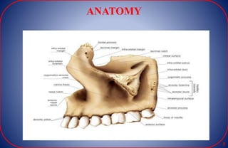
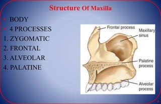
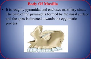
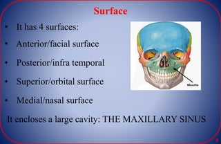
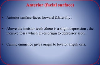
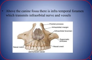
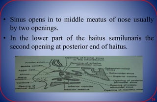
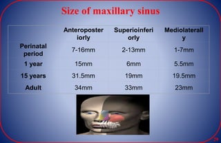
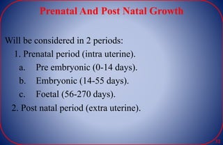
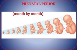
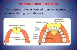
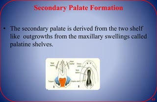
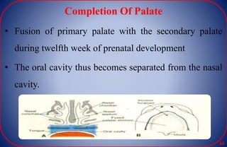
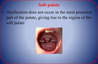
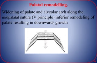
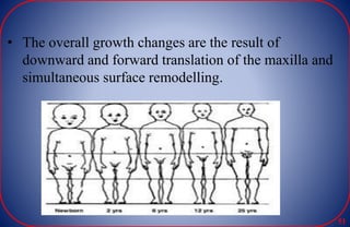
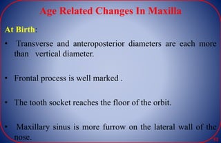
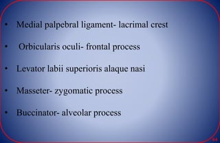
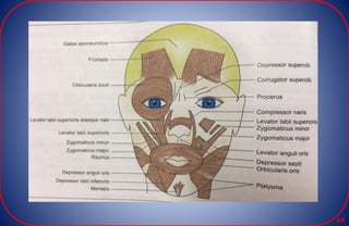
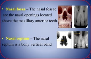
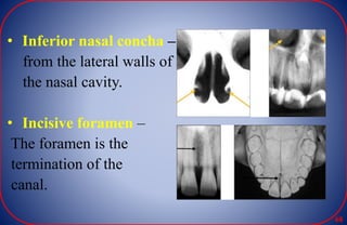
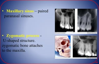
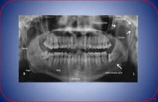
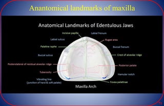
MAXILLA
Presented By
Dr.Akshay Bhojraj
1St yr PG
1
CONTENT
 Introduction
 Anatomy Of Maxilla
 Growth And Development
 Prenatal And Post Natal Growth
 Age Changes
 Radiographic Landmarks
 Anatomical Landmarks
 Clinical And Prosthodontic Consideration
 Conclusion
2
INTRODUCTION
3
-Maxilla, is second largest of facial bones.
It is a paired bone enters into formation of the
 face
 nose
 mouth
 orbit
 Part of the infratemporal bone
 Part of pterygopalatine fossa 4
ANATOMY
5
Structure Of Maxilla
• BODY
• 4 PROCESSES
1. ZYGOMATIC
2. FRONTAL
3. ALVEOLAR
4. PALATINE
6
Body Of Maxilla
• It is roughly pyramidal and encloses maxillary sinus.
The base of the pyramid is formed by the nasal surface
and the apex is directed towards the zygomatic
process
7
Surface
• It has 4 surfaces:
• Anterior/facial surface
• Posterior/infra temporal
• Superior/orbital surface
• Medial/nasal surface
It encloses a large cavity: THE MAXILLARY SINUS
8
Anterior (facial surface)
9
• Anterior surface-faces forward &laterally .
• Above the incisior teeth ,there is a slight depression , the
incisive fossa which gives origin to depressor septi.
• Canine eminence gives origin to levator anguli oris.
• Above the canine fossa there is infra temporal foramen
which transmits infraorbital nerve and vessels
10
Posterior (Infratemporal Surface)
• Convex and directed backwards and laterally.
• forms anterior wall of the infratemporal fossa.
• It seperates from anterior surface by zygomatic
process.
11
• 2-3 alveolar canals for posterior superior alveolar
nerve and vessels
12
Superior (Orbital Surface)
• Smooth and triangular , forms greater part of the floor
of orbit.
• It provide attachment to lacrimal bone medially.
13
Medial (nasal surface)
• Part of lateral wall of nose
• Posterosuperiorly attaches to part of the inferior
meatus.
• Encloses greater and
lesser palatine nerve
14
PROCESSES OF MAXILLA
15
Frontal Process
• It projects postero-superiorly between the nasal
and lacrimal bones.
• The frontal process apically joins with the nasal
notch of frontal bone at fronto -maxillary suture.
16
• Anterior border articulates with lateral border of nasal
bone and the posterior with lacrimal bone.
17
Zygomatic Process
It is a pyramidal projection where anterior, infra
temporal and orbital surfaces converge.
• It articulates with the maxillary process of
zygomatic bone.
18
Alveolar Process
• Thick, arched, wide behind .
• socketed for tooth roots -
- Eights sockets on either side
-Canine: deepest
- Molars: widest and subdivided into three by
septa
- Incisors and Second premolar: single
- first premolar: sometimes double
19
• Maxillary torus may be occasionally present
• Buccinator muscle aries upto the first molar tooth
20
Palatine process
• Thick, strong and horizontal projecting medially
• Its inferior surface is concave and uneven
• Displays numerous vascular foramina
• depressions for palatine glands
21
• Posterolaterally two grooves
• Incisive fossa and incisive canal
• Intermaxillary palatal suture
• Superior surface, concave and smooth, forms most of the
nasal floor
22
• Lateral border continuous with maxillary body
• Medial border, raised into nasal crest
• forms a groove for the vomer
• Posterior border is serrated
23
24
MAXILLARY SINUS
25
• Pyramidal shaped.
• Maxillary sinus is first to develop (at 4 month)
• ROOF: floor of the orbit transvered by the infraorbital
canal.
• FLOOR: by the alveolar process of maxilla. Lies about
half inch below the level of the floor of the nose.
• APEX : Directed laterally towards zygomatic bone.
• MEDIAL WALL OR BASE : Partly by the lateral wall of
the nose and by palate
26
27
• Sinus opens in to middle meatus of nose usually
by two openings.
• In the lower part of the haitus semilunaris the
second opening at posterior end of haitus.
28
Size of maxillary sinus
29
Anteroposter
iorly
Superioinferi
orly
Mediolaterall
y
Perinatal
period
7-16mm 2-13mm 1-7mm
1 year 15mm 6mm 5.5mm
15 years 31.5mm 19mm 19.5mm
Adult 34mm 33mm 23mm
GROWTH AND DEVELOPMENT
30
• To determine the growth deviation of particular individual,
we study normal health variations
31
Prenatal And Post Natal Growth
Will be considered in 2 periods:
1. Prenatal period (intra uterine).
a. Pre embryonic (0-14 days).
b. Embryonic (14-55 days).
c. Foetal (56-270 days).
2. Post natal period (extra uterine).
32
PRENATAL PERIOD
33
• Prenatal period of development is a dynamic phase in
the development of human body.it is divided into three
periods
• 1.During this period cleavage of ovum and its
attachment to intra-uterine wall occurs.
• 2.Major part of development of the facial and cranial
region occurs.
• 3.Acclerated growth of craniofacial structures occur
resulting in increase in their size
34
• Morula – Series of cell division give rise to egg cell
mass.
• Blastula – inner cell mass form two layer by
embryonic dics called epiblast and hypoblast
35
Formation Of Germ Layers
• The inner cell mass or embryoblast differentiates into
the epiblast and hypoblast to form a bilaminar disc
36
Derivatives of first pharyngeal arch
Pharyngeal
arch
Nerve Muscles Skeleton
Mandibular
arch
Trigeminal N. MASTICATI
ON,
Mylohyoid,
Ant. Belly of
digastric
Premaxilla,
MAXILA,
Zygoma,
Temporal
bone
Mandi.
37
Prenatal Growth Of Maxilla
Maxilla is formed from 1st pharyngeal arch.
1st pharyngeal arch lying lateral to the stomadeum
divided in 2 processes.
Dorsal process – Maxillary process.
Ventral process – Mandibular process.
38
• Maxillary process, extending forward beneath the
region of the eye and subsequently gives rise to the:
• Premaxilla
• Maxilla
• Zygomatic bone and part of the temporal bone.
39
Develoment Of the maxilla starts around the 4 week
 Maxillary prominence
 Mandibular prominence
 Olfactory placodes
 Lat. Nasal prominence
 Med. Nasal prominence
 Stomodeum
40
Development Of Palate
• The palate is formed from two separate embryonic
structures:
*the primary palate and * the secondary palate.
• The formation begins in fifth week of prenatal
development, within the embryonic period.
• The palate is then completed during the twelfth week,
within the fetal period.
41
Primary Palate Formation
• The primary palate is derived from the intermaxillary
segment during the fifth week.
42
Secondary Palate Formation
• The secondary palate is derived from the two shelf
like outgrowths from the maxillary swellings called
palatine shelves.
43
Completion Of Palate
• Fusion of primary palate with the secondary palate
during twelfth week of prenatal development
• The oral cavity thus becomes separated from the nasal
cavity.
44
Soft palate
• Ossification does not occur in the most posterior
part of the palate, giving rise to the region of the
soft palate
45
Postnatal Period
46
• The growth of maxilla depends on influence of
several functional matrices that act upon different
areas of the bone thus allowing its subdivision into
skeletal units
• The alveolar unit provide the functional matrix
for the teeth
47
Maxillary tuberosity and arch lengthening
• Maxillary tuberosity is the major site for maxillary
growth. Arch lengthening in this part takes place by
deposition of new bone on backward facing
periosteum
48
Vertical drift of teeth.
• It is a significant intrinsic growth factor .Eruption
of the teeth causes deposition on the alveolar
margins
49
Palatal remodelling.
Widening of palate and alveolar arch along the
midpalatal suture (V principle) inferior remodeling of
palate resulting in downwards growth
50
• The overall growth changes are the result of
downward and forward translation of the maxilla and
simultaneous surface remodelling.
51
Age Related Changes In Maxilla
At Birth:
• Transverse and anteroposterior diameters are each more
than vertical diameter.
• Frontal process is well marked .
• The tooth socket reaches the floor of the orbit.
• Maxillary sinus is more furrow on the lateral wall of the
nose. 52
In Adults:-
• Vertical diameter of the sinuse closer is greatest
due to development of the alveolar process and
increase in size.
53
In Old Age:-
• Height of the bone is reduced .
• Resorption of the alveolar process or ridges takes
place in a characteristic fashion .
• In addition to this, there is either lingual or vestibular
resorption ,the alveolar ridges frequently do not
develop cortical bone and present sharp and
cancellous bony ridges immediately below the
alveolar mucosa. 54
• The maxillary tooth are inclined labially and
following their loss the labial plate of the bone
resorbs more than the lingual plate.
• It results in smaller alveolar arch and an overall
decrease in necessary support area.
• Vertically the posterior segment tends to shrink
more than the anterior segment.
55
NERVES AND VESSELS
56
• Infraorbital nerve and vessels
• Anterior superior alveolar nerve and vessels- canalis
sinosus
• Middle superior alveolar nerve
57
• Nasopalatine nerve- incisive canal
• Greater palatine artery
• Greater palatine nerve and vessels
• Posterior superior alveolar nerve and vessels
58
NERVES
59
VENOUS DRAINAGE
• The venous drainage : the superficial temporal
vein to the maxillary and retromandibular veins,
the posterior auricular vein to the posterior
retromandibular and external jugular veins, the
maxillary vein to the superficial temporal and
retromandibular vein, and draining veins (external
acoustic meatus) to the pterygoid plexus.
60
61
• ATTACHMENTS AND RELATIONS
62
A. MUSCLES AND LIGAMENTS:
• Levator anguli oris- canine fossa
• Depressor septi- incisive fossa
• Levator labii superioris- infraorbital margin
• Fibers of medial pterygoid- maxillary tuberosity
• Inferior oblique muscle- lacrimal groove
63
• Medial palpebral ligament- lacrimal crest
• Orbicularis oculi- frontal process
• Levator labii superioris alaque nasi
• Masseter- zygomatic process
• Buccinator- alveolar process
64
65
RADIOGRAPHIC LANDMARKS OF
MAXILLA
66
• Nasal fossa – The nasal fossae
are the nasal openings located
above the maxillary anterior teeth.
• Nasal septum – The nasal
septum is a bony vertical band
67
• Inferior nasal concha –
from the lateral walls of
the nasal cavity.
• Incisive foramen –
The foramen is the
termination of the
canal.
68
• Maxillary sinus – paired
paranasal sinuses.
• Zygomatic process -
U-shaped structure.
zygomatic bone attaches
to the maxilla.
69
70
Anantomical landmarks of maxilla
71
PROSTHODONTICS
CONSIDERATION
72
 Zygomatico-Alveolar crest:
The mucosal covering is very thin, mucosa not
considered desirable for stress bearing and sometimes
should be relieved. If not, it results in poor retention of the
denture.
 Maxillary tuberosity:
• Provides resistance to horizontal movements. Posterior
wall resists Movement in anterior direction.
• The denture base should cover the tuberosity and fill the
hamular notch.
73
Mid palatine suture:
• If prominent, becomes fulcrum point around which
the denture rotates causing discomfort and damage to
soft tissues.
Incisive foramen:
• Nasopalatine nerve makes its exit to the palate. The
foramen should be relieved in the denture.
Cleft Palate:
• Feeding plate and Obturator
74
Cleft lip :
• Missing lateral incisors are replaced with RPD or
FPD or Implants
 Palatal torus:
• Hyperplastic growth of bone which needs to be
relieved or removed surgically.
Torus Palatinus: LARGE- Surgery
SMALL- Relief in denture
75
DEVELOPMENTAL
ANOMALIES
76
CLEFT LIP & PALATE
77
These disorders can result in feeding problems, speech
problems, hearing problems, and frequent ear
infections Cleft lip and cleft palate, also known
as orofacial cleft, is a group of conditions that
includes cleft lip, cleft palate, and both together.
A cleft lip contains an opening in the upper lip that
may extend into the nose. The opening may be on one
side, both sides, or in the middle. A cleft palate occurs
when the roof of the mouth contains an opening into
the nose. These disorders can result in feeding
problems, speech problems, hearing problems, and
frequent ear infection
78
Cleft lip & Palate
79
Torus Palatinus.
Torus palatinus is a harmless, painless bony growth
located on the roof of the mouth
80
ANATOMICAL CONSIDERATION
OF MAXILLA IN
IMPLANTOLOGY
81
• The implant should be at least 1.5 mm away from the
adjacent teeth
• The implant should be at least 3 mm away from an
adjacent implant
• A wider diameter implant should be selected for molar
teeth.
• Distance of implant from nasal cavity is 1mm
• Distance of implant from maxillary sinus is 1 mm.
• Success rate of mandible is more , compare to maxilla
as more bone density and blood supply.
82
83
84
85
CONCLUSION
86
• In order to construct a prosthesis a dentist requires
an understanding of the foundation, it’s
components, its properties and qualities must be
analysed to assure proper support for the proposed
prosthesis.
87
REFERENCES
88
• Chaurasia BD. Human anatomy. CBS Publisher;
2004.
• An Introduction To Human Embryology For
Medical Students – Inderbir Singh
• Dental Impalnt Posthetics-Carl E Misch;2nd
edition
• Oral Anatomy,histology And Embryology ;3rd Ed;
Berkovitz
89

Maxilla

  • 1.
  • 2.
    CONTENT  Introduction  AnatomyOf Maxilla  Growth And Development  Prenatal And Post Natal Growth  Age Changes  Radiographic Landmarks  Anatomical Landmarks  Clinical And Prosthodontic Consideration  Conclusion 2
  • 3.
  • 4.
    -Maxilla, is secondlargest of facial bones. It is a paired bone enters into formation of the  face  nose  mouth  orbit  Part of the infratemporal bone  Part of pterygopalatine fossa 4
  • 5.
  • 6.
    Structure Of Maxilla •BODY • 4 PROCESSES 1. ZYGOMATIC 2. FRONTAL 3. ALVEOLAR 4. PALATINE 6
  • 7.
    Body Of Maxilla •It is roughly pyramidal and encloses maxillary sinus. The base of the pyramid is formed by the nasal surface and the apex is directed towards the zygomatic process 7
  • 8.
    Surface • It has4 surfaces: • Anterior/facial surface • Posterior/infra temporal • Superior/orbital surface • Medial/nasal surface It encloses a large cavity: THE MAXILLARY SINUS 8
  • 9.
    Anterior (facial surface) 9 •Anterior surface-faces forward &laterally . • Above the incisior teeth ,there is a slight depression , the incisive fossa which gives origin to depressor septi. • Canine eminence gives origin to levator anguli oris.
  • 10.
    • Above thecanine fossa there is infra temporal foramen which transmits infraorbital nerve and vessels 10
  • 11.
    Posterior (Infratemporal Surface) •Convex and directed backwards and laterally. • forms anterior wall of the infratemporal fossa. • It seperates from anterior surface by zygomatic process. 11
  • 12.
    • 2-3 alveolarcanals for posterior superior alveolar nerve and vessels 12
  • 13.
    Superior (Orbital Surface) •Smooth and triangular , forms greater part of the floor of orbit. • It provide attachment to lacrimal bone medially. 13
  • 14.
    Medial (nasal surface) •Part of lateral wall of nose • Posterosuperiorly attaches to part of the inferior meatus. • Encloses greater and lesser palatine nerve 14
  • 15.
  • 16.
    Frontal Process • Itprojects postero-superiorly between the nasal and lacrimal bones. • The frontal process apically joins with the nasal notch of frontal bone at fronto -maxillary suture. 16
  • 17.
    • Anterior borderarticulates with lateral border of nasal bone and the posterior with lacrimal bone. 17
  • 18.
    Zygomatic Process It isa pyramidal projection where anterior, infra temporal and orbital surfaces converge. • It articulates with the maxillary process of zygomatic bone. 18
  • 19.
    Alveolar Process • Thick,arched, wide behind . • socketed for tooth roots - - Eights sockets on either side -Canine: deepest - Molars: widest and subdivided into three by septa - Incisors and Second premolar: single - first premolar: sometimes double 19
  • 20.
    • Maxillary torusmay be occasionally present • Buccinator muscle aries upto the first molar tooth 20
  • 21.
    Palatine process • Thick,strong and horizontal projecting medially • Its inferior surface is concave and uneven • Displays numerous vascular foramina • depressions for palatine glands 21
  • 22.
    • Posterolaterally twogrooves • Incisive fossa and incisive canal • Intermaxillary palatal suture • Superior surface, concave and smooth, forms most of the nasal floor 22
  • 23.
    • Lateral bordercontinuous with maxillary body • Medial border, raised into nasal crest • forms a groove for the vomer • Posterior border is serrated 23
  • 24.
  • 25.
  • 26.
    • Pyramidal shaped. •Maxillary sinus is first to develop (at 4 month) • ROOF: floor of the orbit transvered by the infraorbital canal. • FLOOR: by the alveolar process of maxilla. Lies about half inch below the level of the floor of the nose. • APEX : Directed laterally towards zygomatic bone. • MEDIAL WALL OR BASE : Partly by the lateral wall of the nose and by palate 26
  • 27.
  • 28.
    • Sinus opensin to middle meatus of nose usually by two openings. • In the lower part of the haitus semilunaris the second opening at posterior end of haitus. 28
  • 29.
    Size of maxillarysinus 29 Anteroposter iorly Superioinferi orly Mediolaterall y Perinatal period 7-16mm 2-13mm 1-7mm 1 year 15mm 6mm 5.5mm 15 years 31.5mm 19mm 19.5mm Adult 34mm 33mm 23mm
  • 30.
  • 31.
    • To determinethe growth deviation of particular individual, we study normal health variations 31
  • 32.
    Prenatal And PostNatal Growth Will be considered in 2 periods: 1. Prenatal period (intra uterine). a. Pre embryonic (0-14 days). b. Embryonic (14-55 days). c. Foetal (56-270 days). 2. Post natal period (extra uterine). 32
  • 33.
  • 34.
    • Prenatal periodof development is a dynamic phase in the development of human body.it is divided into three periods • 1.During this period cleavage of ovum and its attachment to intra-uterine wall occurs. • 2.Major part of development of the facial and cranial region occurs. • 3.Acclerated growth of craniofacial structures occur resulting in increase in their size 34
  • 35.
    • Morula –Series of cell division give rise to egg cell mass. • Blastula – inner cell mass form two layer by embryonic dics called epiblast and hypoblast 35
  • 36.
    Formation Of GermLayers • The inner cell mass or embryoblast differentiates into the epiblast and hypoblast to form a bilaminar disc 36
  • 37.
    Derivatives of firstpharyngeal arch Pharyngeal arch Nerve Muscles Skeleton Mandibular arch Trigeminal N. MASTICATI ON, Mylohyoid, Ant. Belly of digastric Premaxilla, MAXILA, Zygoma, Temporal bone Mandi. 37
  • 38.
    Prenatal Growth OfMaxilla Maxilla is formed from 1st pharyngeal arch. 1st pharyngeal arch lying lateral to the stomadeum divided in 2 processes. Dorsal process – Maxillary process. Ventral process – Mandibular process. 38
  • 39.
    • Maxillary process,extending forward beneath the region of the eye and subsequently gives rise to the: • Premaxilla • Maxilla • Zygomatic bone and part of the temporal bone. 39
  • 40.
    Develoment Of themaxilla starts around the 4 week  Maxillary prominence  Mandibular prominence  Olfactory placodes  Lat. Nasal prominence  Med. Nasal prominence  Stomodeum 40
  • 41.
    Development Of Palate •The palate is formed from two separate embryonic structures: *the primary palate and * the secondary palate. • The formation begins in fifth week of prenatal development, within the embryonic period. • The palate is then completed during the twelfth week, within the fetal period. 41
  • 42.
    Primary Palate Formation •The primary palate is derived from the intermaxillary segment during the fifth week. 42
  • 43.
    Secondary Palate Formation •The secondary palate is derived from the two shelf like outgrowths from the maxillary swellings called palatine shelves. 43
  • 44.
    Completion Of Palate •Fusion of primary palate with the secondary palate during twelfth week of prenatal development • The oral cavity thus becomes separated from the nasal cavity. 44
  • 45.
    Soft palate • Ossificationdoes not occur in the most posterior part of the palate, giving rise to the region of the soft palate 45
  • 46.
  • 47.
    • The growthof maxilla depends on influence of several functional matrices that act upon different areas of the bone thus allowing its subdivision into skeletal units • The alveolar unit provide the functional matrix for the teeth 47
  • 48.
    Maxillary tuberosity andarch lengthening • Maxillary tuberosity is the major site for maxillary growth. Arch lengthening in this part takes place by deposition of new bone on backward facing periosteum 48
  • 49.
    Vertical drift ofteeth. • It is a significant intrinsic growth factor .Eruption of the teeth causes deposition on the alveolar margins 49
  • 50.
    Palatal remodelling. Widening ofpalate and alveolar arch along the midpalatal suture (V principle) inferior remodeling of palate resulting in downwards growth 50
  • 51.
    • The overallgrowth changes are the result of downward and forward translation of the maxilla and simultaneous surface remodelling. 51
  • 52.
    Age Related ChangesIn Maxilla At Birth: • Transverse and anteroposterior diameters are each more than vertical diameter. • Frontal process is well marked . • The tooth socket reaches the floor of the orbit. • Maxillary sinus is more furrow on the lateral wall of the nose. 52
  • 53.
    In Adults:- • Verticaldiameter of the sinuse closer is greatest due to development of the alveolar process and increase in size. 53
  • 54.
    In Old Age:- •Height of the bone is reduced . • Resorption of the alveolar process or ridges takes place in a characteristic fashion . • In addition to this, there is either lingual or vestibular resorption ,the alveolar ridges frequently do not develop cortical bone and present sharp and cancellous bony ridges immediately below the alveolar mucosa. 54
  • 55.
    • The maxillarytooth are inclined labially and following their loss the labial plate of the bone resorbs more than the lingual plate. • It results in smaller alveolar arch and an overall decrease in necessary support area. • Vertically the posterior segment tends to shrink more than the anterior segment. 55
  • 56.
  • 57.
    • Infraorbital nerveand vessels • Anterior superior alveolar nerve and vessels- canalis sinosus • Middle superior alveolar nerve 57
  • 58.
    • Nasopalatine nerve-incisive canal • Greater palatine artery • Greater palatine nerve and vessels • Posterior superior alveolar nerve and vessels 58
  • 59.
  • 60.
    VENOUS DRAINAGE • Thevenous drainage : the superficial temporal vein to the maxillary and retromandibular veins, the posterior auricular vein to the posterior retromandibular and external jugular veins, the maxillary vein to the superficial temporal and retromandibular vein, and draining veins (external acoustic meatus) to the pterygoid plexus. 60
  • 61.
  • 62.
    • ATTACHMENTS ANDRELATIONS 62
  • 63.
    A. MUSCLES ANDLIGAMENTS: • Levator anguli oris- canine fossa • Depressor septi- incisive fossa • Levator labii superioris- infraorbital margin • Fibers of medial pterygoid- maxillary tuberosity • Inferior oblique muscle- lacrimal groove 63
  • 64.
    • Medial palpebralligament- lacrimal crest • Orbicularis oculi- frontal process • Levator labii superioris alaque nasi • Masseter- zygomatic process • Buccinator- alveolar process 64
  • 65.
  • 66.
  • 67.
    • Nasal fossa– The nasal fossae are the nasal openings located above the maxillary anterior teeth. • Nasal septum – The nasal septum is a bony vertical band 67
  • 68.
    • Inferior nasalconcha – from the lateral walls of the nasal cavity. • Incisive foramen – The foramen is the termination of the canal. 68
  • 69.
    • Maxillary sinus– paired paranasal sinuses. • Zygomatic process - U-shaped structure. zygomatic bone attaches to the maxilla. 69
  • 70.
  • 71.
  • 72.
  • 73.
     Zygomatico-Alveolar crest: Themucosal covering is very thin, mucosa not considered desirable for stress bearing and sometimes should be relieved. If not, it results in poor retention of the denture.  Maxillary tuberosity: • Provides resistance to horizontal movements. Posterior wall resists Movement in anterior direction. • The denture base should cover the tuberosity and fill the hamular notch. 73
  • 74.
    Mid palatine suture: •If prominent, becomes fulcrum point around which the denture rotates causing discomfort and damage to soft tissues. Incisive foramen: • Nasopalatine nerve makes its exit to the palate. The foramen should be relieved in the denture. Cleft Palate: • Feeding plate and Obturator 74
  • 75.
    Cleft lip : •Missing lateral incisors are replaced with RPD or FPD or Implants  Palatal torus: • Hyperplastic growth of bone which needs to be relieved or removed surgically. Torus Palatinus: LARGE- Surgery SMALL- Relief in denture 75
  • 76.
  • 77.
    CLEFT LIP &PALATE 77
  • 78.
    These disorders canresult in feeding problems, speech problems, hearing problems, and frequent ear infections Cleft lip and cleft palate, also known as orofacial cleft, is a group of conditions that includes cleft lip, cleft palate, and both together. A cleft lip contains an opening in the upper lip that may extend into the nose. The opening may be on one side, both sides, or in the middle. A cleft palate occurs when the roof of the mouth contains an opening into the nose. These disorders can result in feeding problems, speech problems, hearing problems, and frequent ear infection 78
  • 79.
    Cleft lip &Palate 79
  • 80.
    Torus Palatinus. Torus palatinusis a harmless, painless bony growth located on the roof of the mouth 80
  • 81.
  • 82.
    • The implantshould be at least 1.5 mm away from the adjacent teeth • The implant should be at least 3 mm away from an adjacent implant • A wider diameter implant should be selected for molar teeth. • Distance of implant from nasal cavity is 1mm • Distance of implant from maxillary sinus is 1 mm. • Success rate of mandible is more , compare to maxilla as more bone density and blood supply. 82
  • 83.
  • 84.
  • 85.
  • 86.
  • 87.
    • In orderto construct a prosthesis a dentist requires an understanding of the foundation, it’s components, its properties and qualities must be analysed to assure proper support for the proposed prosthesis. 87
  • 88.
  • 89.
    • Chaurasia BD.Human anatomy. CBS Publisher; 2004. • An Introduction To Human Embryology For Medical Students – Inderbir Singh • Dental Impalnt Posthetics-Carl E Misch;2nd edition • Oral Anatomy,histology And Embryology ;3rd Ed; Berkovitz 89