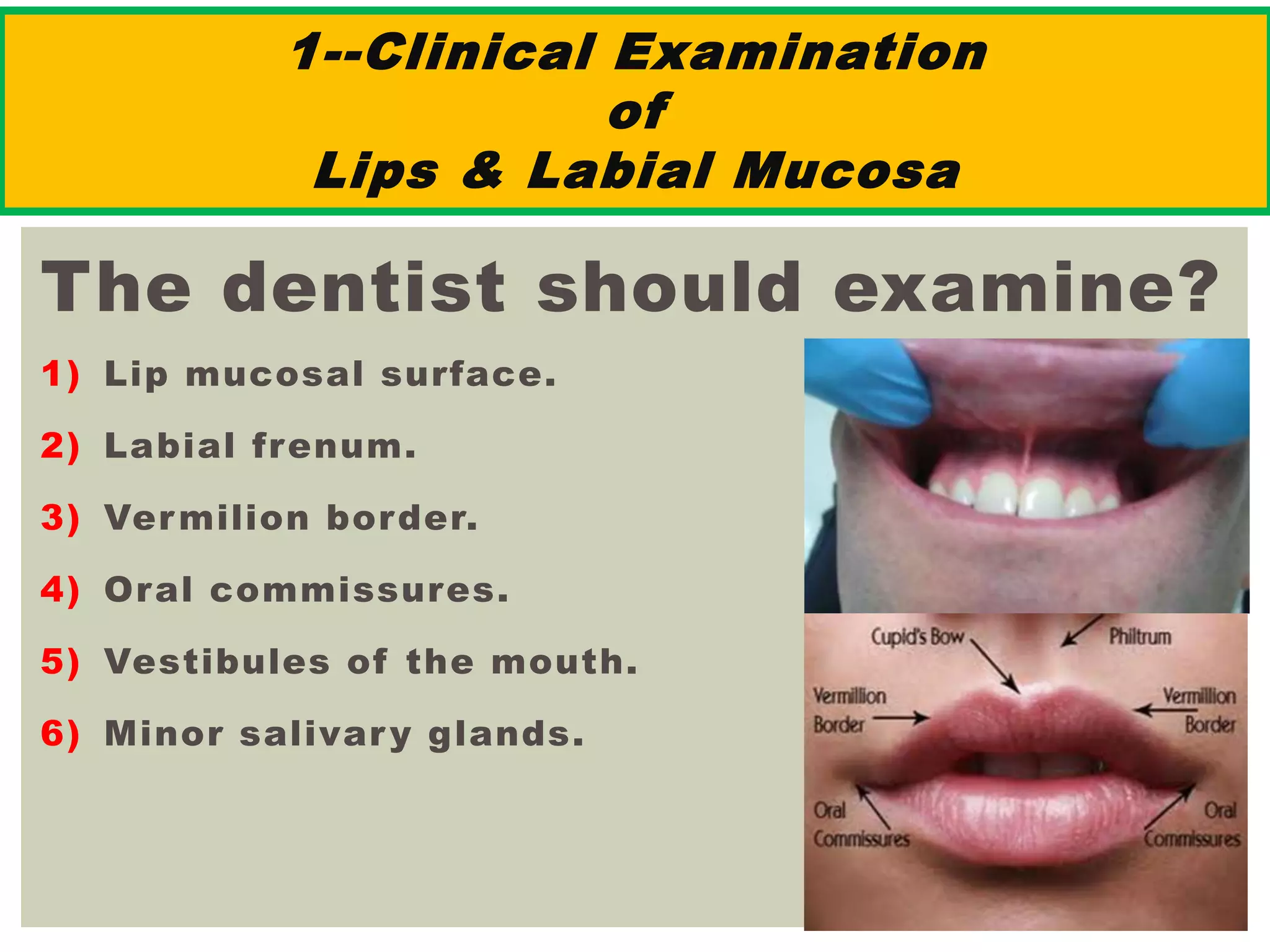
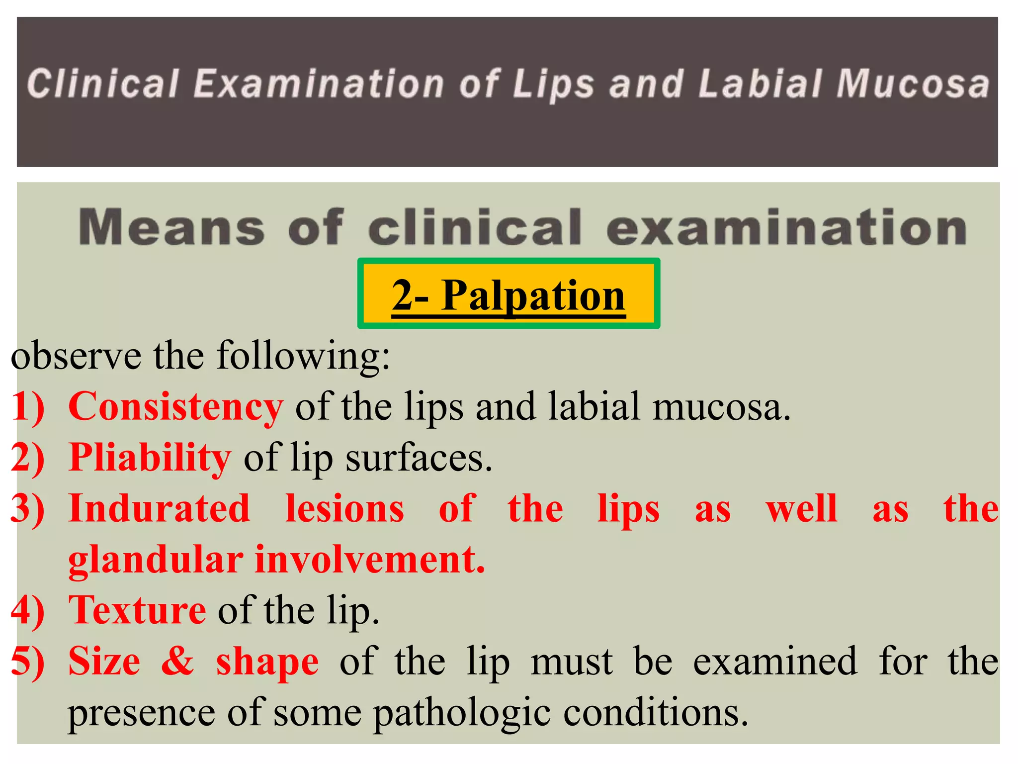
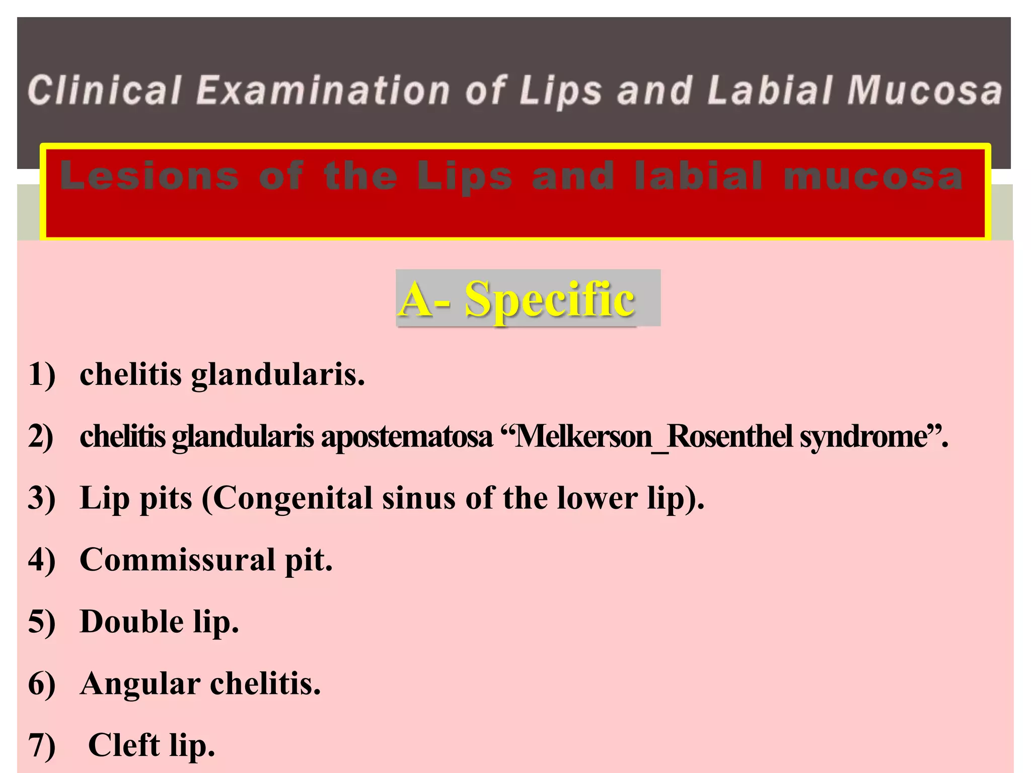
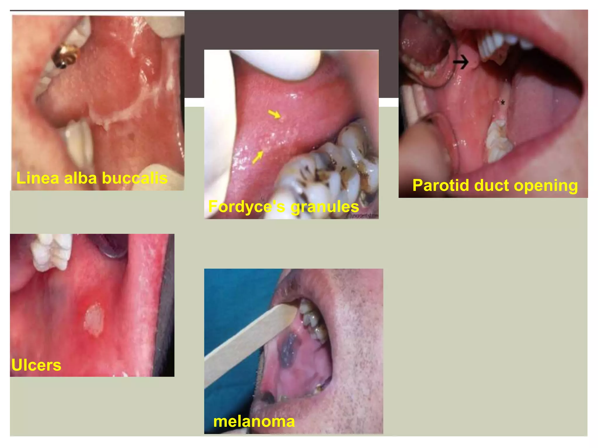
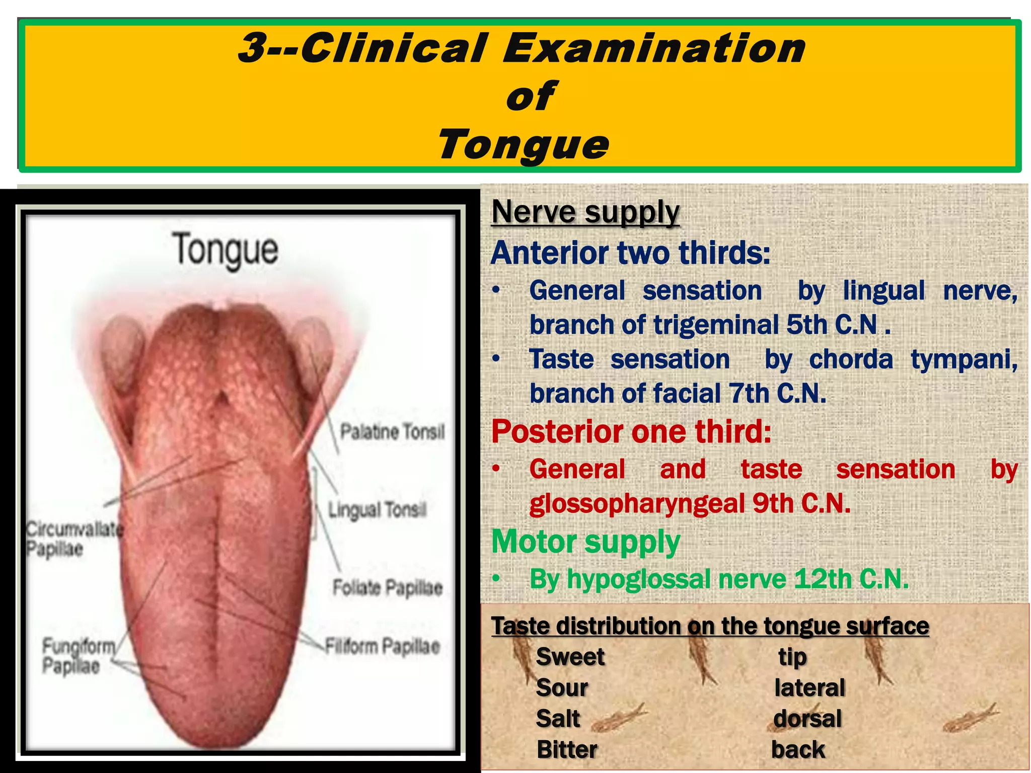
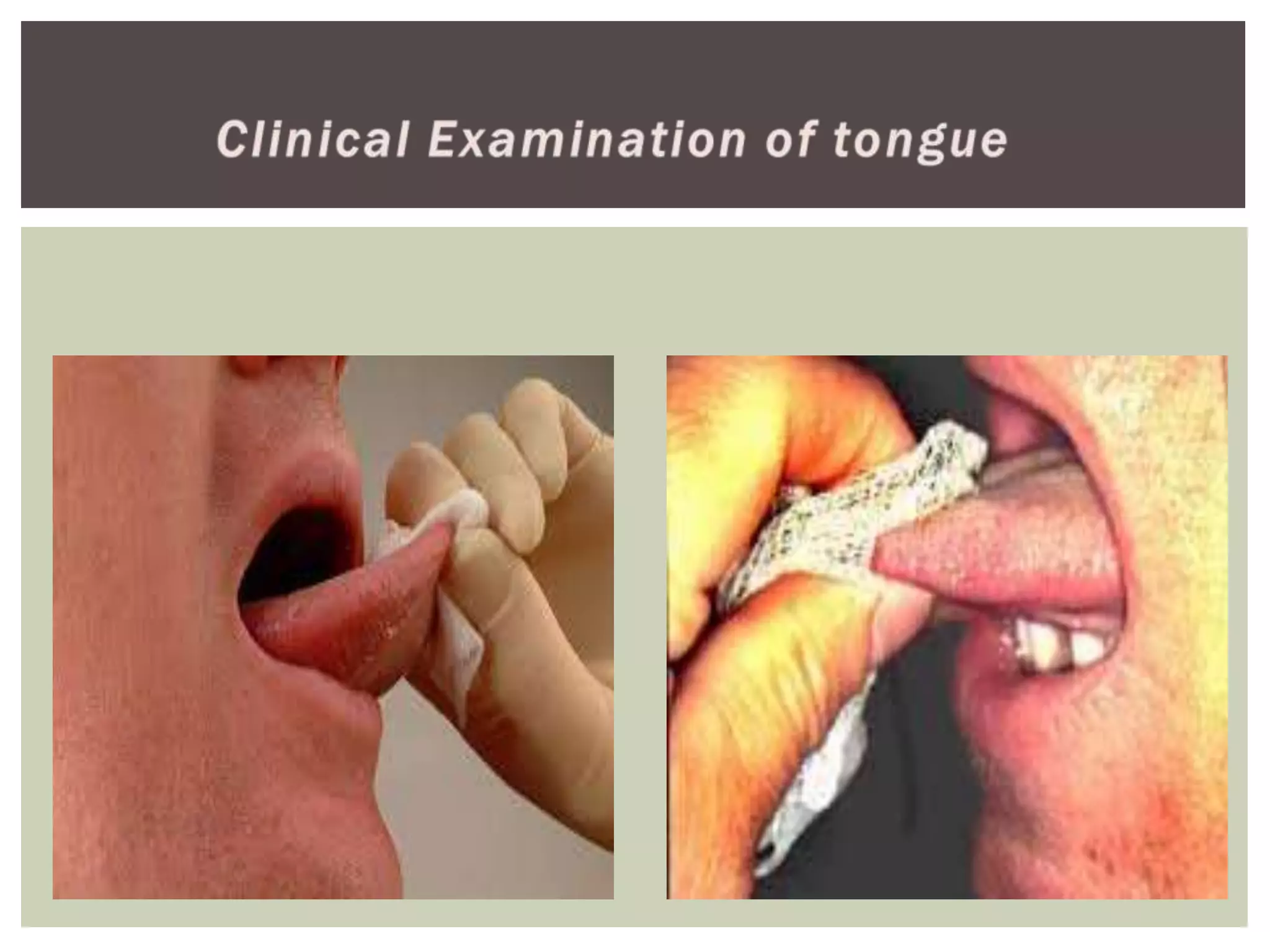
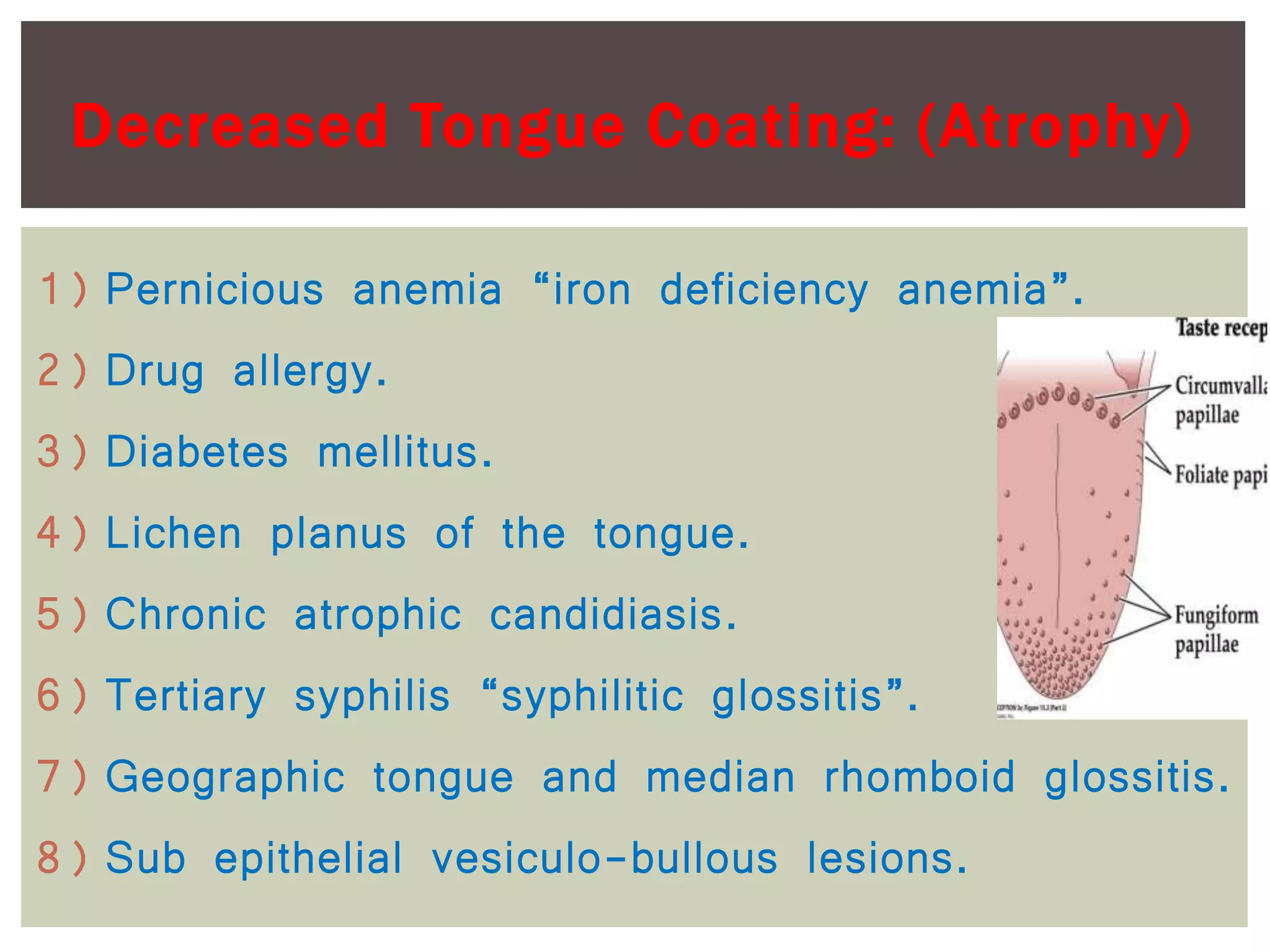
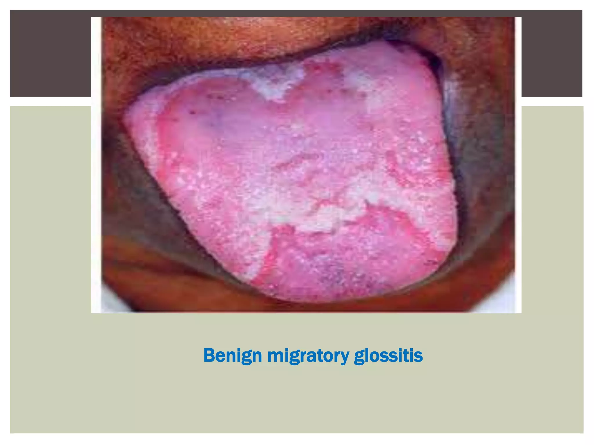
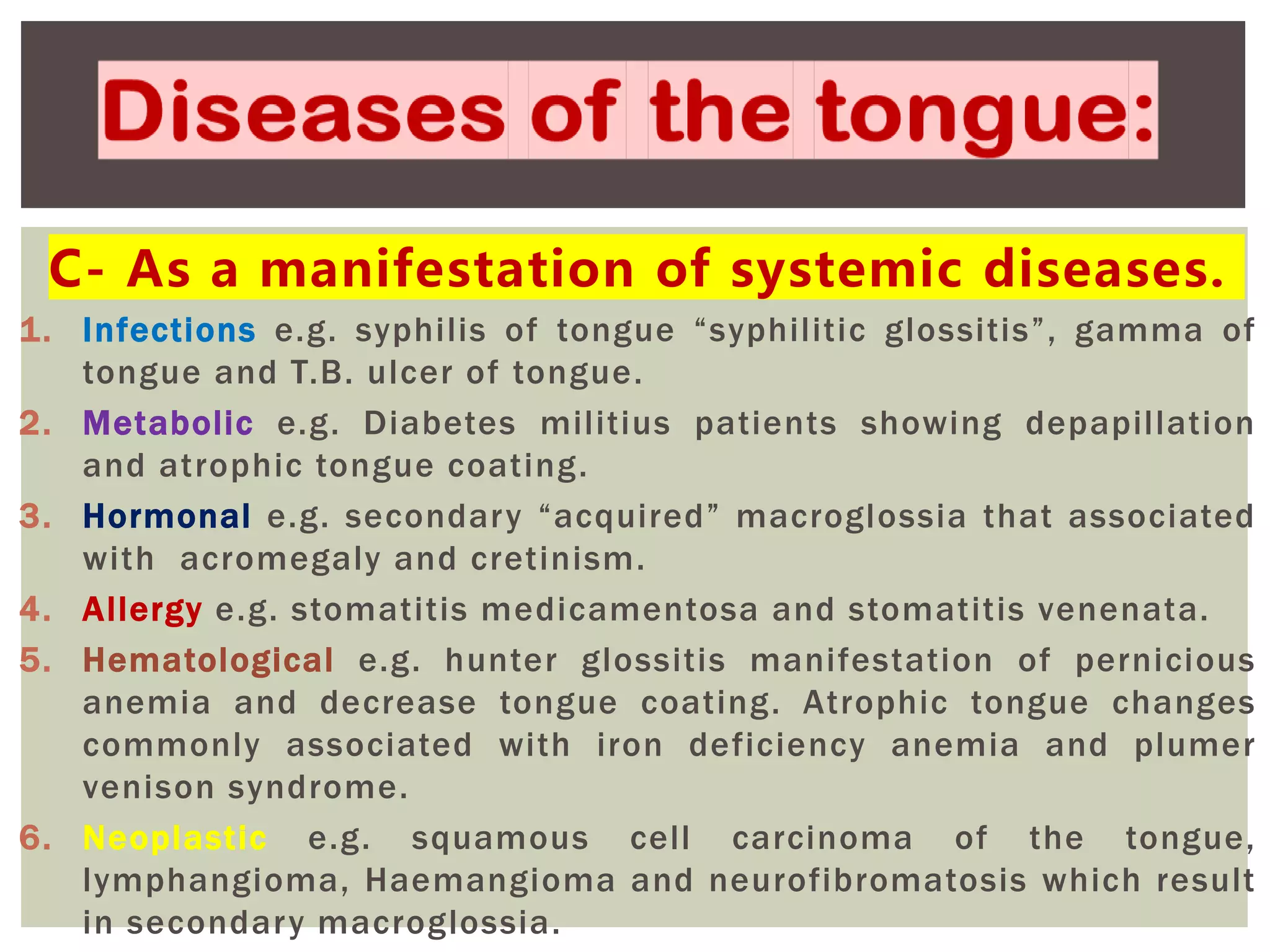
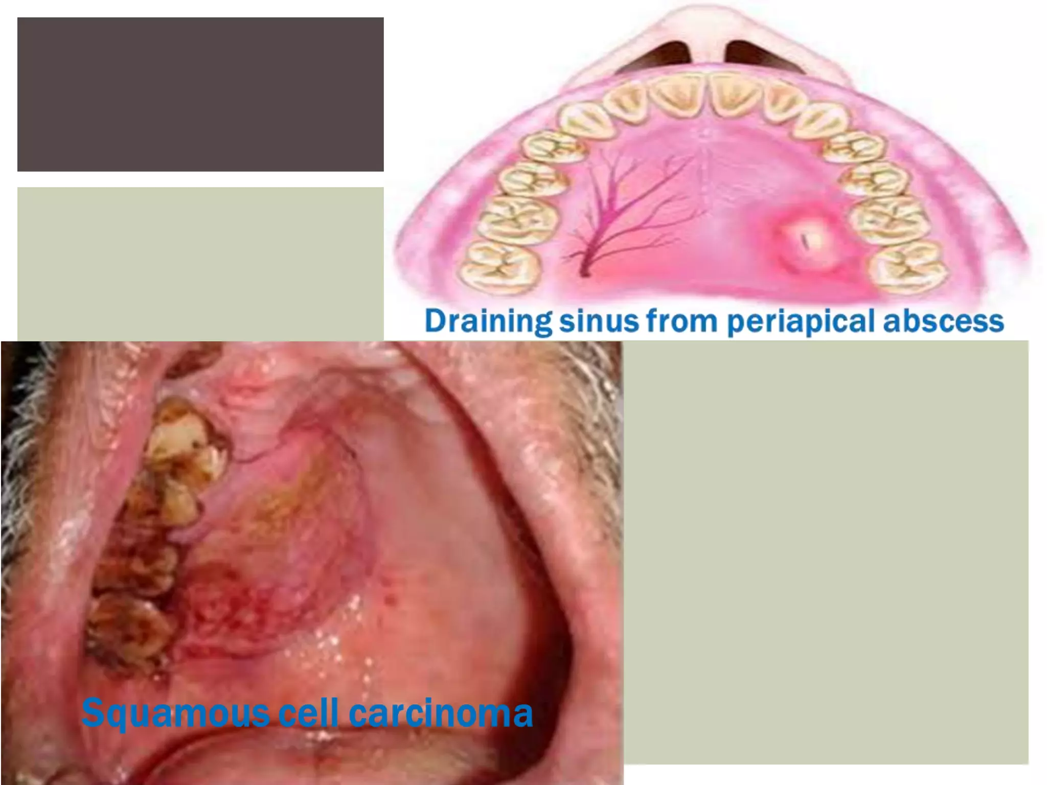
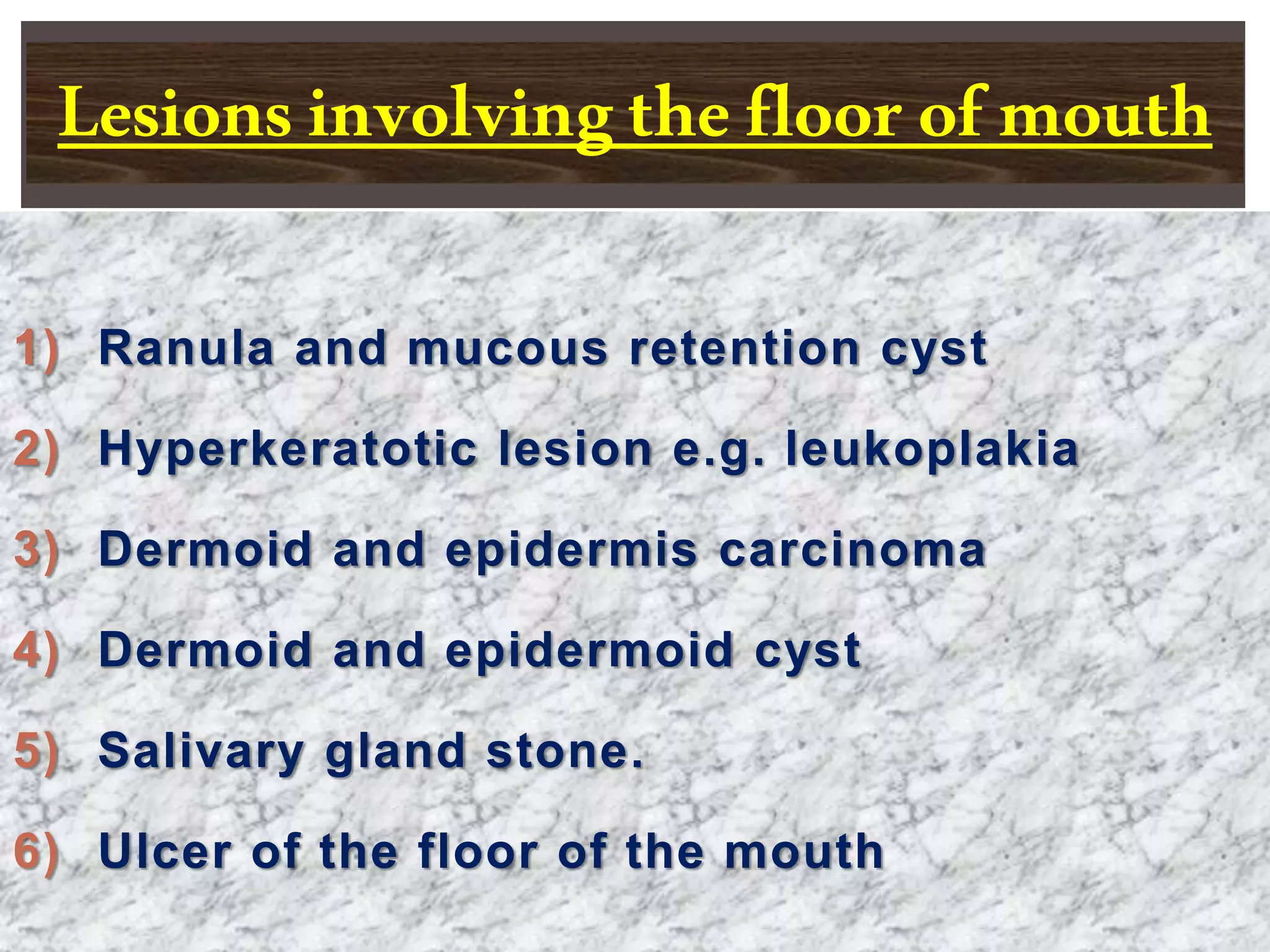
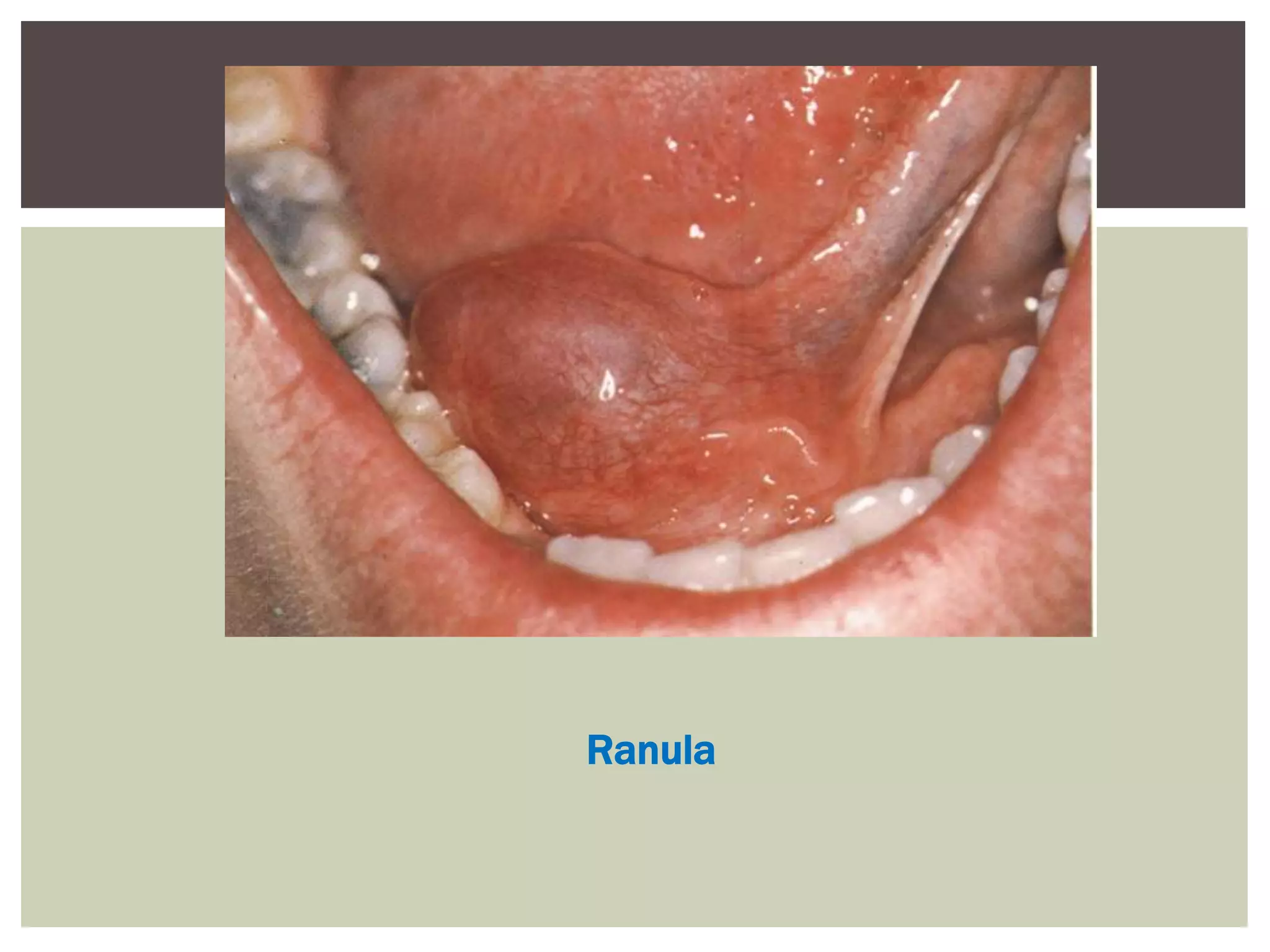
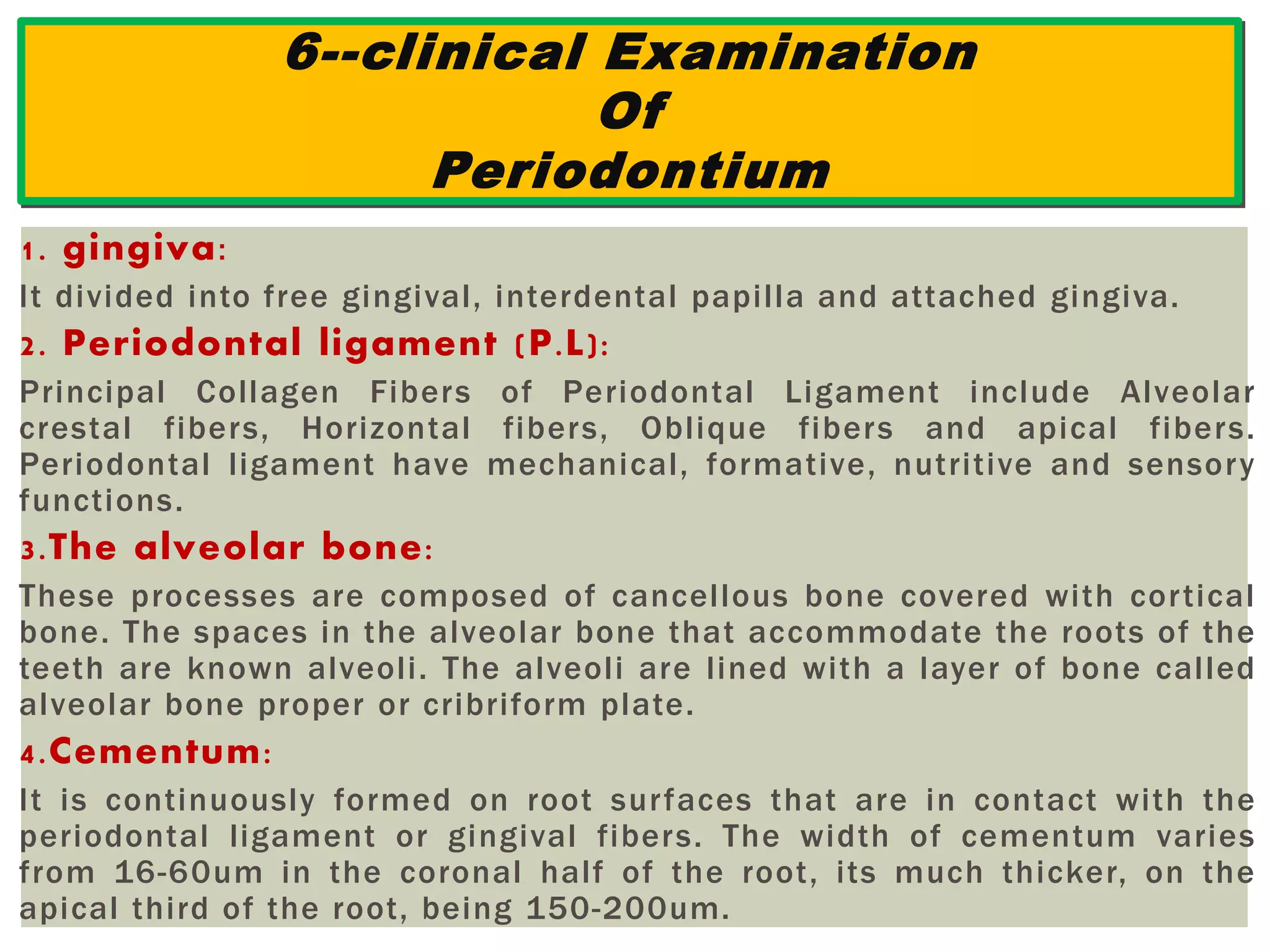
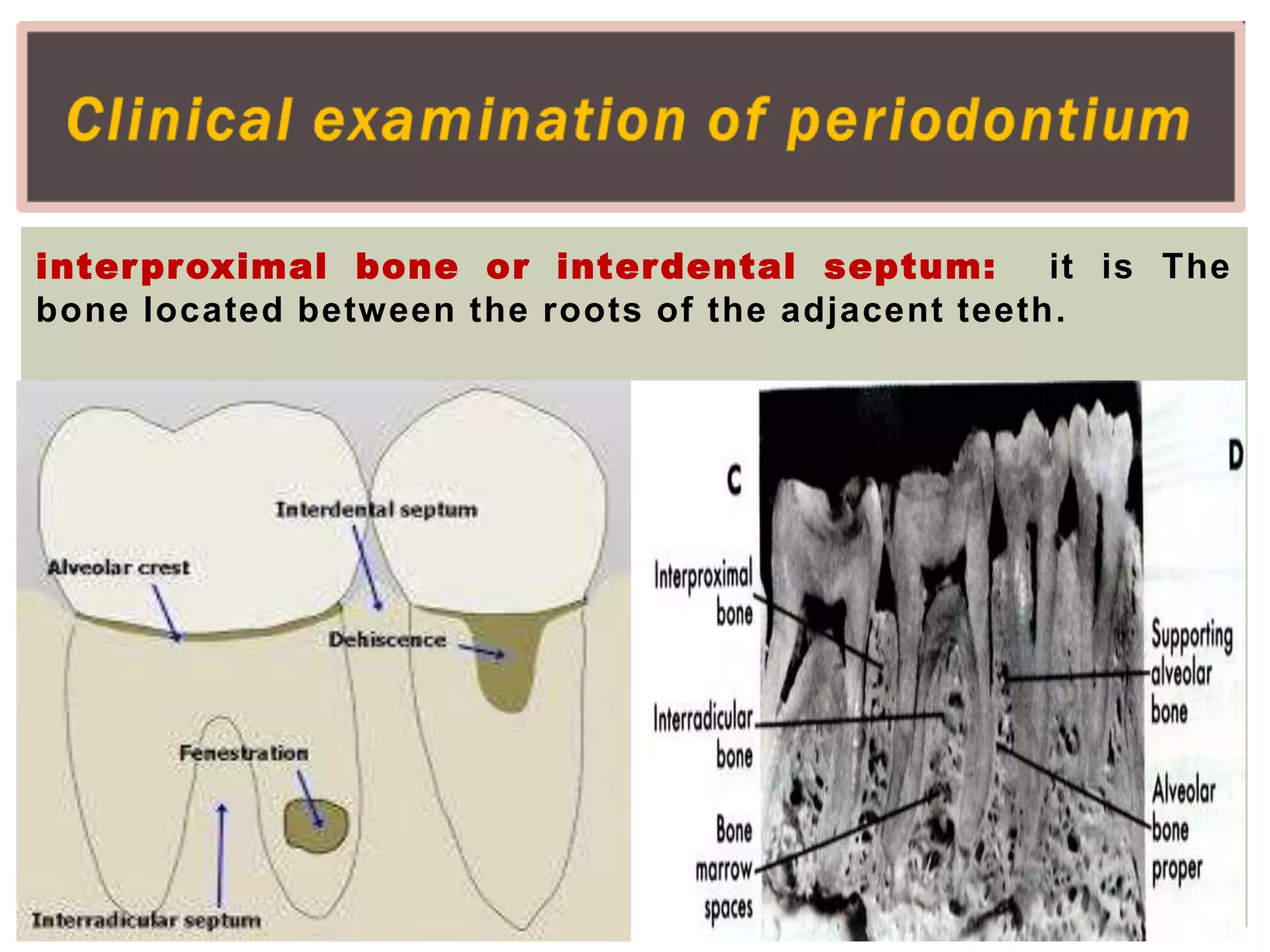
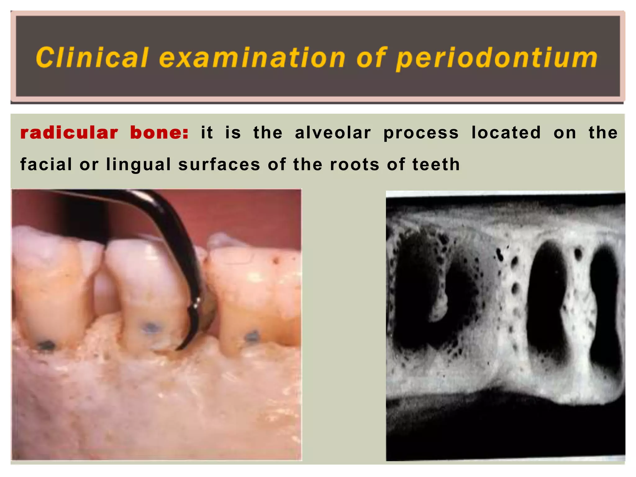
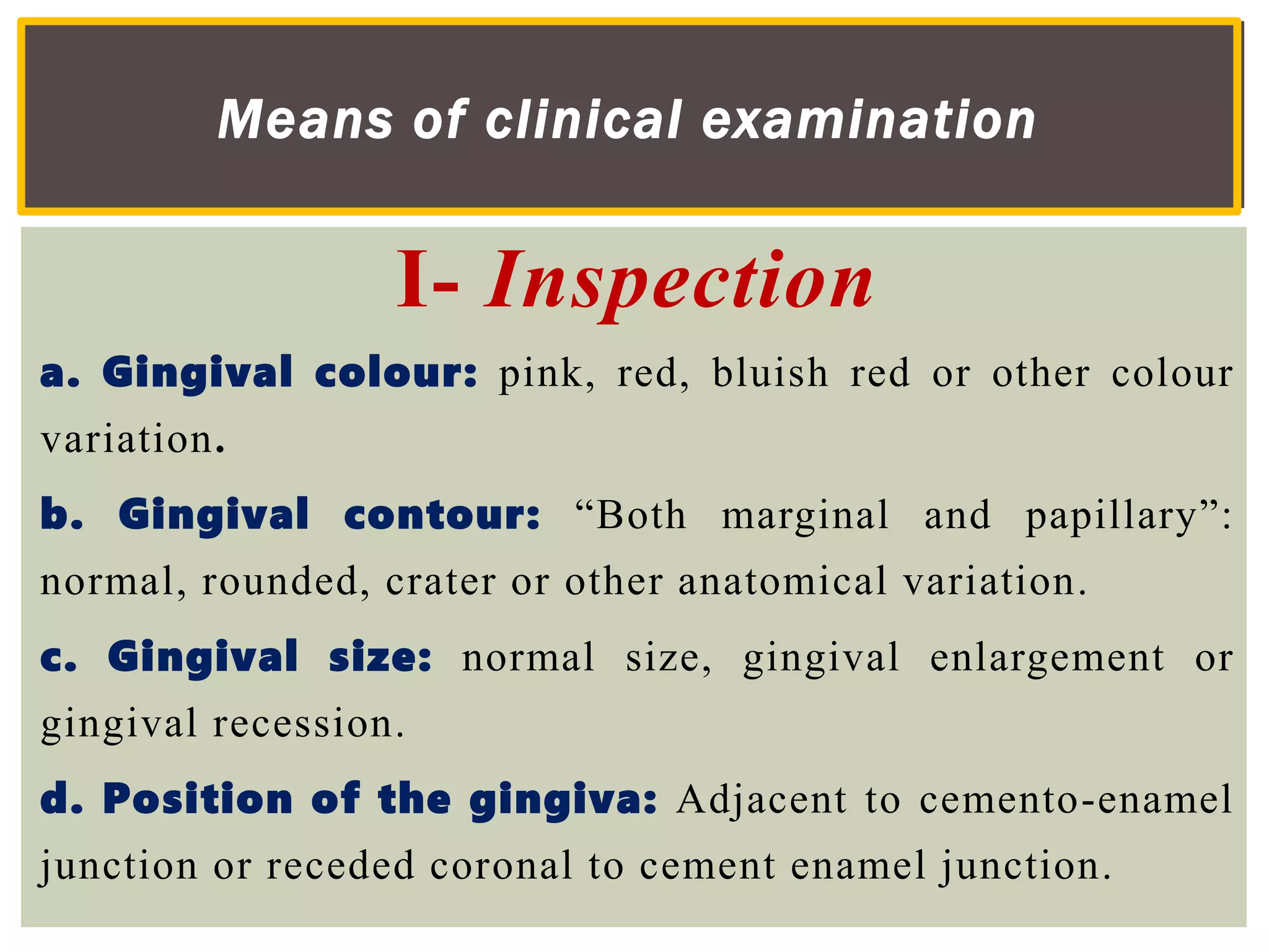
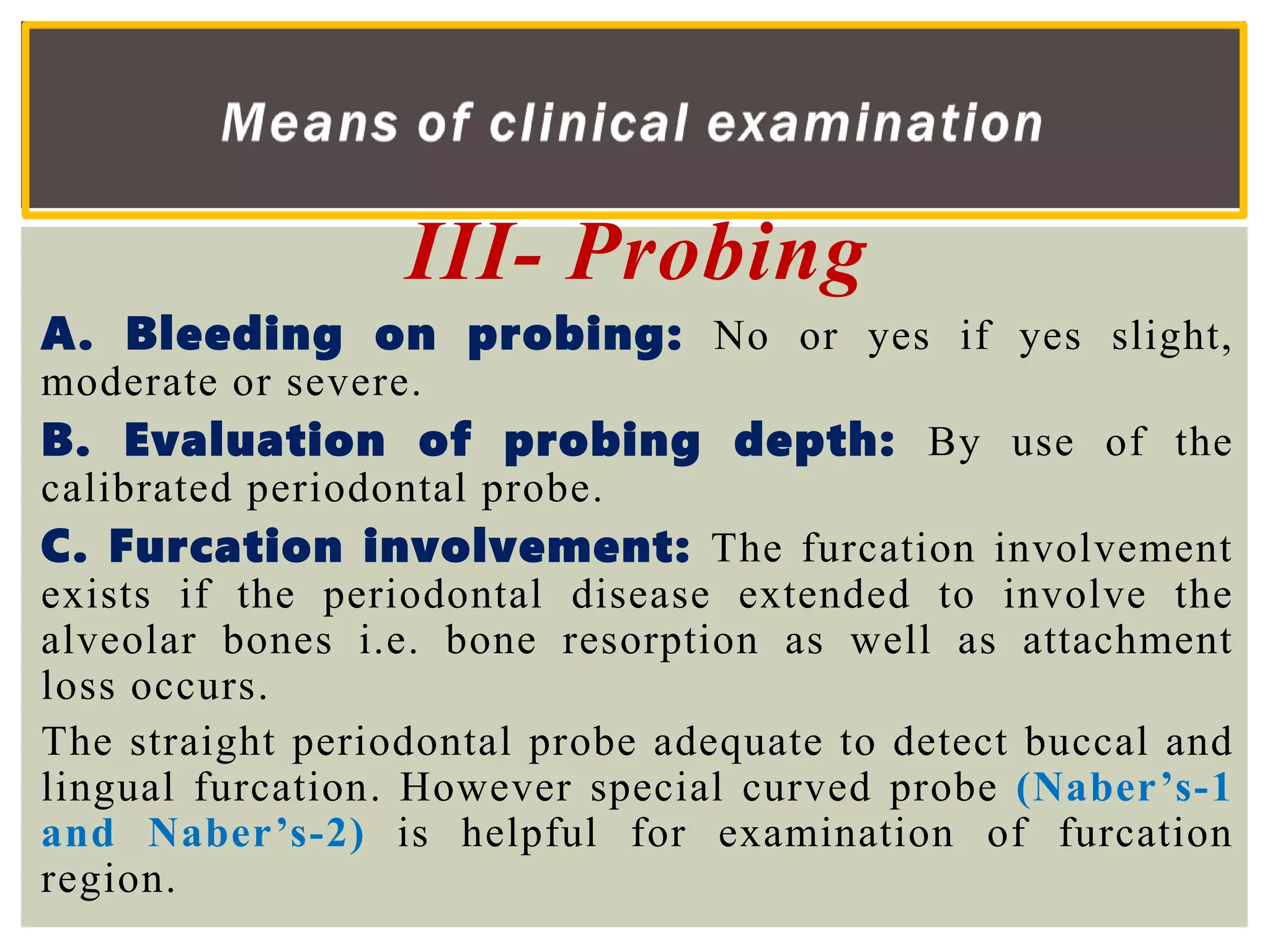
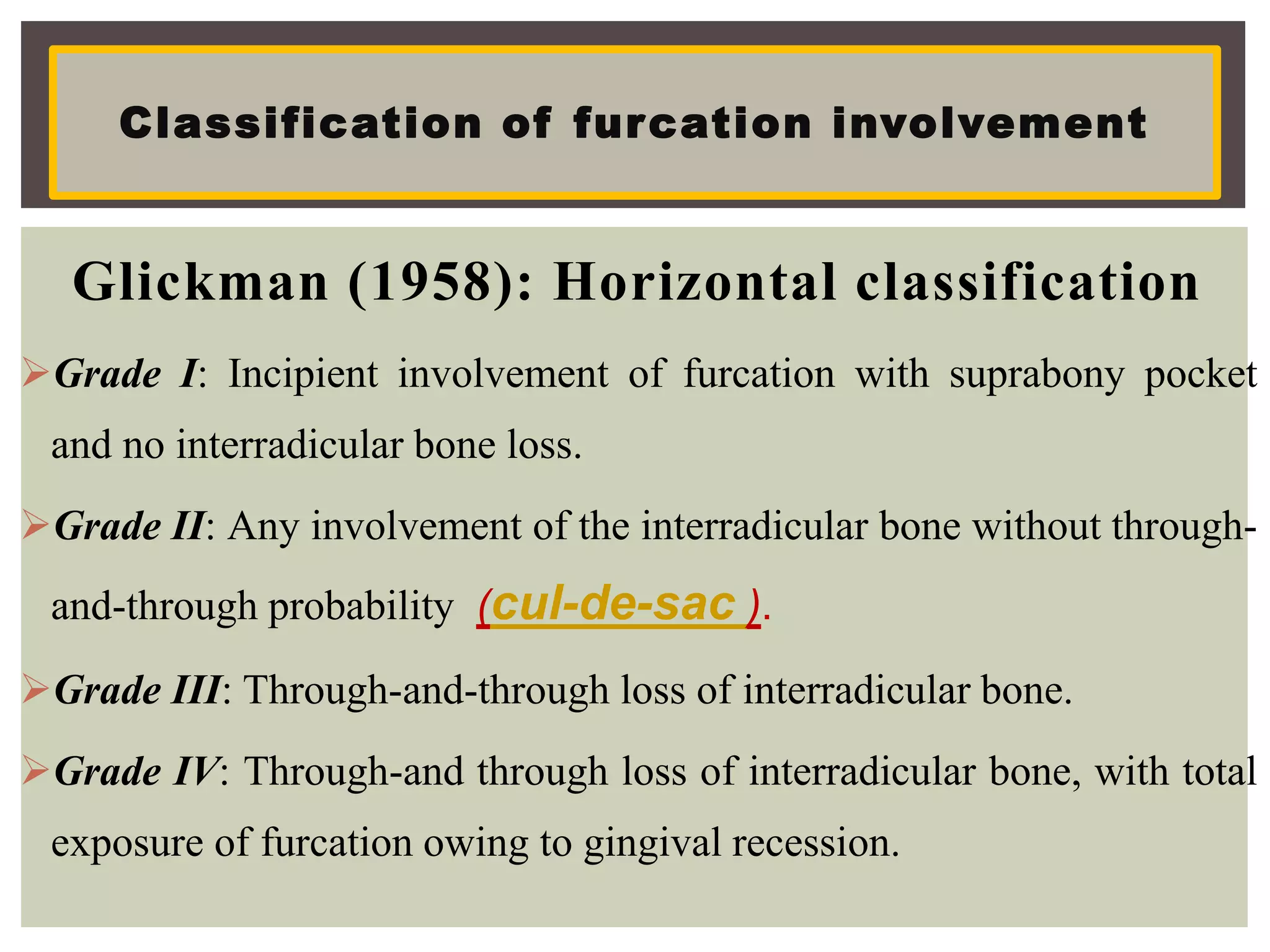
Dr. Asem Mohammed Kamel
Intraoral Examination
The dentist should examine?
1) Lip mucosal surface.
2) Labial frenum.
3) Vermilion border.
4) Oral commissures.
5) Vestibules of the mouth.
6) Minor salivary glands.
1--Clinical Examination
of
Lips & Labial Mucosa
Means of clinical examination
1- Inspection
Color of the lips and labial mucosa,
morphology, function, texture and the
presence of abnormal lesion such as
ulceration, anatomic or developmental
abnormalities as cleft lip, double lip, lip pit
and short lip.
observe the following:
1) Consistency of the lips and labial mucosa.
2) Pliability of lip surfaces.
3) Indurated lesions of the lips as well as the
glandular involvement.
4) Texture of the lip.
5) Size & shape of the lip must be examined for the
presence of some pathologic conditions.
2- Palpation
Lesions of the Lips and labial mucosa
A- Specific
1) chelitis glandularis.
2) chelitisglandularisapostematosa “Melkerson_Rosenthel syndrome”.
3) Lip pits (Congenital sinus of the lower lip).
4) Commissural pit.
5) Double lip.
6) Angular chelitis.
7) Cleft lip.
B-common
1) Recurrent herpes labialis.
2) Erythema multiform “Steven Johnson” syndrome.
3) Stomatitis medicamentosa and venenata and angioedema.
4) Smoker’s patch of the lip.
5) Condyloma latum and Condyloma acuminatum .
6) Lichen planus showing white lines “Wickham's stria” on the vermilion border of the
lip.
7) Focal epithelial hyperplasia, characterized by appearance of slightly raised multiple
papules that could involve the lip, caused by human papilloma virus (Heck’s disease).
8) Malignant lesions of the lip as spindle cell carcinoma and epidermoid carcinoma.
Recurrent herpes labialis
Erythema multiform
Stomatitis medicamentosa
Smoker’s patch of the lip
Condyloma latum
Condyloma acuminatum
Focal epithelial hyperplasia
Means of clinical examination
to see any abnormalities e.g.
- Linea alba buccalis
- Fordyce's granules
- Parotid duct opening
- Ulcers
- Pigmentation e.g. melanoma
2--Clinical examination
of
buccal mucosa
Linea alba buccalis
Fordyce's granules
Parotid duct opening
Ulcers
melanoma
Means of clinical examination
to feel the consistency and texture of some pathologic conditions as:-
 leukodema
 leukoplakia
 White spongy nevus
 keratotic lichen planus “papule type”
 Specked leukoplakia
 Frictional keratosis
 Smoker keratosis
 Indurated ulcers e.g. malignant ulcer
Clinical examination of buccal mucosa
leukodema
leukoplakia
White spongy nevus
keratotic lichen planus
malignant ulcer
Frictional keratosis Specked leukoplakia
3--Clinical Examination
of
Tongue
Taste distribution on the tongue surface
Sweet tip
Sour lateral
Salt dorsal
Bitter back
Nerve supply
Anterior two thirds:
• General sensation by lingual nerve,
branch of trigeminal 5th C.N .
• Taste sensation by chorda tympani,
branch of facial 7th C.N.
Posterior one third:
• General and taste sensation by
glossopharyngeal 9th C.N.
Motor supply
• By hypoglossal nerve 12th C.N.
1- Active movement
Technique:
The clinician ask
- the patient to raise his
tongue against the
palate to see the
ventral surface.
- the patient move the
tongue anteriorly and
laterally to see the
dorsal surface.
Movements of the tongue
2- Passive movement
Technique:
examiner holds tongue
with a piece of gaze
and moves it up and
down and from side
to side to detect any
abnormalities or any
associated pain.
3- Abnormal
movement:
This
movement
usually
performed
by mental
patient.
Decreased Tongue Coating: (Atrophy)
1) Pernicious anemia “iron deficiency anemia”.
2) Drug allergy.
3) Diabetes mellitus.
4) Lichen planus of the tongue.
5) Chronic atrophic candidiasis.
6) Tertiary syphilis “syphilitic glossitis”.
7) Geographic tongue and median rhomboid glossitis.
8) Sub epithelial vesiculo-bullous lesions.
Pernicious anemia
Drug allergyLichen planus
Chronic atrophic
candidiasis Syphilitic glossitis Geographic tongue
median rhomboid glossitisErythema multiform
1) Hairy leukoplakia “AIDs patients”.
2) Mouth breather patients.
3) Vomiting and stomach upset.
4) Smoker patients.
5) Xerostomia.
6) Some tongue lesions as black hairy tongue.
Increase Tongue Coating
Hairy leukoplakia
Black hairy tongue
By bidigital palpation to examine
- The consistency of tongue lesion.
- The resiliency and texture.
- The presence of induration, scars and
lesions.
Palpation of the tongue
A-- Congenital anomalies
1. Microglossia and aglossia.
2. Macroglossia.
3. Cleft tongue “bifid tongue”.
4. Fissural tongue .
5. Lingual thyroid nodule.
6. Thyroglossal tract cyst.
7. Ankyloglossia
8. Median Rhomboid glossitis.
Diseases of the tongue:
Microglossia
Aglossia
Macroglossia
Cleft tongue
Fissural tongue
Lingual thyroid nodule
AnkylogossiaMedian Rhomboid glossitis
Diseases of the tongue:
B-- Acquired tongue disease
1. Benign migratory glossitis (geographic tongue).
2. Hairy tongue.
3. Depapillation and atrophic tongue
4. Glossodynia and glossopyrosis.
5. Acquired macroglossia
Benign migratory glossitis
C- As a manifestation of systemic diseases.
1. Infections e.g. syphilis of tongue “syphilitic glossitis”, gamma of
tongue and T.B. ulcer of tongue.
2. Metabolic e.g. Diabetes militius patients showing depapillation
and atrophic tongue coating.
3. Hormonal e.g. secondary “acquired” macroglossia that associated
with acromegaly and cretinism.
4. Allergy e.g. stomatitis medicamentosa and stomatitis venenata.
5. Hematological e.g. hunter glossitis manifestation of pernicious
anemia and decrease tongue coating. Atrophic tongue changes
commonly associated with iron deficiency anemia and plumer
venison syndrome.
6. Neoplastic e.g. squamous cell carcinoma of the tongue,
lymphangioma, Haemangioma and neurofibromatosis which result
in secondary macroglossia.
 H a r d p a l a t e
 S o f t p a l a t e
 U v u l a
By using direct and indirect light, visual examination
can be performed by observing the following: contour,
height, rugae area, incisive papillae and width of the
arch. The soft palate should be inspected for color
changes, which usually associated with different types
of anemia, and the mobility of soft palate must be
observed.
4--Clinical examination of Palate
1- Inspection
By using one finger of one hand i.e. “Bidigital
palpation” the palate can be palpated starting
from the anterior region and proceeding
posteriorly and laterally.
The junction between soft and hard palate
should be palpated gently. Any abnormal
texture such as egg shell, cracking or
fluctuation and swelling must be recorded.
The movement of soft palate should be
observed by asking the patient to say ahh,
this giving idea about the integrity of the 9th
and 10th cranial nerves. If paralysis of these
nerves occurred the uvula will be shifted
toward the unaffected side.
The uvula is varying considerably in size
from individual to another and examined by
means of inspection. It may be congenitally
absent or lost during surgery or may appear
bifid and represent a form of cleft palate.
Cystsofthepalate
-Incisivecanalcyst -Cystofpalatinepapilla
-Median-palatinecyst -Globulo-maxillary cyst
-Naso-palatine cyst
Whitelesionscommonly involvingthepalate
-Thermalburnsuchaspizzaburn -Stomatitisnicotina
Congenitalabnormality ofthepalate
-Cleftpalate. -Toruspalatinus.
Common infectionsinvolvingthehardpalate e.g.
-Syphiliticinfectionduringsecondaryandtertiarystagesuchas “gamma”
-Fungalinfectionsuchasmoniliasisespecially inAIDSpatient
-Drainingsinusfromperiapicalabscess.
Tumorsofhardpalate:
-Adenocarcinoma. -Squamouscellcarcinoma
Lesionsofthehardpalate
Syphilitic perforation of the palate
Fungal infection of the palate
Incisive canal cyst Cyst of palatine papilla
Globulo-maxillary cyst
Median-palatine cyst
Naso-palatine cyst
 Herpetic infections of herpes simplex
virus (primary and secondary lesions)
 Herpes zoster
 Herpangina
 Recurrent aphthous ulcers
 Diphtheria.
Lesions of the soft palate
Secondary herpetic stomatitisHerpes zoster
Herpangina
Recurrent aphthous ulcers Diphtheria
floor of mouth can be inspected by asking the
patient to raise his tongue to reveal the
structures and landmarks such as lingual frenum,
orifices of the sublingual and submaxillary salivary
gland and lingual vein.
The color of the mucosa covering the floor of the
mouth must be observed for any pathological lesions
such as rannula, mucocele, ulceration and swelling.
5--Clinical examination
of
Floor of mouth
1- Inspection
The Clinician fix the tissues extra-orally by four
finger of one hand and the other hand running intra-
orally to help in the identification of any pathologic
condition involving the floor of mouth such as
salivary gland calculi and sub-maxillary lymph node
involvement.
2- Bimanual palpation
1) Ranula and mucous retention cyst
2) Hyperkeratotic lesion e.g. leukoplakia
3) Dermoid and epidermis carcinoma
4) Dermoid and epidermoid cyst
5) Salivary gland stone.
6) Ulcer of the floor of the mouth
Lesionsinvolvingthefloorofmouth
Ranula
1. gingiva:
It divided into free gingival, interdental papilla and attached gingiva.
2. Periodontal ligament (P.L):
Principal Collagen Fibers of Periodontal Ligament include Alveolar
crestal fibers, Horizontal fibers, Oblique fibers and apical fibers.
Periodontal ligament have mechanical, formative, nutritive and sensory
functions.
3.The alveolar bone:
These processes are composed of cancellous bone covered with cortical
bone. The spaces in the alveolar bone that accommodate the roots of the
teeth are known alveoli. The alveoli are lined with a layer of bone called
alveolar bone proper or cribriform plate.
4.Cementum:
It is continuously formed on root surfaces that are in contact with the
periodontal ligament or gingival fibers. The width of cementum varies
from 16-60um in the coronal half of the root, its much thicker, on the
apical third of the root, being 150-200um.
6--clinical Examination
Of
Periodontium
Lamina Dura: it is alveolar bone proper or cribriform plate
which appears as white line on the radiograph.
crestal lamina Dura: layer of bone covers the crest of the
interproximal bone
supporting alveolar bone: the cancellous and cortical
bone that surrounds the alveolar bone proper
interproximal bone or interdental septum: it is The
bone located between the roots of the adjacent teeth.
inter-radicular bone: it is located between the roots of
multirooted teeth.
radicular bone: it is the alveolar process located on the
facial or lingual surfaces of the roots of teeth
Fenestration: it is isolated areas which the root denuded of bone
and root surface is covered only by periosteum and overlying
gingiva.
Dehiscence: it is denuded areas extend through the marginal
bone.
to determine the following:
1) Whether the patient periodontium is healthy or
diseased.
2) The extent of the tissue damaged if
pathological change is present.
3) The characteristics of the periodontal disease
that well enable the determination of the
diagnosis, etiology, prognosis and treatment
plan.
importance of periodontium examination
I- Inspection
a. Gingival colour: pink, red, bluish red or other colour
variation.
b. Gingival contour: “Both marginal and papillary”:
normal, rounded, crater or other anatomical variation.
c. Gingival size: normal size, gingival enlargement or
gingival recession.
d. Position of the gingiva: Adjacent to cemento-enamel
junction or receded coronal to cement enamel junction.
Means of clinical examination
II- Palpation
a. Gingival consistency: Normal, edematous, fibrotic or
fiber edematous.
b. Gingival texture “stippling”: normal, decrease,
increase or lack of it.
c. Tooth mobility: by using the handles of two dental
instruments apply alternate pressure on the buccal or lingual
surface of each tooth.
d. Migration: migration is a pathological movement of the
teeth in labial, distal, mesial or supra occlusion. Migration is
common feature of some periodontal disease e.g. aggressive
periodontal, it may relate to some habit e.g. tongue
thrusting.
III- Probing
A. Bleeding on probing: No or yes if yes slight,
moderate or severe.
B. Evaluation of probing depth: By use of the
calibrated periodontal probe.
C. Furcation involvement: The furcation involvement
exists if the periodontal disease extended to involve the
alveolar bones i.e. bone resorption as well as attachment
loss occurs.
The straight periodontal probe adequate to detect buccal and
lingual furcation. However special curved probe (Naber’s-1
and Naber’s-2) is helpful for examination of furcation
region.
• The graduated periodontal probe should not
penetrate apically beyond cemento-enamel
junction
Gingival
pocket
• The graduated periodontal probe should
penetrate beyond the cemento-enamel
junction but not pass apically to the crest of
the alveolar bone
Suprabony
pocket
• the periodontal probe should penetrate the
cement enamel junction and pass apically to
the crest of the alveolar bone.
infrabony
pocket
Types of periodontal pockets
Bi or Tri furcation involvement
Glickman (1958): Horizontal classification
Grade I: Incipient involvement of furcation with suprabony pocket
and no interradicular bone loss.
Grade II: Any involvement of the interradicular bone without through-
and-through probability (cul-de-sac ).
Grade III: Through-and-through loss of interradicular bone.
Grade IV: Through-and through loss of interradicular bone, with total
exposure of furcation owing to gingival recession.
Classification of furcation involvement
Tarnow and Fletcher (1984): vertical classification
loss is measured in mm from the roof of the furcation
Grade I: Vertical loss of to 3 mm.
Grade II: Vertical loss of 4 to 6 mm.
Grade III: Vertical loss of 7 mm.
The mesial furcation should be probed from the
palatal aspect of the tooth.
The distal furcation can be probed from either the
buccal or the palatal aspect of the tooth.
Maxillary molars furcation
involvement
D. Assessment of attachment level:
The attachment level is measured by means of
graduated periodontal probe “ mm
measurement” from the cemento-enamel
junction to the bottom of the gingival sulcus.
Probing of attachment levels gives an accurate
estimation of the degree of the periodontal
tissue destruction and indirectly reflect the
levels of the alveolar bony defects.
If there is apparent recession clinically i.e. the cemento-
enamel junction visible
the attachment level is measured directly from the
cemento-enamel junction to the bottom of the pocket.
In cases of gingival recession the attachment level is
greater than pocket depth.
Assessment of attachment level
If there is no apparent recession i.e. the cemento-enamel junction is
covered by the free gingiva.
Slide the probe along the tooth surface into the pocket until the
CEJ is felt. Then Record in mm from CEJ to the gingival margin.
Measure in mm the distance from the gingival margin to the bottom
of the pocket.
Subtract the mm distance from CEJ to the gingival margin from the
total pocket depth to obtain attachment level.
E. Bone involvement
by using periodontal probe the amount of
bone resorption can evaluated by
measuring the distance from the dento-
gingival attachment to the level of
cemento-enamel junction.
The patient is anesthetized and the periodontal
probe is placed in the sulcus and pushed through
the attachment apparatus until the tip of the
probe engages alveolar bone. The measurements
are made on anterior teeth mid-facially and at the
facial/interproximal line angles.
Bone sounding procedure
Mucogingival assessment
It is determined by subtracting the sulcus or
pocket depth from total width of gingiva.
Width Of Attached Gingiva
1. Mucogingival junction assessed as a
scalloped line separating attached
gingiva from the alveolar mucosa.
Assessed as a borderline between
movable and immovable tissue.
2. Tissue mobility is assessed by
running a horizontally positioned
probe from the vestibule toward the
gingival margin using light force.
3. Assessed visually after staining the
Mucogingival junction with iodine
solution. Attached Gingiva –
Keratinized – No glycogen in the
superficial layer – Iodine Reactive
Negative
Inspection of the dentition must be include the following:
1. Discoloration and staining.
2. number.
3. shape and structures.
4. size.
5. Dental caries.
6. Attrition, Abrasion and erosion.
7. Functional relation, proximal contact & occlusion.
8. Teeth vitality.
9. Other problems e.g. tooth fracture.
10. Mobility of the teeth.
7--clinical Examination
Of
The Teeth
I. Inspection
Causes of extrinsic discoloration.
1. - Dental plaque formation.
2. - Food pigments.
3. - Tobacco and smoking.
4. - Chromogenic bacteria.
5. - Medications by drugs.
Causes of intrinsic discoloration of the teeth
1.Amelogenesis imperfecta.
2.Dentinogenesis imperfecta.
3.Fluorine intoxication, it appears “chalky white”.
4.Severe jaundice, the teeth appear blue or green.
5.Porphyria the teeth appear dark red.
6.Tetracycline administration appear dark brown .
7.Teeth with necrotic pulp, caries or injuries
Fluorine intoxicationjaundice
Tetracycline stain
Necrotic pulp
1- Anodontia (Complete or Partial)
2- Supernumaray teeth
3- Predeciduous dentition
4- Post permanent dentition
Size of tooth
Number of teeth
- Macrodontia - Microdontia
( true generalized, relative generalized or localized)
MacrodontiaMicrodontia
Supernumerary teeth
Supernumaray teeth
Predeciduous dentition
a- Amelogenesis imperfecta
b- Enamel hypoplasia due to
- Nutritional deficiency.
- Congenital syphilis.
- Hypoplasia due to hypocalcaemia.
- Hypoplasia due to tooth injuries.
- Hypoplasia due to local infection and trauma.
- Hypoplasia due to fluoride (Mottled enamel) .
c- Enamel and dentin aplasia
d- Enamel and dentin hypocalcification.
Abnormality of tooth structure.
Enamel hypoplasia
Anomalies of growth “eruption of the teeth”
Attrition, erosion and abrasion of the teeth
Function relationship between teeth and jaws:
1- Pathologic migration
2- Open contact (proximal contact)
3- Occlusion abnormalities
- Open bite - Cross bite
- Edge to edge - Deep over jet
- Deep over bite
1- Delayed eruption.
2- Multiple unerupted teeth.
3- Embedded and impacted teeth.
Pathologic migration
Open contact
Open bite
Cross bite
Edge to edge bite
Deep overjet bite
Deep overbite
II. Palpation
tooth mobility
Causes of tooth mobility:
1- Periodontal inflammation
2- Bone loss “bone resorption”.
3- The presence of occlusal trauma
Classification of tooth mobility:
Grade I: Slight mobility but the patient is not aware of it.
Grade II: Moderate mobility but the patient know it and not feel
discomfort during eating.
Grade III: Severe mobility and the patient feel discomfort
during eating.
III. Probing “Exploration of the teeth”
detect pulpal exposure, worn teeth and dental caries.
Exploration of teeth must be fine and sharp, the
clinician should be considered that many areas of the
occlusal and proximal portions of the teeth which may
be over looked. Exploration is performed with explorer
No. (17).
All surface of the tooth must be examined before
proceeding to the next one.
by using light instrument e.g. explorer no. 17
1- Solid sound (vital tooth)
2- Dull sound (non-vital tooth)
IV. Percussion of the teeth
1- Dental caries
2- Gemination
3- Concrescence
4- Dilaceration
5- Dens in dent
6- Supernumerary root
7- Ankylosed decidous tooth
8- Impeded and impacted teeth
V. Radiographic examination
Dental caries
Gemination
Concrescence
Dilaceration
Dens in dent
Supernumerary root
Ankylosed decidous tooth
Impacted teeth
General indication of vitality test:
1) Teeth discoloration.
2) Fractured tooth.
3) Deep carious lesion.
4) Dental restoration.
Non vital teeth appear clinically as gray, black or
bluish black.
Causes of non-vital tooth: Trauma, Caries,
Accidental exposure
VI.Pulp vitality test
Vitality pulp examined by
1- Electric pulp testing
2- Thermal pulp test
3- Periapical x- ray film
4- Percussion
Indication;
 It is indicated to determine the presence or absence of
vital nerve within the pulp chamber.
Test result;
 The result of electric pulp testing is painful response
following stimulation is an evidence of tooth vitality.
Advantage of electric pulp testing
 control stimulation can be applied in gradual increase
of the degree so the clinician avoid excessive pain to
patient.
Electric pulp testing
Indication:
Painful tooth in which the cause of pain is not clear.
Procedures:
1. The tooth surface should be dry.
2. Small piece of cotton sprayed with ethyl chloride is applied on
the tooth surface.
3. A hot cylindrical stick of gutta percha may be used.
The result of thermal pulp testing
Tooth with painful pulpitis will give severe painful response either
with hot or cold application than adjacent normal teeth.
Thermal pulp testing
THANK YOU

Intra oral examination

  • 1.
    Dr. Asem MohammedKamel Intraoral Examination
  • 3.
    The dentist shouldexamine? 1) Lip mucosal surface. 2) Labial frenum. 3) Vermilion border. 4) Oral commissures. 5) Vestibules of the mouth. 6) Minor salivary glands. 1--Clinical Examination of Lips & Labial Mucosa
  • 4.
    Means of clinicalexamination 1- Inspection Color of the lips and labial mucosa, morphology, function, texture and the presence of abnormal lesion such as ulceration, anatomic or developmental abnormalities as cleft lip, double lip, lip pit and short lip.
  • 5.
    observe the following: 1)Consistency of the lips and labial mucosa. 2) Pliability of lip surfaces. 3) Indurated lesions of the lips as well as the glandular involvement. 4) Texture of the lip. 5) Size & shape of the lip must be examined for the presence of some pathologic conditions. 2- Palpation
  • 6.
    Lesions of theLips and labial mucosa A- Specific 1) chelitis glandularis. 2) chelitisglandularisapostematosa “Melkerson_Rosenthel syndrome”. 3) Lip pits (Congenital sinus of the lower lip). 4) Commissural pit. 5) Double lip. 6) Angular chelitis. 7) Cleft lip.
  • 8.
    B-common 1) Recurrent herpeslabialis. 2) Erythema multiform “Steven Johnson” syndrome. 3) Stomatitis medicamentosa and venenata and angioedema. 4) Smoker’s patch of the lip. 5) Condyloma latum and Condyloma acuminatum . 6) Lichen planus showing white lines “Wickham's stria” on the vermilion border of the lip. 7) Focal epithelial hyperplasia, characterized by appearance of slightly raised multiple papules that could involve the lip, caused by human papilloma virus (Heck’s disease). 8) Malignant lesions of the lip as spindle cell carcinoma and epidermoid carcinoma.
  • 9.
    Recurrent herpes labialis Erythemamultiform Stomatitis medicamentosa Smoker’s patch of the lip Condyloma latum Condyloma acuminatum Focal epithelial hyperplasia
  • 10.
    Means of clinicalexamination to see any abnormalities e.g. - Linea alba buccalis - Fordyce's granules - Parotid duct opening - Ulcers - Pigmentation e.g. melanoma 2--Clinical examination of buccal mucosa
  • 11.
    Linea alba buccalis Fordyce'sgranules Parotid duct opening Ulcers melanoma
  • 12.
    Means of clinicalexamination to feel the consistency and texture of some pathologic conditions as:-  leukodema  leukoplakia  White spongy nevus  keratotic lichen planus “papule type”  Specked leukoplakia  Frictional keratosis  Smoker keratosis  Indurated ulcers e.g. malignant ulcer Clinical examination of buccal mucosa
  • 13.
    leukodema leukoplakia White spongy nevus keratoticlichen planus malignant ulcer Frictional keratosis Specked leukoplakia
  • 14.
    3--Clinical Examination of Tongue Taste distributionon the tongue surface Sweet tip Sour lateral Salt dorsal Bitter back Nerve supply Anterior two thirds: • General sensation by lingual nerve, branch of trigeminal 5th C.N . • Taste sensation by chorda tympani, branch of facial 7th C.N. Posterior one third: • General and taste sensation by glossopharyngeal 9th C.N. Motor supply • By hypoglossal nerve 12th C.N.
  • 15.
    1- Active movement Technique: Theclinician ask - the patient to raise his tongue against the palate to see the ventral surface. - the patient move the tongue anteriorly and laterally to see the dorsal surface. Movements of the tongue 2- Passive movement Technique: examiner holds tongue with a piece of gaze and moves it up and down and from side to side to detect any abnormalities or any associated pain. 3- Abnormal movement: This movement usually performed by mental patient.
  • 17.
    Decreased Tongue Coating:(Atrophy) 1) Pernicious anemia “iron deficiency anemia”. 2) Drug allergy. 3) Diabetes mellitus. 4) Lichen planus of the tongue. 5) Chronic atrophic candidiasis. 6) Tertiary syphilis “syphilitic glossitis”. 7) Geographic tongue and median rhomboid glossitis. 8) Sub epithelial vesiculo-bullous lesions.
  • 18.
    Pernicious anemia Drug allergyLichenplanus Chronic atrophic candidiasis Syphilitic glossitis Geographic tongue median rhomboid glossitisErythema multiform
  • 19.
    1) Hairy leukoplakia“AIDs patients”. 2) Mouth breather patients. 3) Vomiting and stomach upset. 4) Smoker patients. 5) Xerostomia. 6) Some tongue lesions as black hairy tongue. Increase Tongue Coating
  • 20.
  • 21.
    By bidigital palpationto examine - The consistency of tongue lesion. - The resiliency and texture. - The presence of induration, scars and lesions. Palpation of the tongue
  • 22.
    A-- Congenital anomalies 1.Microglossia and aglossia. 2. Macroglossia. 3. Cleft tongue “bifid tongue”. 4. Fissural tongue . 5. Lingual thyroid nodule. 6. Thyroglossal tract cyst. 7. Ankyloglossia 8. Median Rhomboid glossitis. Diseases of the tongue:
  • 23.
    Microglossia Aglossia Macroglossia Cleft tongue Fissural tongue Lingualthyroid nodule AnkylogossiaMedian Rhomboid glossitis
  • 24.
    Diseases of thetongue: B-- Acquired tongue disease 1. Benign migratory glossitis (geographic tongue). 2. Hairy tongue. 3. Depapillation and atrophic tongue 4. Glossodynia and glossopyrosis. 5. Acquired macroglossia
  • 25.
  • 26.
    C- As amanifestation of systemic diseases. 1. Infections e.g. syphilis of tongue “syphilitic glossitis”, gamma of tongue and T.B. ulcer of tongue. 2. Metabolic e.g. Diabetes militius patients showing depapillation and atrophic tongue coating. 3. Hormonal e.g. secondary “acquired” macroglossia that associated with acromegaly and cretinism. 4. Allergy e.g. stomatitis medicamentosa and stomatitis venenata. 5. Hematological e.g. hunter glossitis manifestation of pernicious anemia and decrease tongue coating. Atrophic tongue changes commonly associated with iron deficiency anemia and plumer venison syndrome. 6. Neoplastic e.g. squamous cell carcinoma of the tongue, lymphangioma, Haemangioma and neurofibromatosis which result in secondary macroglossia.
  • 27.
     H ar d p a l a t e  S o f t p a l a t e  U v u l a By using direct and indirect light, visual examination can be performed by observing the following: contour, height, rugae area, incisive papillae and width of the arch. The soft palate should be inspected for color changes, which usually associated with different types of anemia, and the mobility of soft palate must be observed. 4--Clinical examination of Palate 1- Inspection
  • 28.
    By using onefinger of one hand i.e. “Bidigital palpation” the palate can be palpated starting from the anterior region and proceeding posteriorly and laterally. The junction between soft and hard palate should be palpated gently. Any abnormal texture such as egg shell, cracking or fluctuation and swelling must be recorded.
  • 29.
    The movement ofsoft palate should be observed by asking the patient to say ahh, this giving idea about the integrity of the 9th and 10th cranial nerves. If paralysis of these nerves occurred the uvula will be shifted toward the unaffected side. The uvula is varying considerably in size from individual to another and examined by means of inspection. It may be congenitally absent or lost during surgery or may appear bifid and represent a form of cleft palate.
  • 30.
    Cystsofthepalate -Incisivecanalcyst -Cystofpalatinepapilla -Median-palatinecyst -Globulo-maxillarycyst -Naso-palatine cyst Whitelesionscommonly involvingthepalate -Thermalburnsuchaspizzaburn -Stomatitisnicotina Congenitalabnormality ofthepalate -Cleftpalate. -Toruspalatinus. Common infectionsinvolvingthehardpalate e.g. -Syphiliticinfectionduringsecondaryandtertiarystagesuchas “gamma” -Fungalinfectionsuchasmoniliasisespecially inAIDSpatient -Drainingsinusfromperiapicalabscess. Tumorsofhardpalate: -Adenocarcinoma. -Squamouscellcarcinoma Lesionsofthehardpalate
  • 31.
    Syphilitic perforation ofthe palate Fungal infection of the palate
  • 33.
    Incisive canal cystCyst of palatine papilla Globulo-maxillary cyst Median-palatine cyst Naso-palatine cyst
  • 34.
     Herpetic infectionsof herpes simplex virus (primary and secondary lesions)  Herpes zoster  Herpangina  Recurrent aphthous ulcers  Diphtheria. Lesions of the soft palate
  • 35.
    Secondary herpetic stomatitisHerpeszoster Herpangina Recurrent aphthous ulcers Diphtheria
  • 36.
    floor of mouthcan be inspected by asking the patient to raise his tongue to reveal the structures and landmarks such as lingual frenum, orifices of the sublingual and submaxillary salivary gland and lingual vein. The color of the mucosa covering the floor of the mouth must be observed for any pathological lesions such as rannula, mucocele, ulceration and swelling. 5--Clinical examination of Floor of mouth 1- Inspection
  • 37.
    The Clinician fixthe tissues extra-orally by four finger of one hand and the other hand running intra- orally to help in the identification of any pathologic condition involving the floor of mouth such as salivary gland calculi and sub-maxillary lymph node involvement. 2- Bimanual palpation
  • 39.
    1) Ranula andmucous retention cyst 2) Hyperkeratotic lesion e.g. leukoplakia 3) Dermoid and epidermis carcinoma 4) Dermoid and epidermoid cyst 5) Salivary gland stone. 6) Ulcer of the floor of the mouth Lesionsinvolvingthefloorofmouth
  • 40.
  • 41.
    1. gingiva: It dividedinto free gingival, interdental papilla and attached gingiva. 2. Periodontal ligament (P.L): Principal Collagen Fibers of Periodontal Ligament include Alveolar crestal fibers, Horizontal fibers, Oblique fibers and apical fibers. Periodontal ligament have mechanical, formative, nutritive and sensory functions. 3.The alveolar bone: These processes are composed of cancellous bone covered with cortical bone. The spaces in the alveolar bone that accommodate the roots of the teeth are known alveoli. The alveoli are lined with a layer of bone called alveolar bone proper or cribriform plate. 4.Cementum: It is continuously formed on root surfaces that are in contact with the periodontal ligament or gingival fibers. The width of cementum varies from 16-60um in the coronal half of the root, its much thicker, on the apical third of the root, being 150-200um. 6--clinical Examination Of Periodontium
  • 42.
    Lamina Dura: itis alveolar bone proper or cribriform plate which appears as white line on the radiograph.
  • 43.
    crestal lamina Dura:layer of bone covers the crest of the interproximal bone
  • 44.
    supporting alveolar bone:the cancellous and cortical bone that surrounds the alveolar bone proper
  • 45.
    interproximal bone orinterdental septum: it is The bone located between the roots of the adjacent teeth.
  • 46.
    inter-radicular bone: itis located between the roots of multirooted teeth.
  • 47.
    radicular bone: itis the alveolar process located on the facial or lingual surfaces of the roots of teeth
  • 48.
    Fenestration: it isisolated areas which the root denuded of bone and root surface is covered only by periosteum and overlying gingiva. Dehiscence: it is denuded areas extend through the marginal bone.
  • 50.
    to determine thefollowing: 1) Whether the patient periodontium is healthy or diseased. 2) The extent of the tissue damaged if pathological change is present. 3) The characteristics of the periodontal disease that well enable the determination of the diagnosis, etiology, prognosis and treatment plan. importance of periodontium examination
  • 51.
    I- Inspection a. Gingivalcolour: pink, red, bluish red or other colour variation. b. Gingival contour: “Both marginal and papillary”: normal, rounded, crater or other anatomical variation. c. Gingival size: normal size, gingival enlargement or gingival recession. d. Position of the gingiva: Adjacent to cemento-enamel junction or receded coronal to cement enamel junction. Means of clinical examination
  • 52.
    II- Palpation a. Gingivalconsistency: Normal, edematous, fibrotic or fiber edematous. b. Gingival texture “stippling”: normal, decrease, increase or lack of it. c. Tooth mobility: by using the handles of two dental instruments apply alternate pressure on the buccal or lingual surface of each tooth. d. Migration: migration is a pathological movement of the teeth in labial, distal, mesial or supra occlusion. Migration is common feature of some periodontal disease e.g. aggressive periodontal, it may relate to some habit e.g. tongue thrusting.
  • 53.
    III- Probing A. Bleedingon probing: No or yes if yes slight, moderate or severe. B. Evaluation of probing depth: By use of the calibrated periodontal probe. C. Furcation involvement: The furcation involvement exists if the periodontal disease extended to involve the alveolar bones i.e. bone resorption as well as attachment loss occurs. The straight periodontal probe adequate to detect buccal and lingual furcation. However special curved probe (Naber’s-1 and Naber’s-2) is helpful for examination of furcation region.
  • 54.
    • The graduatedperiodontal probe should not penetrate apically beyond cemento-enamel junction Gingival pocket • The graduated periodontal probe should penetrate beyond the cemento-enamel junction but not pass apically to the crest of the alveolar bone Suprabony pocket • the periodontal probe should penetrate the cement enamel junction and pass apically to the crest of the alveolar bone. infrabony pocket Types of periodontal pockets
  • 56.
    Bi or Trifurcation involvement
  • 57.
    Glickman (1958): Horizontalclassification Grade I: Incipient involvement of furcation with suprabony pocket and no interradicular bone loss. Grade II: Any involvement of the interradicular bone without through- and-through probability (cul-de-sac ). Grade III: Through-and-through loss of interradicular bone. Grade IV: Through-and through loss of interradicular bone, with total exposure of furcation owing to gingival recession. Classification of furcation involvement
  • 59.
    Tarnow and Fletcher(1984): vertical classification loss is measured in mm from the roof of the furcation Grade I: Vertical loss of to 3 mm. Grade II: Vertical loss of 4 to 6 mm. Grade III: Vertical loss of 7 mm.
  • 60.
    The mesial furcationshould be probed from the palatal aspect of the tooth. The distal furcation can be probed from either the buccal or the palatal aspect of the tooth. Maxillary molars furcation involvement
  • 61.
    D. Assessment ofattachment level: The attachment level is measured by means of graduated periodontal probe “ mm measurement” from the cemento-enamel junction to the bottom of the gingival sulcus. Probing of attachment levels gives an accurate estimation of the degree of the periodontal tissue destruction and indirectly reflect the levels of the alveolar bony defects.
  • 62.
    If there isapparent recession clinically i.e. the cemento- enamel junction visible the attachment level is measured directly from the cemento-enamel junction to the bottom of the pocket. In cases of gingival recession the attachment level is greater than pocket depth. Assessment of attachment level
  • 63.
    If there isno apparent recession i.e. the cemento-enamel junction is covered by the free gingiva. Slide the probe along the tooth surface into the pocket until the CEJ is felt. Then Record in mm from CEJ to the gingival margin. Measure in mm the distance from the gingival margin to the bottom of the pocket. Subtract the mm distance from CEJ to the gingival margin from the total pocket depth to obtain attachment level.
  • 64.
    E. Bone involvement byusing periodontal probe the amount of bone resorption can evaluated by measuring the distance from the dento- gingival attachment to the level of cemento-enamel junction.
  • 65.
    The patient isanesthetized and the periodontal probe is placed in the sulcus and pushed through the attachment apparatus until the tip of the probe engages alveolar bone. The measurements are made on anterior teeth mid-facially and at the facial/interproximal line angles. Bone sounding procedure
  • 67.
  • 68.
    It is determinedby subtracting the sulcus or pocket depth from total width of gingiva. Width Of Attached Gingiva
  • 69.
    1. Mucogingival junctionassessed as a scalloped line separating attached gingiva from the alveolar mucosa. Assessed as a borderline between movable and immovable tissue. 2. Tissue mobility is assessed by running a horizontally positioned probe from the vestibule toward the gingival margin using light force. 3. Assessed visually after staining the Mucogingival junction with iodine solution. Attached Gingiva – Keratinized – No glycogen in the superficial layer – Iodine Reactive Negative
  • 70.
    Inspection of thedentition must be include the following: 1. Discoloration and staining. 2. number. 3. shape and structures. 4. size. 5. Dental caries. 6. Attrition, Abrasion and erosion. 7. Functional relation, proximal contact & occlusion. 8. Teeth vitality. 9. Other problems e.g. tooth fracture. 10. Mobility of the teeth. 7--clinical Examination Of The Teeth I. Inspection
  • 71.
    Causes of extrinsicdiscoloration. 1. - Dental plaque formation. 2. - Food pigments. 3. - Tobacco and smoking. 4. - Chromogenic bacteria. 5. - Medications by drugs.
  • 72.
    Causes of intrinsicdiscoloration of the teeth 1.Amelogenesis imperfecta. 2.Dentinogenesis imperfecta. 3.Fluorine intoxication, it appears “chalky white”. 4.Severe jaundice, the teeth appear blue or green. 5.Porphyria the teeth appear dark red. 6.Tetracycline administration appear dark brown . 7.Teeth with necrotic pulp, caries or injuries
  • 73.
  • 74.
    1- Anodontia (Completeor Partial) 2- Supernumaray teeth 3- Predeciduous dentition 4- Post permanent dentition Size of tooth Number of teeth - Macrodontia - Microdontia ( true generalized, relative generalized or localized)
  • 75.
  • 76.
    a- Amelogenesis imperfecta b-Enamel hypoplasia due to - Nutritional deficiency. - Congenital syphilis. - Hypoplasia due to hypocalcaemia. - Hypoplasia due to tooth injuries. - Hypoplasia due to local infection and trauma. - Hypoplasia due to fluoride (Mottled enamel) . c- Enamel and dentin aplasia d- Enamel and dentin hypocalcification. Abnormality of tooth structure.
  • 77.
  • 78.
    Anomalies of growth“eruption of the teeth” Attrition, erosion and abrasion of the teeth Function relationship between teeth and jaws: 1- Pathologic migration 2- Open contact (proximal contact) 3- Occlusion abnormalities - Open bite - Cross bite - Edge to edge - Deep over jet - Deep over bite 1- Delayed eruption. 2- Multiple unerupted teeth. 3- Embedded and impacted teeth.
  • 79.
  • 80.
  • 81.
  • 82.
  • 83.
  • 84.
  • 85.
  • 86.
    II. Palpation tooth mobility Causesof tooth mobility: 1- Periodontal inflammation 2- Bone loss “bone resorption”. 3- The presence of occlusal trauma Classification of tooth mobility: Grade I: Slight mobility but the patient is not aware of it. Grade II: Moderate mobility but the patient know it and not feel discomfort during eating. Grade III: Severe mobility and the patient feel discomfort during eating.
  • 87.
    III. Probing “Explorationof the teeth” detect pulpal exposure, worn teeth and dental caries. Exploration of teeth must be fine and sharp, the clinician should be considered that many areas of the occlusal and proximal portions of the teeth which may be over looked. Exploration is performed with explorer No. (17). All surface of the tooth must be examined before proceeding to the next one.
  • 88.
    by using lightinstrument e.g. explorer no. 17 1- Solid sound (vital tooth) 2- Dull sound (non-vital tooth) IV. Percussion of the teeth
  • 89.
    1- Dental caries 2-Gemination 3- Concrescence 4- Dilaceration 5- Dens in dent 6- Supernumerary root 7- Ankylosed decidous tooth 8- Impeded and impacted teeth V. Radiographic examination
  • 90.
  • 91.
  • 92.
  • 93.
  • 94.
  • 95.
  • 96.
  • 97.
  • 98.
    General indication ofvitality test: 1) Teeth discoloration. 2) Fractured tooth. 3) Deep carious lesion. 4) Dental restoration. Non vital teeth appear clinically as gray, black or bluish black. Causes of non-vital tooth: Trauma, Caries, Accidental exposure VI.Pulp vitality test
  • 99.
    Vitality pulp examinedby 1- Electric pulp testing 2- Thermal pulp test 3- Periapical x- ray film 4- Percussion
  • 100.
    Indication;  It isindicated to determine the presence or absence of vital nerve within the pulp chamber. Test result;  The result of electric pulp testing is painful response following stimulation is an evidence of tooth vitality. Advantage of electric pulp testing  control stimulation can be applied in gradual increase of the degree so the clinician avoid excessive pain to patient. Electric pulp testing
  • 102.
    Indication: Painful tooth inwhich the cause of pain is not clear. Procedures: 1. The tooth surface should be dry. 2. Small piece of cotton sprayed with ethyl chloride is applied on the tooth surface. 3. A hot cylindrical stick of gutta percha may be used. The result of thermal pulp testing Tooth with painful pulpitis will give severe painful response either with hot or cold application than adjacent normal teeth. Thermal pulp testing
  • 103.