Tissues
•Download as PPTX, PDF•
0 likes•71 views

1. Tissues are groups of cells that perform specific functions. There are four main types of animal tissues: epithelial, connective, muscular and nervous tissue. In plants, there are meristematic tissues which actively divide and permanent tissues which do not divide. Permanent tissues are further divided into simple parenchyma, collenchyma and sclerenchyma, and complex vascular tissues xylem and phloem. Tissues provide structure and fulfill functions like protection, support, storage, and transport within animals and plants.

Recommended
More Related Content
What's hot
What's hot (20)
Similar to Tissues
Similar to Tissues (20)
Recently uploaded
Recently uploaded (20)
Tissues
- 1. Tissues
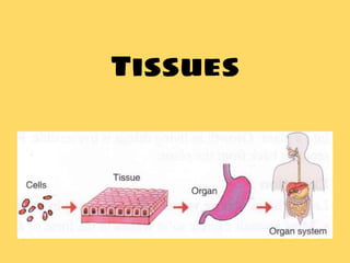
- 2. What are tissues? • A group of cells which are similar in structure and perform a specific function are termed as tissues. • eg. Muscle tissue, Nervous tissue, Blood etc Types of Tissue Animal TissuePlant Tissue
- 3. 1. Meristematic Tissue • actively dividing cells • cells have a large nucleus and no vacuoles • Location: found at the growing parts of plant eg. root tip, shoot tip, nodes • Function: elongation of shoot and root, increase in the diameter of the stem / bark 2. Permanent Tissue • Simple Parenchyma Collenchyma Sclerenchyma Complex Phloem Xylem
- 5. PERMANENT TISSUE • The cells of this tissue have lost their ability to multiply and acquire a definite shape, size and function • These tissues may be living or dead • There are 3 types of permanent tissues based on the function performed: 1.PROTECTIVE TISSUE 1.SUPPORTING TISSUE 1.CONDUCTING TISSUE
- 6. Protective Tissue • Characteristic: Cells with thick walls • Location: Found on the outermost layer of plant body such as roots, stems and leaves • Function: Prevents water loss from the leaves • Protects underlying tissues • Example: – Epidermis of leaves- secretes a waxy water proof material – Cork cells in the bark
- 7. Supporting Tissue • Three important types are – Parenchyma – Collenchyma – Sclerenchyma
- 8. PARENCHYMA
- 9. PARENCHYMA ● The cells are thin walled usually with a single large vacuole ● The cells are living ● Location: Found in soft parts of the plant root and stem ● Function ranging from storage of food ● Some cells contain chloroplast
- 10. COLLENCHYMA 1.Cells are elongated and thick at corners 1.Found in leaf stalks and below the epidermis of stems 1.Function is to provide support and flexibility to parts of plant
- 12. SCLERENCHYMA 1.Cells are long, narrow and thick walled due to deposition of lignin 1.The cells are dead 1.Found in stems, veins of leaves, hard covering of seeds & nuts 1.Also found in the gritty part of the ripe fruits & contribute hardness to the seed coat & nutshells. 1.Function is to provide strength to parts of plant
- 16. CONDUCTING TISSUE (VASCULAR TISSUE) XYLEM PHLOEM 1. Function • Transport of water from roots to all parts of plant • Transport of food leaves to various parts of plant 2. Composition • Consist of: a)Tracheids b)Xylem vessels c)Xylem parenchyma • Consists of: a)Sieve tubes b)Companion cells c)Phloem parenchyma d)Phloem fibres • Cells except for xylem parenchyma are dead • Cells are living except for phloem fibres •Xylem and phloem are found in the veins of leaf, stem and root • They constitute complex tissue
- 19. ANIMAL TISSUES • There are four main type of animal tissues: 1.Epithelial tissue 2.Connective tissue 3.Muscle tissue 4.Nervous tissue The microscopic study of cells and tissue type is called Histology
- 20. Epithelial tissue • Thin, closely packed, continuous sheet of cells • Location: Outermost layer of skin, lines the cavities and surfaces of internal organs • Shape: flat, cuboidal or columnar • Function: Protection (radiation, germs), absorption (nutrient), secretion (sweat)
- 21. Epithelial tissues are of 6 categories depending on their structure (extra information)
- 22. Connective tissue The connective tissue is specialized to connect and anchor various body organs. • Structure: 3 characteristics 1.Abundant matrix 2.Fewer cellular elements 3.Fibres • Location: between different tissues and organs • Function: binding,supporting and packing together different organs of the body
- 24. • It is the simplest and most widely distributed connective tissue. • Structure: It is a loose and cellular connective tissue. Its matrix consists of two kinds of fibres • Location: All over in the body, fills spaces between the organs, around muscles, blood vessels and nerves. • Functions : 1) Binds various tissues together 2) Makes skin elastic 3) Helps to withstand pulling strain Areolar connective tissue (packing)
- 25. Adipose (fat) tissue • Description: Specialised cells (adipocytes) that store fat • Location: below the skin, around organs like kidneys, heart, eye ball etc • Function: 1. It serves as a fat reservoir. 2. It provides shape to the limbs and body. 3. It acts an insulator and conserves body temperature.
- 26. Fibrous connective tissue Fibrous connective tissue is characterized by ordered and densely packed collection of fibres Ligament connect bone to bone
- 27. Supportive connective tissue The skeletal or supporting tissue includes Cartilage and bone which forms the endoskeleton of the vertebrate body. Bone 1. It is a porous and non- flexible 2. It has solid calcified matrix 3. It has good supply of blood vessels and nerves 4. Forms supportive framework of the body. (limbs, skull, ribcage) 5. It protects vital organs such as brains, heart,lungs etc. Cartilage 1. It is non-porous and flexible 2. It has thickened matrix 3. It has no blood vessels or nerves 4. Located at the tip of nose, external part of ear between vertebrae & end of long bones 5. It provides flexibility to the body parts
- 28. CARTILAGE BONE
- 29. Fluid connective tissue Blood 1. Composed of plasma + RBC’s, WBC’s and platelets 2. Flows through arteries, veins and capillaries 3. Transports nutrients, hormones and O2 to tissues and organs Lymph 1. It is a colourless fluid that composes of plasma +WBC’s 2. It is present in lymph vessels all over the body 3. It brings CO2 and nitrogenous wastes from tissue fluid to blood. Both blood and lymph protect the body against infections (provide immunity). It forms the defence system of the body.
- 30. MUSCULAR TISSUE • Muscular tissue is a contractile tissue made up of muscle cells • Description: elongated cells called muscle fibres • Ability to contract and relax • Function: movement and locomotion of body
- 31. Types of Muscle tissue
- 32. Striated muscles • Also known as voluntary, striped or skeletal muscles • Structure: elongated, cylindrical and unbranched muscle fibres • Multinucleated • Alternate light and dark striations • Location: muscles of arms, legs, neck, face, diaphragm etc • Function: Movement and locomotion ⮚ Muscles undergo rapid contractions which are voluntary ⮚ These muscles get tired and need rest
- 33. Unstriated muscles • Also known as involuntary, smooth or visceral muscles. • Structure: long, narrow, spindle shaped and tapering towards the end • Uninucleated • Smooth with NO striations • Location: iris of the eye, lining of blood vessels, intestine, urinary bladder, uterus • Function: Involuntary movements (passage of food) ⮚ Muscles undergo slow contractions which are involuntary ⮚ These muscles do not get tired
- 34. Cardiac muscles • Also known as heart muscles. • Structure: short, striated and branched muscle fibres • Location: Only in walls of the heart • Function: pumping of blood throughout the body ⮚ Muscles undergo continuous involuntary rhythmic contractions and relaxations throughout life ⮚ These muscles do not get tired
- 35. Neural tissue • Description: Consists of specialised elongated cells called neurons or nerve cells • Location: nervous system of the body (brain, spinal cord and nerves) • Function: conduction of nerve impulse and response to stimuli
- 36. Structure of Neuron • 3 distinct parts: 1. Cyton / perikaryon or cell body 2. Dendron 3. Axon or nerve fibre ❑Cyton contains the nucleus, granular cytoplasm
- 37. ❑ Dendrons are short cytoplasmic extensions arising from the cyton & further branch into dendrites ❑ Axons are long, cylindrical cytoplasmic extensions which form fine branches terminally The Dendrites receive impulses and the axon takes impulses away from the cell body
- 38. SUMMARY
- 39. Quiz 1. A group of cells performing a particular function is __________ • A) Organ • B. Body • C Tissue • D organ system
- 40. 2. Group of dividing cells of a plant a) Permanent tissue b) Meristematic tissue c) Vascular tissue d) None
- 41. 3. Non dividing cells produced to meristem are called • A) Permanent tissue • B Bark tissue • C) Epidermis • D Parenchyma
- 42. 4. There are two types of permanent tissue. One is simple permanent tissue and another is: • a. Double permanent tissue • b. Special • c. Complex • d. None
- 43. 5. Food storing simple permanent tissue is: • a. Parenchyma • b. Collenchyma c • . Scleroid • d. None
- 44. 6. Chlorenchyma is a type of: • a. Parenchyma • b. Vascular bundle • c. Xylem • d. Phloem.
- 45. 7. Simple permanent tissue has only a single kind of permanent cells while Complex permanent tissue has: a. At least four b. b. At least two c. c. All cells are different d. d. Does not matter
- 46. 8. Tissues in tendrils of a climber plant and leaf stalk of a plant are examples of which tissue? • a. Parenchyma • b. Collenchyma • c. Vascular • d. Bark
- 47. 9. Lignified thick cell wall with no cytoplasmic space in a cell is a charecteristic of : a. Parenchyma b. b. Collenchyma c. c. Sclerenchyma d. d. Epidermisis
- 48. 10. The pore through which transpiration in plant occurs is: • a. Stoma • b. Vacuole • c. Plastid • d. None
- 49. • 11. One of the tissues in the given options is dead tissue. Identify it. • a. Parenchyma • b. Collenchyma • c. Sclerenchyma • d. Xylem
- 50. • 12 Phloem and xylems are: • a. Simple permanent tissue • b. Complex permanent tissue • c. Vascular tissue • d. both b & c