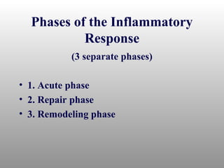
Tissue Response to Injury
Inflammatory Response
• Acute Inflammation
  – Short onset and duration
  – Production of exudate, leukocytes
• Chronic Inflammation
  – Long onset and duration
  – Presence of extensive scar tissue
Cardinal Signs of
             Inflammation
•   Rubor (redness)
•   Tumor (swelling)
•   Color (heat)
•   Dolor (pain)
•   Functio laesa (loss of function)
Phases of the Inflammatory
           Response
           (3 separate phases)

• 1. Acute phase
• 2. Repair phase
• 3. Remodeling phase
Phase I: Acute Phase
• Initial reaction to an injury occurring 3
  hours to 2 days following injury
• Goal
  –   Protect
  –   Localize
  –   Decrease injurious agents
  –   Prepare for healing and repair
• Caused by trauma, chemical agents,
  thermal extremes, pathogenic organisms
• External and internal injury result in
  tissue death and cell death
• Decreased oxygen to area increases cell
  death
• Rest, ice, compression & elevation are
  critical to limiting cell death
• First hour
  – Vasoconstriction and coagulation occur to
    seal blood vessels and chemical mediators
    are released
  – Immediately followed by vasodilation or
    blood vessel
• Second hour
  – Vasodilation decreases blood flow, increased
    blood viscosity resulting in edema (swelling)
• Second hour (continued)
  – Exudate increases (high concentration of
    RBC’s) due to increased vessel permeability
  – Permeability changes generally occur in
    capillary and venules
• Cellular response
  – Mast cells (connective tissue cells) and
    leukocytes (basophils, monocytes,
    neutrophils) enter area
  – Mast cells with heparin and histamine serve
    as first line of defense
  – Basophils provide anticoagulant
  – Neutrophils and monocytes are responsible
    for small and large particles undergoing
    phagocytosis - ingestion of debris and
    bacteria
Phase II: Repair Phase
• Phase will extent from 48 hours to 6
  weeks following cleaning of fibrin clot,
  erythrocytes, and debris
• Repaired through 3 phases
  – Resolution (little tissue damage and normal
                restoration)
  – Restoration (if resolution is delayed)
  – Regeneration (replacement of tissue by same
                    tissue)
• Scar formation
  – Less viable than normal tissue, may
    compromise healing
  – Firm, inelastic mass devoid of capillary
    circulation
  – Develops from exudate with high protein
    and debris levels resulting in granulation
    tissue
  – Invaded by fibroblasts and and collagen
    forming a dense scar and while normally
    requiring 3-14 weeks may require 6 months
    to contract
• Primary healing (healing by first intention)
  – Closely approximated edges with little
    granulation tissue production
• Secondary healing (heal by secondary
  intention)
  – Gapping, tissue loss, and development of
    extensive granulation tissue
  – Common in external lacerations and internal
    musculoskeletal injuries
• Regeneration
  – Related to health, nutrition and tissue type
  – Dependent on levels of:
      • debris (phagocytosis)
      • endothelial production (hypoxia and
        macrophages stimulate capillary buds)
      • production of fibroblasts (revascularization
        allows for enhanced fibroblast activity and
        collagen production which is tied to Vitamin C,
        lactic acid, and oxygen
  –
Phase III: Remodeling
• Overlaps repair and regeneration
• First 3-6 weeks involves laying down of
  collagen and strengthening of fibers
• 3 months to 2 years allowed for enhanced
  scar tissue strength
• Balance must be maintained between
  synthesis and lysis
• Take into consideration forces applied
  and immobilization/mobilization time
  frames relative to tissue and healing time
Chronic Inflammation
• Result of failed acute inflammation
  resolution within one month termed
  subacute inflammation
• Inflammation lasting months/years
  termed chronic
  – Results from repeated microtrauma and
    overuse
  – Proliferation of connective tissue and tissue
    degeneration
Characteristics of Chronic
        Inflammation
• Proliferation of connective tissue and tissue
  degeneration
• Presence of lymphocytes, plasma cell,
  macrophages(monocytes) in contrast to
  neutrophils (during acute conditions)
• Major chemicals include
  – Kinins (bradykinin) - responsible for
    vasodilation, permeability and pain
  – Prostaglandin - responsible for vasodilation
    but can be inhibited with aspirin and NSAID’s
Factors That Impede Healing
• Extent of injury       • Corticosteroids
• Edema                  • Keloids and
• Hemorrhage               Hypertrophic Scars
• Poor Vascular          • Infection
  Supply                 • Humidity, Climate,
• Separation of Tissue     Oxygen Tension
• Muscle Spasm           • Health, Age, and
• Atrophy                  Nutrition
Soft Tissue Healing
• Cell structure/function
  – All organisms composed of cells
  – Properties of soft tissue derived from
    structure and function of cells
  – Cells consist of nucleus surrounded by
    cytoplasm and encapsulated by phospholipid
    cell membrane
  – Nucleus contains chromosomes (DNA)
  – Functional elements of cells (organelles)
    include mitochondria, ribosomes, endoplasmic
    reticulum, Golgi apparatus & centrioles
Soft Tissue Adaptations
• Metaplasia - transformation of tissue from one type to
               another that is not normal for that tissue
• Dysplasia - abnormal development of tissue
• Hyperplasia- excessive proliferation of normal cells in
               normal tissue arrangement
• Atrophy- a decrease in the size of tissue due to cell
           death and re-absorption or decreased cell
           proliferation
• Hypertrophy - an increase in the size of tissue without
                necessarily changing the number of cells
Cartilage Healing
• Limited capacity to heal
• Little or no direct blood supply
• Chrondrocyte and matrix disruption
  result in variable healing
• Articular cartilage that fails to clot and
  has no perichondrium heals very slowly
• If area involves subchondral bone
  (enhanced blood supply) granulation
  tissue is present and healing proceeds
  normally
Ligament Healing
• Follows similar healing course as
  vascular tissue
• Proper care will result in acute, repair,
  and remodeling phases in same time
  required by other vascular tissue
• Repair phase will involve random laying
  down of collagen which, as scar forms,
  will mature and realign in reaction to
  joint stresses and strain
• Full healing may require 12 months
Skeletal Muscle Healing
• Skeletal muscle cannot undergo mitotic
  activity to replace injured cells
• New myofibril regeneration is minimal
• Healing and repair follow the same
  course as other soft tissues.
Nerve Healing
• Cannot regenerate after injury
• Regeneration can take place within a nerve
  fiber
• Proximity of injury to nerve cell makes
  regeneration more difficult
• For regeneration, optimal environment is
  required
• Rate of healing occurs at 3-4 mm per day
• Injured central nervous system nerves do
  not heal as well as peripheral nerves
Modifying Soft-Tissue Healing
• Varying issues exist for all soft tissues
  relative to healing (cartilage, muscle,
  nerves)
• Blood supply and nutrients is necessary
  for all healing
• Healing in older athletes or those with
  poor diets may take longer
• Certain organic disorders (blood
  conditions) may slow or inhibit the
  healing process
Management Concepts
• Drug utilization
  – Anitprostaglandin agents used to combat
    inflammation
  – Non-steroidal anti-inflammatory agents
    (NSAID’s)
  – Medications will work to decrease
    vasodilatation and capillary permeability
• Therapeutic Modalities
  – Thermal agents are utilized
     • Heat stimulates acute inflammation (but works
       as a depressant in chronic conditions)
     • Cold is utilized as an inhibitor
  – Electrical modalities
     • Treatment of inflammation
     • Ultrasound, microwave, electrical stimulation
       (includes transcutaneous electrical muscle
       stimulation and electrical muscle stimulation
• Therapeutic Exercise
  – Major aim involves pain free movement, full
    strength power, and full extensibility of
    associated muscles
  – Immobilization, while sometimes necessary,
    can have a negative impact on an injury
     • Adverse biochemical changes can occur in
       collagen
  – Early mobilization (that is controlled) may
    enhance healing
Fracture Healing
• Potential serious bone fractures are part
  of athletics
• Time is necessary for proper bone union
  to occur and is often out of the control of
  a physician
• Conservative treatment will be necessary
  for adequate healing to occur
• Bone undergoes constant remodeling
  through osteocyte activity
• Osteocytes cellular component of bone
  – Osteoblasts are responsible for bone
    formation while osteoclasts resorb bone
• Cambium (periosteum)
  – A fibrous covering involved in bone healing
  – Vascular and very dense
• Inner cambium
  – less vascular and more cellular.
  – Provides attachments for muscle, ligaments
    and tendons
Acute Fracture of Bone
• Follows same three phases of soft tissue
  healing
• Less complex process
• Acute fractures have 5 stages
  –   Hematoma formation
  –   Cellular proliferation
  –   Callus formation
  –   Ossification
  –   Remodeling
Callus Formation
• Soft callus is a random network of woven
  bone
• Osteoblasts fill the internal and external
  calluses to immobilize the site
• Calluses are formed by bone fragments
  that bridge the fracture gap
• The internal callus creates a rigid
  immobilization early
• Hard callus formation occurs after 3-4
  weeks and lasts 3-4 months
• Hard callus is a gradual connection of
  bone filaments to the woven bone
• Less than ideal immobilization produces
  a cartilagenous union instead of a bony
  union
Ossification
• Ossification is complete when bone has
  been laid down and the excess callus has
  been resorbed by osteoclasts.
Remodeling
• Occurs following callus resorption and
  trabecular bone is laid along lines of stress
• Bioelectric stimulation plays a major role
  in completing the remodeling process
• The process is complete when the original
  shape is achieved or the structure can
  withstand imposed stresses
Acute Fracture Management
• Must be appropriately immobilized, until X-
  rays reveal the presence of a hard callus
• Fractures can limit participation for weeks
  or months
• A clinician must be certain that the following
  areas do not interfere with healing
  – Poor blood supply
  – Poor immobilization
  – Infection
• Poor blood supply
  – Bone may die and union/healing will not occur
    (avascular necrosis)
  – Common sites include:
     • Head of femur, navicular of the wrist, talus, and
       isolated bone fragments
  – Relatively rare in healthy, young athletes
    except in navicular of the wrist
• Poor immobilization
  – Result of poor casting allowing for motion
    between bone parts
  – May prevent proper union or result in bony
    deformity
• Infection
  – May interfere with normal healing,
    particularly with compound fractures
  – Severe streptococcal and staphylococcal
    infections
  – Modern antibiotics has reduced the risk of
    infections
  – Closed fractures are not immune to
    infections within the body or blood
• If soft tissue alters bone positioning,
  surgery may be required to ensure
  proper union
• Gate Theory
  – Area in dorsal horn of spinal cord causes
    inhibition of pain impulses ascending to
    cortex
  – T-cells will transmit signals to brain
  – Substantia gelatinosa functions as gate
    determining if stimulus sent to T-cells
  – Pain stimuli exceeding threshold results in
    pain perception
  – Stimulation of large fast nerves can block
    signal of small pain fiber input
  – Rationale for TENS, accupressure/puncture,
    thermal agents and chemical skin irritants
• Pain assessment
  – Self report is the best reflection of pain and
    discomfort
  – Assessment techniques include:
     • visual analog scales (0-10, marked no pain to
       severe pain)
     • verbal descriptor scales (marked none, slight,
       moderate, and severe)
• Pain Treatment
  – Must break pain-spasm-hypoxia-pain cycle
    through treatment
  – Agents used; heat/cold, electrical
    stimulation-induced analgesia,
    pharmacological agents
• Heat/Cold
  – Heat increases circulation, blood vessel
    dilation, reduces nociception and ischemia
    caused by muscle spasm
  – Cold applied for vasoconstriction and
    prevention of extravasation of blood into
    tissue
  – Pain reduced through decrease in swelling
    and spasm
• Induced analgesia
  – Utilize electrical modalities to reduce pain
  – TENS and acupuncture commonly used to
    target Gate Theory
Psychological Aspects of Pain
• Pain can be subjective and psychological
• Pain thresholds vary per individual
• Pain is often worse at night due to solitude and
  absence of external distractions
• Personality differences can also have an impact
• A number of theories relative to pain exist and
  it physiological and psychological components
• Athlete, through conditioning are often able to
  endure pain and block sensations of minor
  injuries

Tissue healing

  • 1.
  • 2.
    Inflammatory Response • AcuteInflammation – Short onset and duration – Production of exudate, leukocytes • Chronic Inflammation – Long onset and duration – Presence of extensive scar tissue
  • 3.
    Cardinal Signs of Inflammation • Rubor (redness) • Tumor (swelling) • Color (heat) • Dolor (pain) • Functio laesa (loss of function)
  • 4.
    Phases of theInflammatory Response (3 separate phases) • 1. Acute phase • 2. Repair phase • 3. Remodeling phase
  • 5.
    Phase I: AcutePhase • Initial reaction to an injury occurring 3 hours to 2 days following injury • Goal – Protect – Localize – Decrease injurious agents – Prepare for healing and repair • Caused by trauma, chemical agents, thermal extremes, pathogenic organisms
  • 6.
    • External andinternal injury result in tissue death and cell death • Decreased oxygen to area increases cell death • Rest, ice, compression & elevation are critical to limiting cell death
  • 7.
    • First hour – Vasoconstriction and coagulation occur to seal blood vessels and chemical mediators are released – Immediately followed by vasodilation or blood vessel • Second hour – Vasodilation decreases blood flow, increased blood viscosity resulting in edema (swelling)
  • 8.
    • Second hour(continued) – Exudate increases (high concentration of RBC’s) due to increased vessel permeability – Permeability changes generally occur in capillary and venules
  • 9.
    • Cellular response – Mast cells (connective tissue cells) and leukocytes (basophils, monocytes, neutrophils) enter area – Mast cells with heparin and histamine serve as first line of defense – Basophils provide anticoagulant – Neutrophils and monocytes are responsible for small and large particles undergoing phagocytosis - ingestion of debris and bacteria
  • 10.
    Phase II: RepairPhase • Phase will extent from 48 hours to 6 weeks following cleaning of fibrin clot, erythrocytes, and debris • Repaired through 3 phases – Resolution (little tissue damage and normal restoration) – Restoration (if resolution is delayed) – Regeneration (replacement of tissue by same tissue)
  • 11.
    • Scar formation – Less viable than normal tissue, may compromise healing – Firm, inelastic mass devoid of capillary circulation – Develops from exudate with high protein and debris levels resulting in granulation tissue – Invaded by fibroblasts and and collagen forming a dense scar and while normally requiring 3-14 weeks may require 6 months to contract
  • 12.
    • Primary healing(healing by first intention) – Closely approximated edges with little granulation tissue production • Secondary healing (heal by secondary intention) – Gapping, tissue loss, and development of extensive granulation tissue – Common in external lacerations and internal musculoskeletal injuries
  • 13.
    • Regeneration – Related to health, nutrition and tissue type – Dependent on levels of: • debris (phagocytosis) • endothelial production (hypoxia and macrophages stimulate capillary buds) • production of fibroblasts (revascularization allows for enhanced fibroblast activity and collagen production which is tied to Vitamin C, lactic acid, and oxygen –
  • 14.
    Phase III: Remodeling •Overlaps repair and regeneration • First 3-6 weeks involves laying down of collagen and strengthening of fibers • 3 months to 2 years allowed for enhanced scar tissue strength • Balance must be maintained between synthesis and lysis • Take into consideration forces applied and immobilization/mobilization time frames relative to tissue and healing time
  • 15.
    Chronic Inflammation • Resultof failed acute inflammation resolution within one month termed subacute inflammation • Inflammation lasting months/years termed chronic – Results from repeated microtrauma and overuse – Proliferation of connective tissue and tissue degeneration
  • 16.
    Characteristics of Chronic Inflammation • Proliferation of connective tissue and tissue degeneration • Presence of lymphocytes, plasma cell, macrophages(monocytes) in contrast to neutrophils (during acute conditions) • Major chemicals include – Kinins (bradykinin) - responsible for vasodilation, permeability and pain – Prostaglandin - responsible for vasodilation but can be inhibited with aspirin and NSAID’s
  • 17.
    Factors That ImpedeHealing • Extent of injury • Corticosteroids • Edema • Keloids and • Hemorrhage Hypertrophic Scars • Poor Vascular • Infection Supply • Humidity, Climate, • Separation of Tissue Oxygen Tension • Muscle Spasm • Health, Age, and • Atrophy Nutrition
  • 18.
    Soft Tissue Healing •Cell structure/function – All organisms composed of cells – Properties of soft tissue derived from structure and function of cells – Cells consist of nucleus surrounded by cytoplasm and encapsulated by phospholipid cell membrane – Nucleus contains chromosomes (DNA) – Functional elements of cells (organelles) include mitochondria, ribosomes, endoplasmic reticulum, Golgi apparatus & centrioles
  • 19.
    Soft Tissue Adaptations •Metaplasia - transformation of tissue from one type to another that is not normal for that tissue • Dysplasia - abnormal development of tissue • Hyperplasia- excessive proliferation of normal cells in normal tissue arrangement • Atrophy- a decrease in the size of tissue due to cell death and re-absorption or decreased cell proliferation • Hypertrophy - an increase in the size of tissue without necessarily changing the number of cells
  • 20.
    Cartilage Healing • Limitedcapacity to heal • Little or no direct blood supply • Chrondrocyte and matrix disruption result in variable healing • Articular cartilage that fails to clot and has no perichondrium heals very slowly • If area involves subchondral bone (enhanced blood supply) granulation tissue is present and healing proceeds normally
  • 21.
    Ligament Healing • Followssimilar healing course as vascular tissue • Proper care will result in acute, repair, and remodeling phases in same time required by other vascular tissue • Repair phase will involve random laying down of collagen which, as scar forms, will mature and realign in reaction to joint stresses and strain • Full healing may require 12 months
  • 22.
    Skeletal Muscle Healing •Skeletal muscle cannot undergo mitotic activity to replace injured cells • New myofibril regeneration is minimal • Healing and repair follow the same course as other soft tissues.
  • 23.
    Nerve Healing • Cannotregenerate after injury • Regeneration can take place within a nerve fiber • Proximity of injury to nerve cell makes regeneration more difficult • For regeneration, optimal environment is required • Rate of healing occurs at 3-4 mm per day • Injured central nervous system nerves do not heal as well as peripheral nerves
  • 24.
    Modifying Soft-Tissue Healing •Varying issues exist for all soft tissues relative to healing (cartilage, muscle, nerves) • Blood supply and nutrients is necessary for all healing • Healing in older athletes or those with poor diets may take longer • Certain organic disorders (blood conditions) may slow or inhibit the healing process
  • 25.
    Management Concepts • Drugutilization – Anitprostaglandin agents used to combat inflammation – Non-steroidal anti-inflammatory agents (NSAID’s) – Medications will work to decrease vasodilatation and capillary permeability
  • 26.
    • Therapeutic Modalities – Thermal agents are utilized • Heat stimulates acute inflammation (but works as a depressant in chronic conditions) • Cold is utilized as an inhibitor – Electrical modalities • Treatment of inflammation • Ultrasound, microwave, electrical stimulation (includes transcutaneous electrical muscle stimulation and electrical muscle stimulation
  • 27.
    • Therapeutic Exercise – Major aim involves pain free movement, full strength power, and full extensibility of associated muscles – Immobilization, while sometimes necessary, can have a negative impact on an injury • Adverse biochemical changes can occur in collagen – Early mobilization (that is controlled) may enhance healing
  • 28.
    Fracture Healing • Potentialserious bone fractures are part of athletics • Time is necessary for proper bone union to occur and is often out of the control of a physician • Conservative treatment will be necessary for adequate healing to occur
  • 29.
    • Bone undergoesconstant remodeling through osteocyte activity • Osteocytes cellular component of bone – Osteoblasts are responsible for bone formation while osteoclasts resorb bone • Cambium (periosteum) – A fibrous covering involved in bone healing – Vascular and very dense • Inner cambium – less vascular and more cellular. – Provides attachments for muscle, ligaments and tendons
  • 30.
    Acute Fracture ofBone • Follows same three phases of soft tissue healing • Less complex process • Acute fractures have 5 stages – Hematoma formation – Cellular proliferation – Callus formation – Ossification – Remodeling
  • 32.
    Callus Formation • Softcallus is a random network of woven bone • Osteoblasts fill the internal and external calluses to immobilize the site • Calluses are formed by bone fragments that bridge the fracture gap • The internal callus creates a rigid immobilization early
  • 33.
    • Hard callusformation occurs after 3-4 weeks and lasts 3-4 months • Hard callus is a gradual connection of bone filaments to the woven bone • Less than ideal immobilization produces a cartilagenous union instead of a bony union
  • 34.
    Ossification • Ossification iscomplete when bone has been laid down and the excess callus has been resorbed by osteoclasts.
  • 35.
    Remodeling • Occurs followingcallus resorption and trabecular bone is laid along lines of stress • Bioelectric stimulation plays a major role in completing the remodeling process • The process is complete when the original shape is achieved or the structure can withstand imposed stresses
  • 36.
    Acute Fracture Management •Must be appropriately immobilized, until X- rays reveal the presence of a hard callus • Fractures can limit participation for weeks or months • A clinician must be certain that the following areas do not interfere with healing – Poor blood supply – Poor immobilization – Infection
  • 37.
    • Poor bloodsupply – Bone may die and union/healing will not occur (avascular necrosis) – Common sites include: • Head of femur, navicular of the wrist, talus, and isolated bone fragments – Relatively rare in healthy, young athletes except in navicular of the wrist • Poor immobilization – Result of poor casting allowing for motion between bone parts – May prevent proper union or result in bony deformity
  • 38.
    • Infection – May interfere with normal healing, particularly with compound fractures – Severe streptococcal and staphylococcal infections – Modern antibiotics has reduced the risk of infections – Closed fractures are not immune to infections within the body or blood • If soft tissue alters bone positioning, surgery may be required to ensure proper union
  • 39.
    • Gate Theory – Area in dorsal horn of spinal cord causes inhibition of pain impulses ascending to cortex – T-cells will transmit signals to brain – Substantia gelatinosa functions as gate determining if stimulus sent to T-cells – Pain stimuli exceeding threshold results in pain perception – Stimulation of large fast nerves can block signal of small pain fiber input – Rationale for TENS, accupressure/puncture, thermal agents and chemical skin irritants
  • 40.
    • Pain assessment – Self report is the best reflection of pain and discomfort – Assessment techniques include: • visual analog scales (0-10, marked no pain to severe pain) • verbal descriptor scales (marked none, slight, moderate, and severe) • Pain Treatment – Must break pain-spasm-hypoxia-pain cycle through treatment – Agents used; heat/cold, electrical stimulation-induced analgesia, pharmacological agents
  • 41.
    • Heat/Cold – Heat increases circulation, blood vessel dilation, reduces nociception and ischemia caused by muscle spasm – Cold applied for vasoconstriction and prevention of extravasation of blood into tissue – Pain reduced through decrease in swelling and spasm • Induced analgesia – Utilize electrical modalities to reduce pain – TENS and acupuncture commonly used to target Gate Theory
  • 42.
    Psychological Aspects ofPain • Pain can be subjective and psychological • Pain thresholds vary per individual • Pain is often worse at night due to solitude and absence of external distractions • Personality differences can also have an impact • A number of theories relative to pain exist and it physiological and psychological components • Athlete, through conditioning are often able to endure pain and block sensations of minor injuries