Chronic
Inflammation
Chronic inflammation is inflammation
of prolonged duration
(weeks or months)
in which
inflammation, tissue injury, and attempts at repair
coexist, in varying combinations.
CAUSES OF CHRONIC
INFLAMMATION
1.Persistent Infection
2.Immune-mediate &
inflammatory diseases
3.Toxic Agents/FOREIGN BODY
• DOES SURGERY ASSOCIATED WITH CHRONIC
INFLAMMATION
• YES
• HOW COME?
• SUTURE MATERIAL
GENERAL FEATURES
• MONONUCLEAR CELL INFILTRATION
• TISSUE DESTRUCTION//NECROSIS
• PROLIFERATIVE CHANGES
FIBROSIS ANGIOGENESIS
• CHRONIC INFLAMMATION ALWAYS ATTEMPTS
TO HEALING
FEATURES
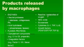
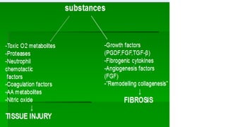
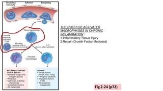
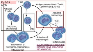
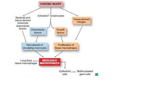
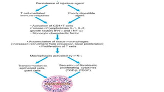
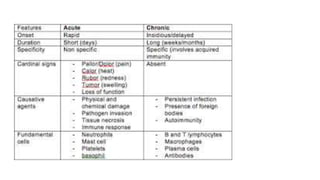
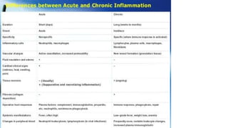
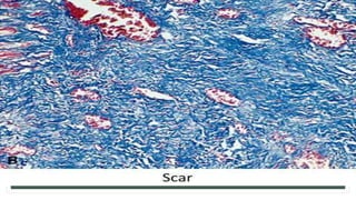
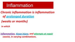
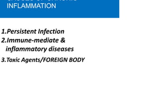
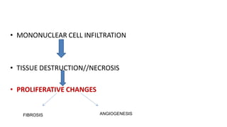
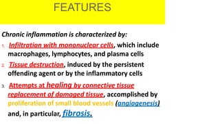
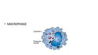
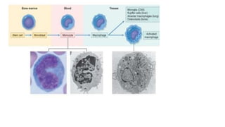
Chronic inflammation is characterized by:
1. Infiltration with mononuclear cells, which include
macrophages, lymphocytes, and plasma cells
2. Tissue destruction, induced by the persistent
offending agent or by the inflammatory cells
3. Attempts at healing by connective tissue
replacement of damaged tissue, accomplished by
proliferation of small blood vessels (angiogenesis)
and, in particular, fibrosis.
• WHAT IS THE KEY PLAYER IN CHRONIC
INFLAMMATION??
• MACROPHAGE
• DOES MACROPHAGE DIRECTLY COMES TO
CHRONIC INFLAMMATION
• NO
• MONOCYTE
• MACROPHAGE
WHAT ARE THE PRODUCTS
SECRETED BY MACROPHAGES??
• CYTOKINES-IL1, IL-2
• INTERFERONS –Y
• GROWTH FACTORS- TGF, PDGF, FGF,EGF
• [TGF, PDGF------- to stimulate fibroblast]
• COMPLEMENT FACTORS[C]
• ENZYMES—Proteases, endonucleases,
elastases
• Chemotactic factors
• NO
• Collagenases
• ANY OTHER??
• LYMPHOKINES
• IS NORMAL LYMPHOCYTE SECRETES
LYMPHOKINES??
• CD4 Lymphocytes[[Th cells]
THE ROLES OF ACTIVATED
MACROPHAGES IN CHRONIC
INFLAMMATION :
1.Inflammatory Tissue Injury
2.Repair (Growth Factor Mediated)
Fig 2-24 (p72)
MACROPHAGE-LYMPHOCYTE
INTERACTIONS IN CHRONIC
INFLAMMATION :
Fig 2-25
(p73)
TYPES
GRANULOMATOUS
INFLAMMATION
Granulomatous inflammation is a distinctive pattern
of chronic inflammation that is encountered in a
limited number of infectious and some
noninfectious conditions. Immune reactions are
usually involved in the development of
granulomas.
A granuloma is a cellular attempt to control an
offending agent that is difficult to eradicate.
.
• In this attempt there is often strong
activation of T lymphocytes leading to
macrophage activation, which can cause
injury to normal tissues
A granuloma is a focus of chronic
inflammation consisting of
a microscopic aggregation of
macrophages
that are transformed into epithelium-
like cells, surrounded by a collar of
mononuclear leukocytes, principally
lymphocytes and occasionally
plasma cells.
Types of
Granulomas
I. Foreign body granulomas
II. Immune granulomas
Foreign body granulomas
Incited by relatively inert foreign bodies.
Typically, foreign body granulomas form around
material that are large enough to preclude
phagocytosis by a single macrophage and do not
incite any specific inflammatory or immune
response.
.
Immune granulomas
Caused by agents that are capable
of inducing an immune response
which produces granulomas
usually when the inciting agent is
poorly degradable or particulate.
Giant
Cells
Older granulomas develop an enclosing rim of
fibroblasts and connective tissue.
Frequently, epithelioid cells fuse to form giant cells
in the periphery or sometimes in the center of
granulomas.
These giant cells may attain diameters of 40 to 50
μm.
peripherally (Langhans-type giant cell) or
haphazardly (foreign body–type giant cell).
• WHAT IS THE SHAPE OF EPITHELOID CELL
• ‘SOLE SHAPE’
SYSTEMIC EFFECTS OF CHRONIC
INFLAMMATION
• Chronic inflammation is associated with following systemic
features:
• 1.Fever. Invariably there is mild fever, often with loss
of weight and weakness.
2.Anaemia:chronic inflammation is accompanied by
anaemia of varying degree.
3.Leucocytosis. As in acute inflammation, chronic
inflammation also has leucocytosis but generally there
is relative lymphocytosis in these cases.
4.ESR. ESR is elevated in all cases of chronic
inflammation.
•
•
•
• Which of the following is always present in
chronic inflammation/
• A. Rubor
• B. Edema
• C. Leucocytosis
• D. Increased ESR
MORPHOLOGY
• DRAW A NEAT LABELLED DIAGRAM OF
GRANULOMA
• PATHOGENSIS OF GRANULOMA
• DIFFERENCE BETWEEN ACUTE &CHRONIC
INFLAMMATION
• Tissue repair: restoration of tissue architecture and function after
an injury
• Occurs in two ways:
• Regeneration of injured tissue.
• Replacement by connective tissue (scarring)
• Usually, tissue repair involves both processes
• Involves cell proliferation, and interaction between cells and
extracellular matrix.
• Many cells proliferate during tissue repair:
• Injured tissue remnants.
• Vascular endothelial cells
• Fibroblasts
Tissues of the body are divided into three
groups:
•Continuously dividing (labile) tissues.
•Stable tissues.
•Permanent tissues
•Continuously dividing (labile) tissues
•Cells are continuously proliferating.
•Can easily regenerate after injury
•Contain a pool of stem cells
•Examples: bone marrow, skin, GIT epithelium.
Stem cells in
skin
Stem cells in GI
epithelium
•Stable tissues
•Cells have limited ability to proliferate.
•Limited ability to regenerate (except liver!)
•Normally in G0, but can proliferate if injured.
•Examples: liver, kidney, pancreas…
• Permeant tissue
• Cells can't proliferate
• Can't regenerate (So injury always lead to scar)
• Examples: Neurons, Cardiac muscles
GROWTH
FACTORS
•Very important in tissue repair.
•Actions:
•Stimulate cell division and proliferation.
•Promote cell survival.
THE EXTRACELLULAR
MATRIX
•ECM is the network that surrounds cells. It is two forms: interstitial
matrix and basement membrane.
•ECM regulates proliferation, movement, and differentiation of the
cells living in it.
REGENERATI
ON
Occurs all the time in labile tissues
Cells are constantly being lost and replaced
If demand increases, supply increases easily
•Occurs in limited form in stable tissues
•Remove one kidney: the other one undergoes hypertrophy and
hyperplasia
•Remove half of the liver: it will grow back
SCARRI
NG
•If injury is severe, regeneration can’t happen
•So, fibrosis (a scar) replaces the injured tissue
•Four components to this process:
•New vessel formation (Angiogenesis).
•Fibroblast proliferation.
•Synthesis of collagen (Scar
formation).
•Remodeling of the scar.
•By 24 hours:
•Endothelial cells start proliferating
•Fibroblasts emigrate
•By 3-5 days:
•Granulation tissue present (blood vessels+ fibroblasts).
•Weeks later:
•Dense fibrosis (scar)
•Scar is remodeled over time
Introduction to wound
healing
• Healing is a complex and dynamic process of restoring cellular structures
and tissue layers.
• The adult wound healing process can be divided into 4 distinct phases:
• The homeostasis phase
• the inflammatory phase
• the proliferative phase
• the remodeling phase.
Sequence of events in
healing
Initial phase - Hemostasis
• Following vasoconstriction,
endothelium and discharge
platelets
adenosine
adhere to
diphosphate
damaged
(ADP),
promoting thrombocyte clumping, which dams the Wound
• The inflammatory phase is initiated by the release of numerous
cytokines by platelets.
• Fibrinogen is cleaved into fibrin and the framework for
completion of the coagulation process is formed.
Second phase - Inflammation
• Within the first 6-8 hours
• polymorph nuclear leukocytes (PMNs) “cleanse” the wound, clearing it of
debris , monocytes also exude from the vessels. These are termed
macrophages. The macrophages continue the cleansing process and
manufacture various growth factors during days 3-4.
Third phase -
Granulation
sub phases are: fibroplasia - matrix deposition - angiogenesis -
and re-epithelialization
• In days 5- 7, fibroblasts have migrated into the wound, laying
down new collagen of the subtypes I and III
• Angiogenesis . The formation of new vasculature requires
migration, mitosis, and maturation of endothelial cells
• Re-epithelization occurs with the migration of cells from the
periphery of the wound . Division of peripheral cells occurs in
hours 48-72, resulting in a thin epithelial cell layer, which bridges
the wound.
Fourth phase - Remodeling After the third week, the wound
undergoes constant alterations, known as remodeling,
• This can last for years after the initial injury occurred. Collagen
is degraded and deposited in an equilibrium-producing fashion
• Contraction of the wound is an ongoing process resulting in part
from the proliferation of the specialized fibroblasts termed
myofibroblasts, which resemble contractile smooth muscle cells.
1. SKIN WOUND
HEALING
• First intention
• Second intention
Healing by First Intention
•Occurs in small wounds that close easily
•Epithelial regeneration predominates over fibrosis
•Healing is fast, with minimal scarring/infection
•Example: Well-approximated surgical incisions
Healing by First Intention: Timeline
• By 24 hours
• Clot forms
• Neutrophils come in
• Epithelium begins to regenerate
• By 3-7 days
• Macrophages come in
• Granulation tissue is formed
 New blood vessels
 Fibroblasts
• Collagen begins to bridge incision
• Epithelium increases in thickness
• Weeks later
• Granulation tissue gone
• Collagen is remodeled
• Epidermis is full and mature, but without dermal
appendages.
Healing by Second Intention
• Occurs in larger wounds that have gaps between wound margins
• Fibrosis predominates over epithelial regeneration
• Healing is slower, with more inflammation and granulation tissue
formation, and more scarring
• Examples:
• Myocardial Infarction
• Large burns and ulcers
Skin ulcer: large gap between edges
FACTORS AFFECTING WOUND HEALING
Local factors
• Infection is the single most important cause of delay in healing.
• Mechanical factors, as early motion of wounds, can delay healing.
• Foreign bodies, such as fragments of steel, glass, or even bone
• Size, location, and type of wound influence healing. Wounds in
richly vascularized areas, such as the face, heal faster than those in
poorly vascularized
General Factors
• Age
• General state of health
• chronic diseases e.g. diabetes, rheumatoid arthritis etc.
• Drugs (steroids) and hormones
• General cardiovascular status
• General dietary deficiencies e.g. protein
• Specific dietary deficiencies
• Vitamin C
• sulphur-containing amino acids
Complications of Repair
• Insufficient fibrosis:
• hernia; ulceration
• Excessive fibrosis:
• Cosmetic scarring; hypertrophic scars; keloid
• Excessive contraction:
• Limitation of joint movement (Contractures); obstruction of tubes
& channels (Strictures)
Keloid scar

CLASS 3 CI.pptx

  • 1.
    Chronic Inflammation Chronic inflammation isinflammation of prolonged duration (weeks or months) in which inflammation, tissue injury, and attempts at repair coexist, in varying combinations.
  • 2.
    CAUSES OF CHRONIC INFLAMMATION 1.PersistentInfection 2.Immune-mediate & inflammatory diseases 3.Toxic Agents/FOREIGN BODY
  • 3.
    • DOES SURGERYASSOCIATED WITH CHRONIC INFLAMMATION • YES • HOW COME? • SUTURE MATERIAL
  • 4.
  • 5.
    • MONONUCLEAR CELLINFILTRATION • TISSUE DESTRUCTION//NECROSIS • PROLIFERATIVE CHANGES FIBROSIS ANGIOGENESIS
  • 6.
    • CHRONIC INFLAMMATIONALWAYS ATTEMPTS TO HEALING
  • 7.
    FEATURES Chronic inflammation ischaracterized by: 1. Infiltration with mononuclear cells, which include macrophages, lymphocytes, and plasma cells 2. Tissue destruction, induced by the persistent offending agent or by the inflammatory cells 3. Attempts at healing by connective tissue replacement of damaged tissue, accomplished by proliferation of small blood vessels (angiogenesis) and, in particular, fibrosis.
  • 9.
    • WHAT ISTHE KEY PLAYER IN CHRONIC INFLAMMATION??
  • 10.
  • 11.
    • DOES MACROPHAGEDIRECTLY COMES TO CHRONIC INFLAMMATION • NO
  • 12.
  • 14.
    WHAT ARE THEPRODUCTS SECRETED BY MACROPHAGES??
  • 15.
    • CYTOKINES-IL1, IL-2 •INTERFERONS –Y • GROWTH FACTORS- TGF, PDGF, FGF,EGF • [TGF, PDGF------- to stimulate fibroblast] • COMPLEMENT FACTORS[C] • ENZYMES—Proteases, endonucleases, elastases
  • 16.
    • Chemotactic factors •NO • Collagenases
  • 17.
  • 18.
    • IS NORMALLYMPHOCYTE SECRETES LYMPHOKINES?? • CD4 Lymphocytes[[Th cells]
  • 21.
    THE ROLES OFACTIVATED MACROPHAGES IN CHRONIC INFLAMMATION : 1.Inflammatory Tissue Injury 2.Repair (Growth Factor Mediated) Fig 2-24 (p72)
  • 22.
  • 24.
  • 27.
    GRANULOMATOUS INFLAMMATION Granulomatous inflammation isa distinctive pattern of chronic inflammation that is encountered in a limited number of infectious and some noninfectious conditions. Immune reactions are usually involved in the development of granulomas. A granuloma is a cellular attempt to control an offending agent that is difficult to eradicate. .
  • 28.
    • In thisattempt there is often strong activation of T lymphocytes leading to macrophage activation, which can cause injury to normal tissues
  • 29.
    A granuloma isa focus of chronic inflammation consisting of a microscopic aggregation of macrophages that are transformed into epithelium- like cells, surrounded by a collar of mononuclear leukocytes, principally lymphocytes and occasionally plasma cells.
  • 34.
    Types of Granulomas I. Foreignbody granulomas II. Immune granulomas
  • 35.
    Foreign body granulomas Incitedby relatively inert foreign bodies. Typically, foreign body granulomas form around material that are large enough to preclude phagocytosis by a single macrophage and do not incite any specific inflammatory or immune response. .
  • 37.
    Immune granulomas Caused byagents that are capable of inducing an immune response which produces granulomas usually when the inciting agent is poorly degradable or particulate.
  • 44.
    Giant Cells Older granulomas developan enclosing rim of fibroblasts and connective tissue. Frequently, epithelioid cells fuse to form giant cells in the periphery or sometimes in the center of granulomas. These giant cells may attain diameters of 40 to 50 μm. peripherally (Langhans-type giant cell) or haphazardly (foreign body–type giant cell).
  • 47.
    • WHAT ISTHE SHAPE OF EPITHELOID CELL • ‘SOLE SHAPE’
  • 48.
    SYSTEMIC EFFECTS OFCHRONIC INFLAMMATION
  • 49.
    • Chronic inflammationis associated with following systemic features: • 1.Fever. Invariably there is mild fever, often with loss of weight and weakness. 2.Anaemia:chronic inflammation is accompanied by anaemia of varying degree. 3.Leucocytosis. As in acute inflammation, chronic inflammation also has leucocytosis but generally there is relative lymphocytosis in these cases. 4.ESR. ESR is elevated in all cases of chronic inflammation. • • •
  • 50.
    • Which ofthe following is always present in chronic inflammation/ • A. Rubor • B. Edema • C. Leucocytosis • D. Increased ESR
  • 51.
  • 56.
    • DRAW ANEAT LABELLED DIAGRAM OF GRANULOMA • PATHOGENSIS OF GRANULOMA • DIFFERENCE BETWEEN ACUTE &CHRONIC INFLAMMATION
  • 58.
    • Tissue repair:restoration of tissue architecture and function after an injury • Occurs in two ways: • Regeneration of injured tissue. • Replacement by connective tissue (scarring)
  • 59.
    • Usually, tissuerepair involves both processes • Involves cell proliferation, and interaction between cells and extracellular matrix.
  • 60.
    • Many cellsproliferate during tissue repair: • Injured tissue remnants. • Vascular endothelial cells • Fibroblasts
  • 61.
    Tissues of thebody are divided into three groups: •Continuously dividing (labile) tissues. •Stable tissues. •Permanent tissues
  • 62.
    •Continuously dividing (labile)tissues •Cells are continuously proliferating. •Can easily regenerate after injury •Contain a pool of stem cells •Examples: bone marrow, skin, GIT epithelium.
  • 63.
  • 64.
    Stem cells inGI epithelium
  • 65.
    •Stable tissues •Cells havelimited ability to proliferate. •Limited ability to regenerate (except liver!) •Normally in G0, but can proliferate if injured. •Examples: liver, kidney, pancreas…
  • 66.
    • Permeant tissue •Cells can't proliferate • Can't regenerate (So injury always lead to scar) • Examples: Neurons, Cardiac muscles
  • 67.
    GROWTH FACTORS •Very important intissue repair. •Actions: •Stimulate cell division and proliferation. •Promote cell survival.
  • 68.
    THE EXTRACELLULAR MATRIX •ECM isthe network that surrounds cells. It is two forms: interstitial matrix and basement membrane. •ECM regulates proliferation, movement, and differentiation of the cells living in it.
  • 69.
    REGENERATI ON Occurs all thetime in labile tissues Cells are constantly being lost and replaced If demand increases, supply increases easily
  • 70.
    •Occurs in limitedform in stable tissues •Remove one kidney: the other one undergoes hypertrophy and hyperplasia •Remove half of the liver: it will grow back
  • 71.
    SCARRI NG •If injury issevere, regeneration can’t happen •So, fibrosis (a scar) replaces the injured tissue
  • 72.
    •Four components tothis process: •New vessel formation (Angiogenesis). •Fibroblast proliferation. •Synthesis of collagen (Scar formation). •Remodeling of the scar.
  • 73.
    •By 24 hours: •Endothelialcells start proliferating •Fibroblasts emigrate •By 3-5 days: •Granulation tissue present (blood vessels+ fibroblasts). •Weeks later: •Dense fibrosis (scar) •Scar is remodeled over time
  • 76.
    Introduction to wound healing •Healing is a complex and dynamic process of restoring cellular structures and tissue layers. • The adult wound healing process can be divided into 4 distinct phases: • The homeostasis phase • the inflammatory phase • the proliferative phase • the remodeling phase.
  • 77.
    Sequence of eventsin healing Initial phase - Hemostasis • Following vasoconstriction, endothelium and discharge platelets adenosine adhere to diphosphate damaged (ADP), promoting thrombocyte clumping, which dams the Wound
  • 78.
    • The inflammatoryphase is initiated by the release of numerous cytokines by platelets. • Fibrinogen is cleaved into fibrin and the framework for completion of the coagulation process is formed.
  • 79.
    Second phase -Inflammation • Within the first 6-8 hours • polymorph nuclear leukocytes (PMNs) “cleanse” the wound, clearing it of debris , monocytes also exude from the vessels. These are termed macrophages. The macrophages continue the cleansing process and manufacture various growth factors during days 3-4.
  • 80.
    Third phase - Granulation subphases are: fibroplasia - matrix deposition - angiogenesis - and re-epithelialization • In days 5- 7, fibroblasts have migrated into the wound, laying down new collagen of the subtypes I and III
  • 81.
    • Angiogenesis .The formation of new vasculature requires migration, mitosis, and maturation of endothelial cells • Re-epithelization occurs with the migration of cells from the periphery of the wound . Division of peripheral cells occurs in hours 48-72, resulting in a thin epithelial cell layer, which bridges the wound.
  • 82.
    Fourth phase -Remodeling After the third week, the wound undergoes constant alterations, known as remodeling, • This can last for years after the initial injury occurred. Collagen is degraded and deposited in an equilibrium-producing fashion
  • 83.
    • Contraction ofthe wound is an ongoing process resulting in part from the proliferation of the specialized fibroblasts termed myofibroblasts, which resemble contractile smooth muscle cells.
  • 84.
    1. SKIN WOUND HEALING •First intention • Second intention
  • 86.
    Healing by FirstIntention •Occurs in small wounds that close easily •Epithelial regeneration predominates over fibrosis •Healing is fast, with minimal scarring/infection •Example: Well-approximated surgical incisions
  • 87.
    Healing by FirstIntention: Timeline • By 24 hours • Clot forms • Neutrophils come in • Epithelium begins to regenerate
  • 88.
    • By 3-7days • Macrophages come in • Granulation tissue is formed  New blood vessels  Fibroblasts • Collagen begins to bridge incision • Epithelium increases in thickness
  • 89.
    • Weeks later •Granulation tissue gone • Collagen is remodeled • Epidermis is full and mature, but without dermal appendages.
  • 93.
    Healing by SecondIntention • Occurs in larger wounds that have gaps between wound margins • Fibrosis predominates over epithelial regeneration • Healing is slower, with more inflammation and granulation tissue formation, and more scarring
  • 94.
    • Examples: • MyocardialInfarction • Large burns and ulcers
  • 97.
    Skin ulcer: largegap between edges
  • 98.
    FACTORS AFFECTING WOUNDHEALING Local factors • Infection is the single most important cause of delay in healing. • Mechanical factors, as early motion of wounds, can delay healing. • Foreign bodies, such as fragments of steel, glass, or even bone
  • 99.
    • Size, location,and type of wound influence healing. Wounds in richly vascularized areas, such as the face, heal faster than those in poorly vascularized
  • 100.
    General Factors • Age •General state of health • chronic diseases e.g. diabetes, rheumatoid arthritis etc. • Drugs (steroids) and hormones • General cardiovascular status
  • 101.
    • General dietarydeficiencies e.g. protein • Specific dietary deficiencies • Vitamin C • sulphur-containing amino acids
  • 102.
    Complications of Repair •Insufficient fibrosis: • hernia; ulceration • Excessive fibrosis: • Cosmetic scarring; hypertrophic scars; keloid
  • 103.
    • Excessive contraction: •Limitation of joint movement (Contractures); obstruction of tubes & channels (Strictures)
  • 104.