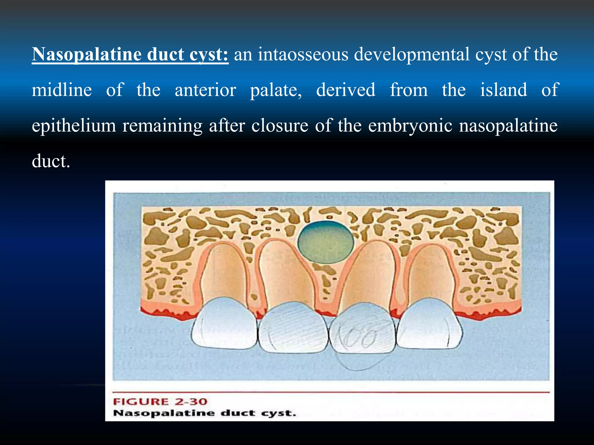
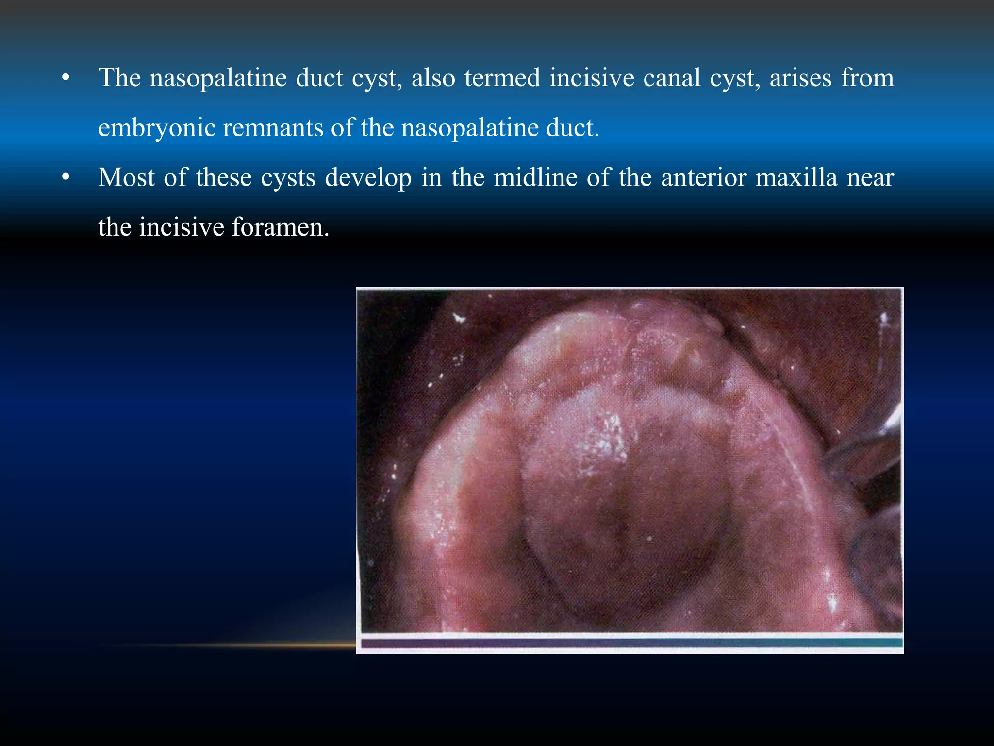
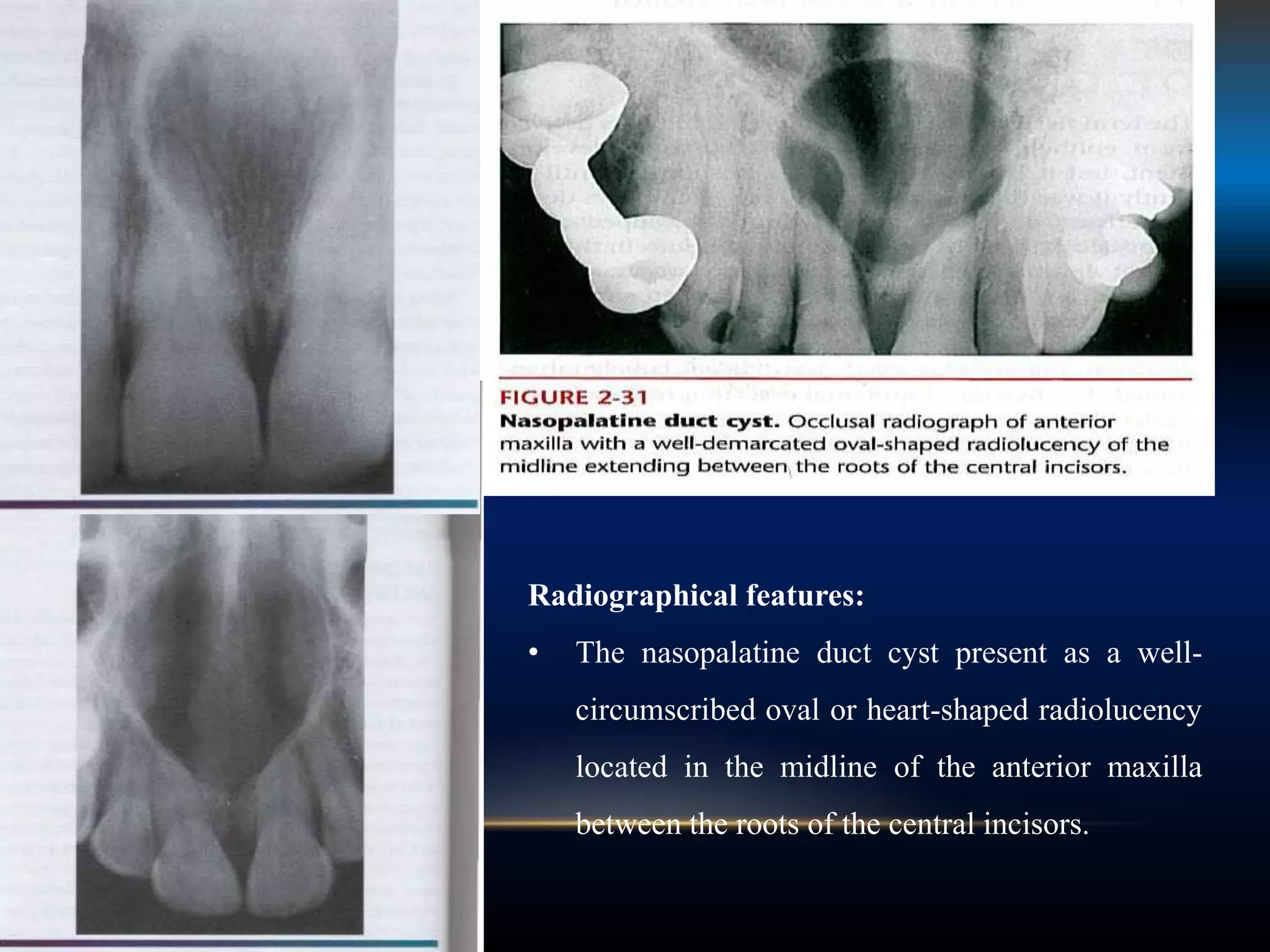
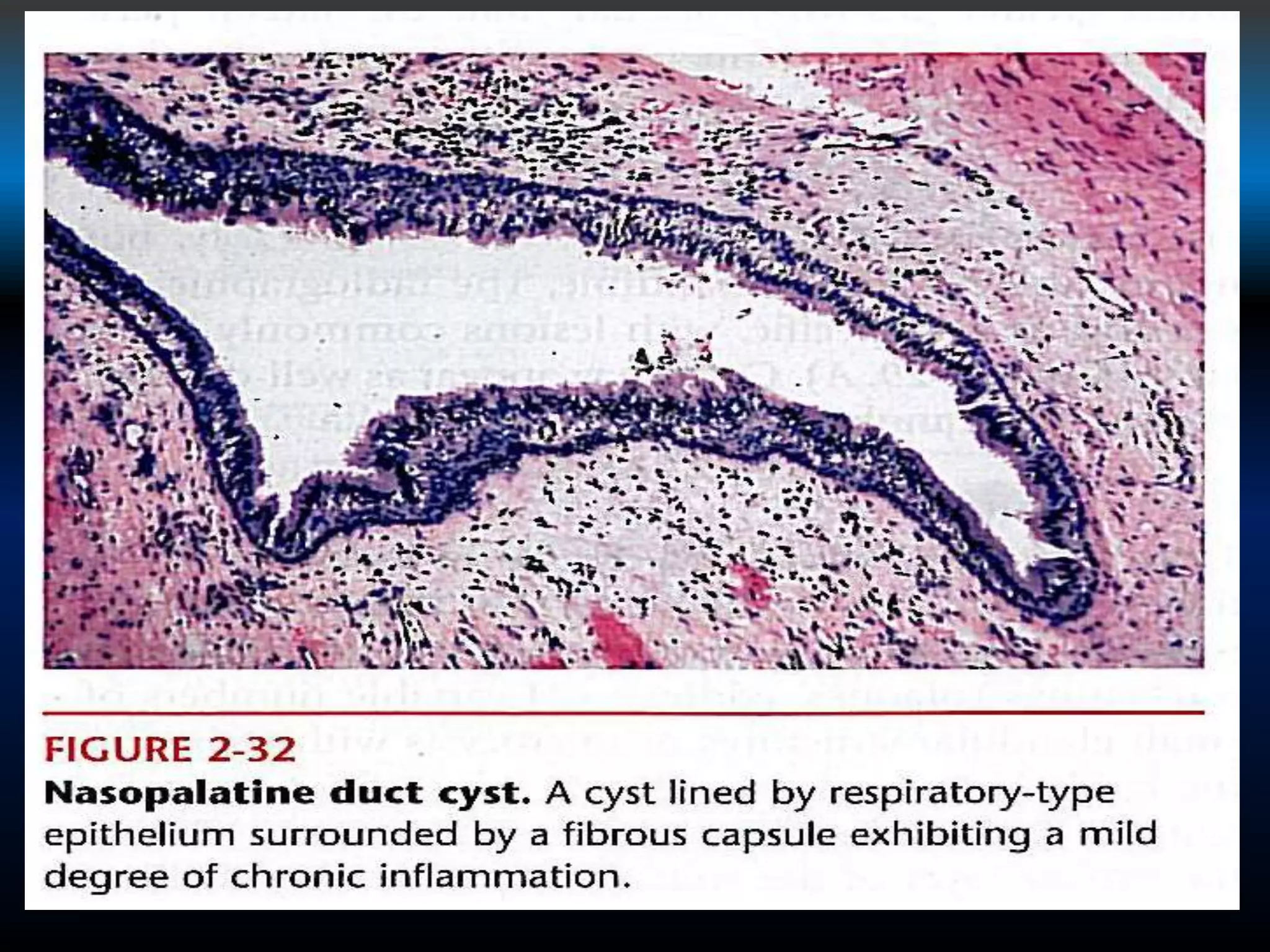
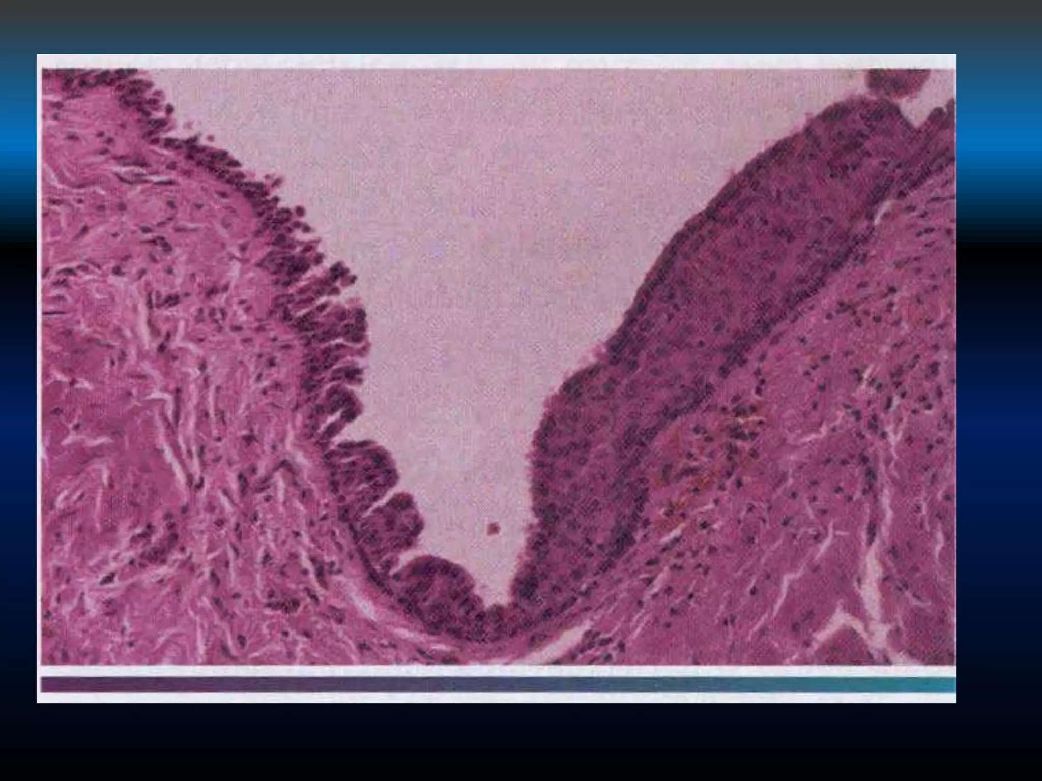
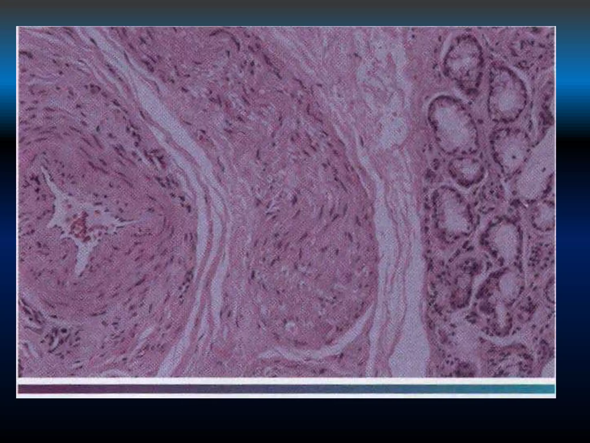
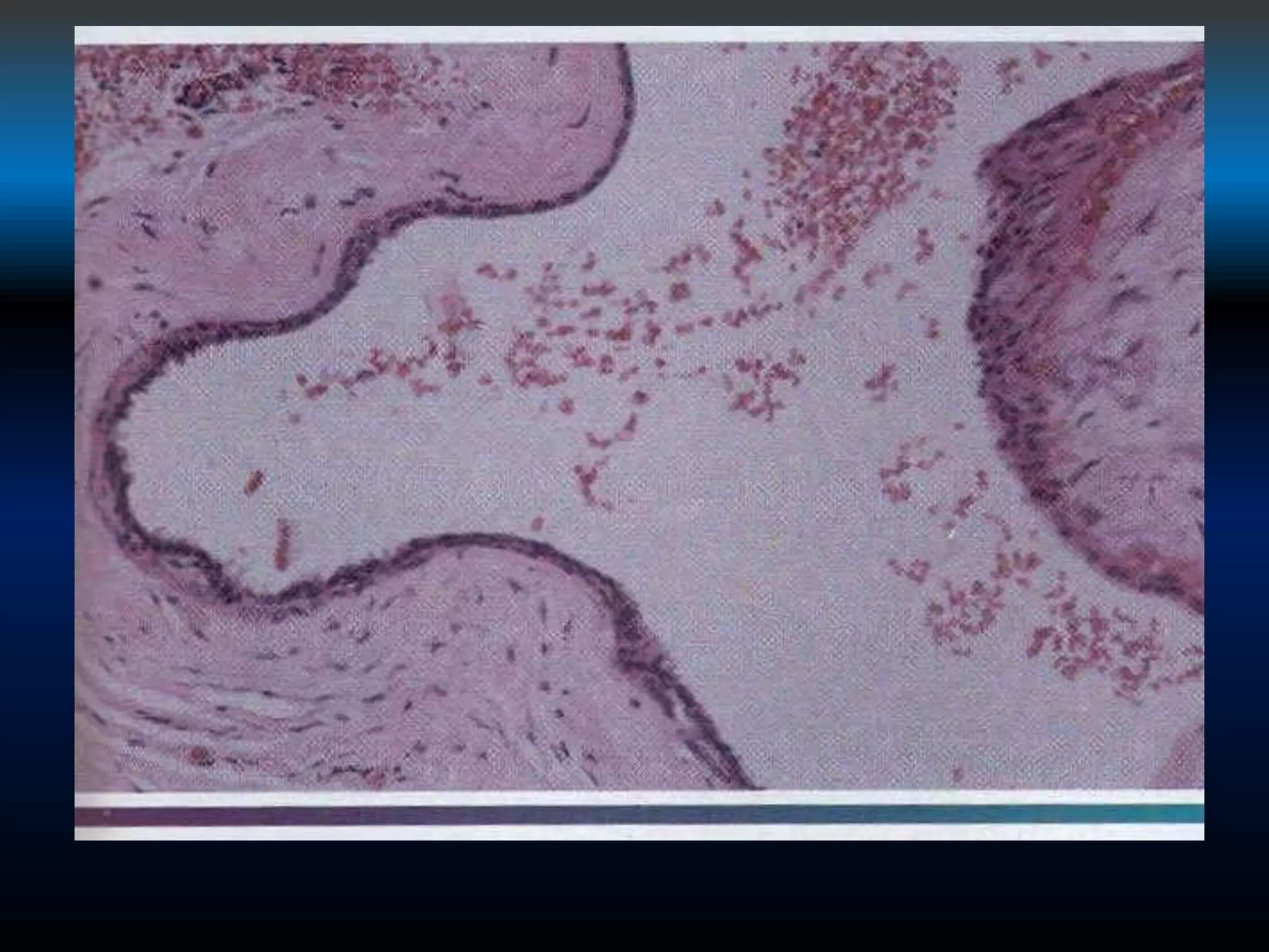
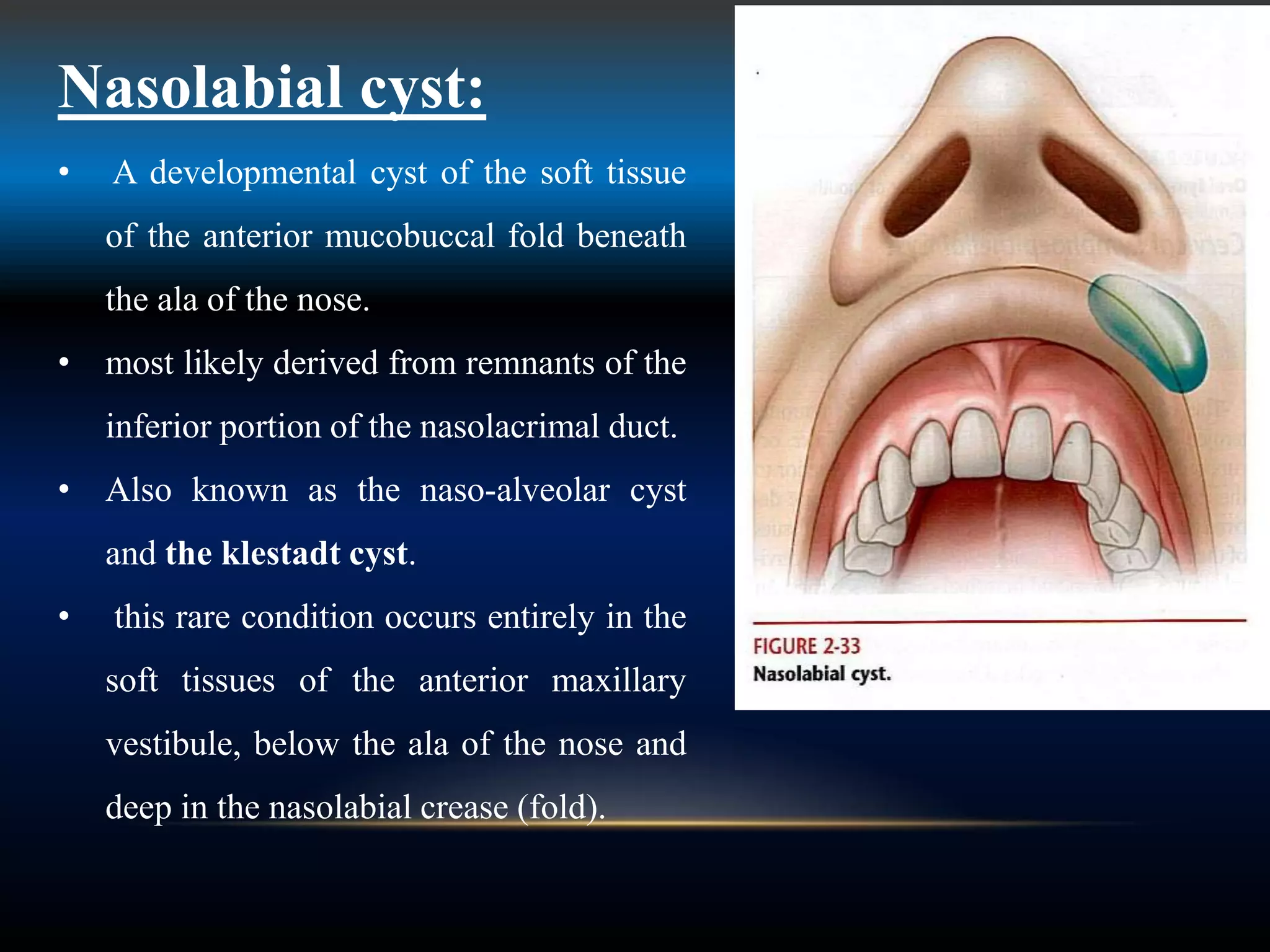
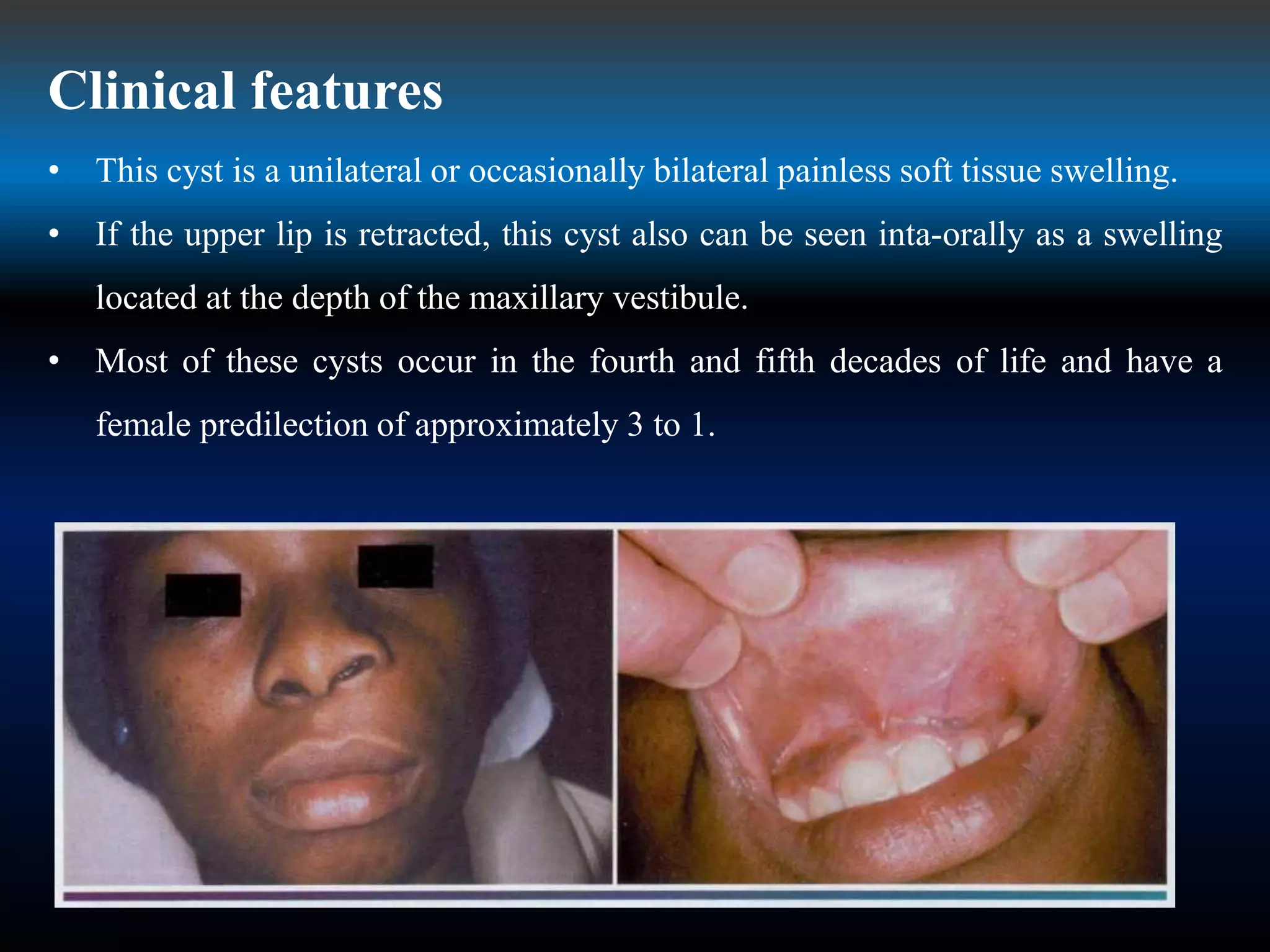
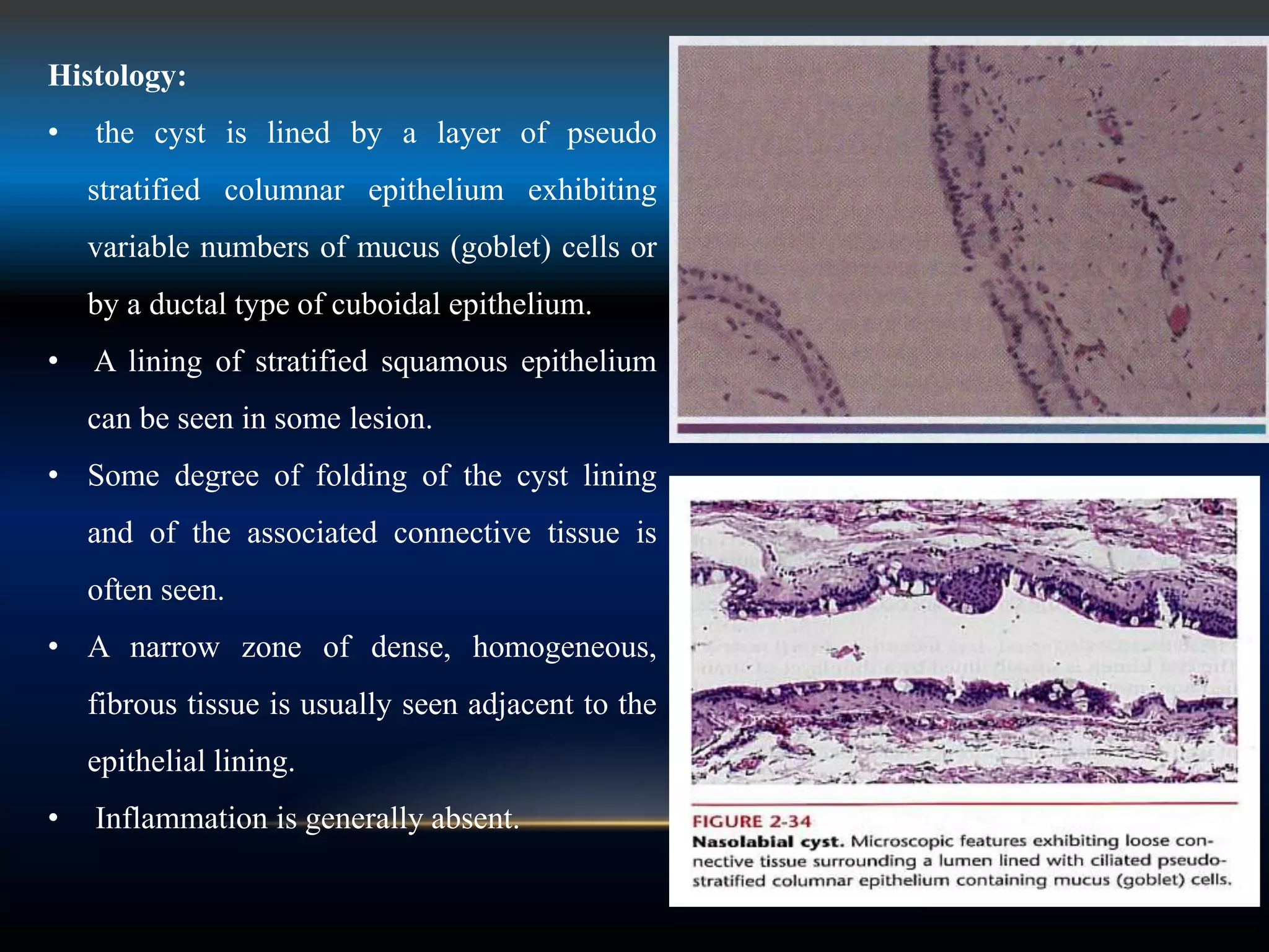
Lecture 15
Developmental cyst
Developmental cysts
Some of these have been considered historically as "fissural"
cysts because they were thought to arise from epithelium
entrapped along embryonal lines of fusion.
• Regardless of their origin, once cysts develop in the oral and
maxillofacial region, they tend to slowly increase in size,
possibly in response to a slightly elevated hydrostatic luminal
pressure.
Nasopalatine duct cyst: an intaosseous developmental cyst of the
midline of the anterior palate, derived from the island of
epithelium remaining after closure of the embryonic nasopalatine
duct.
• The nasopalatine duct cyst, also termed incisive canal cyst, arises from
embryonic remnants of the nasopalatine duct.
• Most of these cysts develop in the midline of the anterior maxilla near
the incisive foramen.
Radiographical features:
• The nasopalatine duct cyst present as a well-
circumscribed oval or heart-shaped radiolucency
located in the midline of the anterior maxilla
between the roots of the central incisors.
Histopathology
• These cyst are lined by a layer of ciliated columnar (respiratory), cuboidal , or
stratified squamous epithelium or by a mixture of these epithelial types.
• If inflammation is present, it usually consists of an infiltrate of plasma cells and
lymphocyte.
• The cyst capsule typically exhibits the prominent component of blood vessels and
peripheral nerves that comprise normal incisive canal contents. The presence of these
neurovascular elements can be helpful in reaching a microscopic diagnosis.
• In addition, occasional small lobules of salivary type mucus glands may be seen in the
cyst wall .premalignant or malignant transformation of the epithelial lining in this cyst
has not been reported.
• The histologic features of the cyst of the incisive papilla closely resemble those of the
nasopalatine duct cyst.
Treatment:
Treatment of the nasopalatine duct cyst is by surgical
enucleation , using a palatal approach. Recurrence of
this cyst is rare.
Nasolabial cyst:
• A developmental cyst of the soft tissue
of the anterior mucobuccal fold beneath
the ala of the nose.
• most likely derived from remnants of the
inferior portion of the nasolacrimal duct.
• Also known as the naso-alveolar cyst
and the klestadt cyst.
• this rare condition occurs entirely in the
soft tissues of the anterior maxillary
vestibule, below the ala of the nose and
deep in the nasolabial crease (fold).
Clinical features
• This cyst is a unilateral or occasionally bilateral painless soft tissue swelling.
• If the upper lip is retracted, this cyst also can be seen inta-orally as a swelling
located at the depth of the maxillary vestibule.
• Most of these cysts occur in the fourth and fifth decades of life and have a
female predilection of approximately 3 to 1.
• Because this cyst is located entirely within soft tissue, it is not
readily apparent radiographically unless contrast medium is
injected into the cystic lumen to facilitate visualization.
• Focal pressure-induced bone resorption of the anterior maxilla
can be occasionally demonstrated on radiographs and is most
readily seen in the edentulous patients.
Histology:
• the cyst is lined by a layer of pseudo
stratified columnar epithelium exhibiting
variable numbers of mucus (goblet) cells or
by a ductal type of cuboidal epithelium.
• A lining of stratified squamous epithelium
can be seen in some lesion.
• Some degree of folding of the cyst lining
and of the associated connective tissue is
often seen.
• A narrow zone of dense, homogeneous,
fibrous tissue is usually seen adjacent to the
epithelial lining.
• Inflammation is generally absent.
Treatment:
A nasolabial cyst is treated by surgical enucleation
with particular care being exercised to prevent the
lesion's perforation and collapse. Recurrence is rare.
Lymphoepithelial cysts:
• A cyst with a lumen lined by a keratinized stratified squamous
epithelium and a capsule containing multiple normal lymphoid follicles
and a dense accumulation of normal lymphocytes.
• Lymphoepithelial cysts are relatively uncommon lesion that occurs in
several areas of the head and neck, most commonly in the floor of the
mouth and on the lateral aspect of the neck.
• Those occurring intraorally are termed oral lymphoepithelial cysts, and
those occurring on the lateral aspect of the neck are termed cervical
lymphoepithelial cysts. Although cysts in the oral cavity are
considerable smaller than those on the lateral aspect of the neck, their
histopathologic features are essentially the same.
Oral lymphoepithelial cyst
• A lymphoepithelial cyst commonly located intraorally on the posterior
lateral tongue and the anterior floor of the mouth.
• The Oral lymphoepithelial cyst , also termed benign lymphoepithelial
cyst, most commonly develops where extratonsillar lymphoid tissue (oral
tonsil) is found.
• The most common sites are the anterior floor of the mouth and the
posterior lateral border of the tongue.
• It appears to develop from epithelial invaginations (crypts) that become
detached from the surface mucosa and entrapped within the lymphoid
tissue; cyst formation ensues.
Clinical features
• The oral lymphoepithelial cyst is most commonly found on the
anterior floor of the mouth and on the posterior lateral borders
of the tongue. However, it can also occur on the ventral surface
of the tongue, soft palate, tonsillar pillars, and oropharynx.
• It is a symptomatic, yellow or tan, superficial submucosal mass
that usually measures less than 1 cm in diameter .
Histopathology
• The cyst is lined by a relatively thin layer of parakeratinized squamous
epithelium surrounded by a well-defined mass of normal lymphoid tissue
exhibiting variable numbers of germinal centers.
• The cystic lumen is usually filled with desquamated parakeratin.
Occasionally, the pore or crypt that communicates between the surface
mucosa and the cystic lumen can be seen microscopically. The presence of
bacteria within the cystic lumen in some of these cysts is also evidence that
communication with the oral cavity is present.
Cervical lymphoepithelial cyst:
• The cervical lymphoepithelial cyst, also commonly termed
branchial cleft cyst or benign cystic lymph node, occurs on the
lateral aspect of the neck, usually anterior to the
sternocleidomastoid muscle.
• It is thought to be derived from epithelium entrapped within
lymphoid tissues of the neck during embryologic
development.
Clinical features
• The cyst becomes apparent in late childhood or early
adulthood as a painless swelling on the lateral aspect of
the neck anterior to the sternomastoid muscle.
• A draining fistula that communicates between the cyst
and the overlying skin surface occasionally develops.
Histopathology
The cyst lumen is usually lined by a thin layer of
stratified squamous epithelium and contain
desquamated orthokeratin , the capsule wall is
thickened , consisting of a fibrous connective tissue
containing large numbers of well-formed lymphoid
follicles.
CERVICALLYMPHOEPITHELIAL CYST.
MEDIUM-POWERED VIEW SHOWING A CYST LINED BY STRATIFIED
SQUAMOUS EPITHELIUM. NOTE THE LYMPHOID TISSUE IN THE CYST
WALL.
Treatment:
As with its intraoral counterpart, the cervical
lymphoepithelial cyst is treated by conservative
surgical excision; recurrence is rare.
Thyroglossal tract cyst:
• A cyst located above the thyroid gland and beneath the base of the tongue,
with a lumen lined by a mixture of epithelial cell types derived from
remnants of the embryonic thyroglossal tract and often containing thyroid
tissue in the capsule.
• The thyroglossal tract cyst is a relatively uncommon lesion derived from
embryologic remnant of the thyroglossal tract. This tract extends from the
foramen caecum on the middorsum of the tongue to the thyroid gland.
Clinical features:
• This cyst occurs primarily in children and young adults.
• presents as an asymptomatic, slowly enlarging, and mobile
swelling involving the midline of the anterior neck above the
thyroid gland.
• A small percentage of these cysts occur within the tongue,
where they can cause dysphagia.
THYROGLOSSAL DUCT CYST. SWELLING
(ARROW) OF THE ANTERIOR MIDLINE OF
THE NECK.
Histopathology:
• This cyst is lined by stratified squamous epithelium, ciliated
columnar epithelium, transitional epithelium, or a mixture of
epithelium types.
• The cyst capsule can exhibit a number of additional findings
including lymphoid aggregates, thyroid tissue mucus glands,
and sebaceous glands.
• Carcinoma can occasionally develop from the lining of
thyroglossal tract cysts and from remnants of the thyroglossal
tract.
THYROGLOSSAL DUET CYST . CYST (TOP) LINED BY STRATIFIED
SQUAMOUS EPITHELIUM. THYROID FOLLICLES CAN BE SEEN IN
THE CYST WALL (BOTTOM).
Treatment:
The treatment of the thyroglossal tract cyst requires
complete surgical excision, because recurrence is a distinct
possibility. In an effort to minimize recurrence of cysts
involving the hyoid area, it is recommended that the central
portion of the hyoid bone and its associated remnants of
thyroglossal tract be removed.
Dermoid cyst:
• a cyst of the midline of the upper neck or the anterior floor of
the mouth of young patients, derived from remnants of
embryonic skin, consisting of a lumen lined by a keratinized
stratified squamous epithelium, and containing one or more
skin appendages such as hair, sweat, or sebaceous glands.
• Most of these cysts occur in the head and neck region ,
primarily in the skin around the eye and the anterior upper
neck, extending superiorly into the floor of the mouth.
DERMOID CYST. FLUCTUANT MIDLINE
SWELLING IN THE
FLOOR OF THE MOUTH.
Clinical features:
• The dermoid cyst is a lesion of young adults (teenagers).
• no gender predilection is seen.
• Cysts of the anterior upper neck or floor of the mouth present as painless
swellings.
• Cysts that develop above the mylohyoid muscle present as a midline
swelling in the sublingual (floor of the mouth) area. In this location the cyst
results in elevation of the tongue and can interfere with eating and speaking.
• Cysts that develop below the mylohyoid muscle appear as a midline
swelling in the submandibular and submental region.
• The size of these cysts is variable, but most are 2 cm or less in diameter.
Histopathology:
• A layer of orthokeratinized squamous epithelium exhibiting
variable number of dermal appendages, including hair
follicle , sebaceous glands, and associated erector pili
muscles lines the cyst.
• The cystic lumen is generally filled with a mixture of
desquamated keratin, sebum, and hair shafts. The cyst
capsule is composed of generally free of inflammation.
DERMOID CYST. SQUAMOUS EPITHELIA L LINING (TOP).
WITH HAIR FOLLICLE (F) AND SEBACEOUSGLANDS (S) IN THE
CYST WALL.
Treatment:
This cyst is best treated by surgical enucleation or excision.
Recurrence is uncommon.
Epidermoid cyst:
• A cyst of skin with a lumen lined by keratinizing stratified squamous
epithelium, usually filled with keratin and without skin appendages in the
capsule wall.
• The epidermoid cyst closely resembles a dermoid cyst, except that the
former exhibit no dermal appendages.
• They are lined by orthokeratinized squamous epithelium and exhibit a
lumen that is generally filled with desquamated keratin. The cyst wall
consists of a narrow zone of compressed fibrous connective tissue that is
generally free of inflammation.
• This cyst is treated by surgical enucleation or excision.
• Recurrence is uncommon.
EPIDERMOID CYST . FLUCTUANT NODULE AT
THE LATERAL
EDGE OF TH EEYEBROW.
EPIDERMOID CYST. INFANT.
EPIDERMOID CYST. A. L OW-POWER VIEW SHOWING A
KERATIN-FILLED CYSTIC CAVITY. B, HIGHPOWERED VIEW
SHOWING STRAT IFIED SQUAMOUS EPITHELIAL LINING
WITH ORTHOKERATIN PRODUCTION.
Heterotopic oral Gastrointestinal cyst:
• The heterotopic oral gastrointestinal cyst is a rare and unusual developmental
entity .
• most commonly found in the tongue or the floor of the mouth of infants or young
children.
• The theory of undifferentiated endodermal entrapment during the fourth or fifth
week of embryonic development, followed by gastrointestinal differentiation.
• This cyst is treated by surgical excision; recurrence is uncommon.
"GLOBULOMAXILLARY CYST":
• fissural cyst that arise from epithelium entrapped during fusion of the globular
portion of the medial nasal process with the maxillary process.
• Current theory holds that most (if not all) cysts that develop in the
globulomaxillary area are actually of odontogenic origin.
Clinical and Radiographic Features
• The "globulomaxillary cyst" classically develops between the maxillary
lateral incisor and cuspid teeth.
• Although occasional globulomaxillary lesions have been reported 'between
the central a lateral Radiographs typically demonstrate a well-circumscribed
unilocular radiolucency between and apical to the teeth because this
radiolucency often is constricted as it extends down between the teeth. It may
resemble an inverted pear. As the lesion expands, tipping of the tooth roots
may occur.
Histopathologic Features: Virtually cysts in the globule-maxiliary
region can be explained on an odontogenic basis.
• Many are lined by inflamed stratified squamous epithelium and are consistent
with periapical cysts.
• some exhibit specific histopathologic features of an odontogenic keratocyst
or developmental lateral periodontal cyst. It also has been theorized that some
of these lesions may arise from inflammation of the reduced enamel
epithelium at the time of eruption of the teeth.
• On rare occasions cysts in the globulomaxiliary area may be lined by
pseudostratified ciliated columnar epithelium.
Treatment and Prognosis
• Treatment of cysts in the globulomaxillary area usually
consists of surgical enucleation. If the lesion can be related to
an adjacent non vital tooth, then endodontic therapy may be
appropriate. Prognosis depends on the specific histopathologic
type of cyst. Except for the odontogenic keratocyst, the
recurrence potential should below.
PALATALCYSTS OFTHE NEWBORN
(EPSTEIN'SPEARLS; BOHN'S NODULES)
• Small developmental cysts are a common finding on the palate of newborn
infants.
• It has been theorized that these "inclusion" cysts may arise in one of two
ways:
• First, as the palatal shelves meet and fuse in the midline during embryonic
life to form the secondary palate, small islands of epithelium may become
entrapped below the surface along the median palatal raphe and form cysts.
• Second, these cysts may arise from epithelial remnants derived tram the
development of the minor salivary glands of the palate.
EPSTEIN'S PEARL S. SMALL KERATIN -FILLED
CYSTS AT THE
JUNCTION OF THE HARD AND SOFT PALATES.
Clinical Features
• Palatal cysts of the newborn are quite common and have
been reported in 65% to 85% of neonates.
• The cysts are small, 1 to 3 mm white or yellowish-white
papules that appear most often along the midline near the
junction of the hard and soft palates,
• Occasionally, they may occur in a more anterior location
along the raphe or on the posterior palate lateral to the
midline.
Histopathologic Features
• Microscopic examination reveals keratin-filled cysts that
are lined by stratified squamous epithelium; sometimes
these cysts demonstrate a communication with the mucosal
surface.
Treatment and Prognosis
Palatal cysts of the newborn are innocuous lesions, and no
treatment is required. They are self-healing and rarely
observable several weeks after birth. Presumably the
epithelium degenerates, or the cysts rupture onto the mucosal
surface and eliminate their keratin contents.
MEDIAN PALATAL (PALATINE) CYST:
The median palatal cyst is a rare fissural cyst that theoretically
develops from epithelium entrapped along the embryonic line of
fusion of the lateral palatal shelves of the maxilla. This cyst may be
difficult to distinguish from a nasopalatine duct cyst. In fact most
"median palatal cysts" may represent posteriorly positioned
nasopalatine duct cysts. Because the nasopalatine ducts course
posteriorly and superiorly as they extend from the incisive canal to
the nasal cavity, a nasopalatine duct cyst that arises from posterior
remnants of this duct near the nasal cavity might be mistaken for a
median palatal cyst.
MEDIAN PALATAL CYST. WELL-CIRCUMSCRIBED RADIOLUCENCY
APICAL TO THE MAXILLARY INCISORS IN THE MIDLINE. AT
SURGERY THE LESION WAS UNRELATED TO THE INCISIVE CANAL.
On the other hand if a true median palatal cyst was to
develop toward the anterior portion of the hard palate. It
could easily be mistaken for a nasopalatine duct cyst.
Clinical and Radiographic Features
• The median palatal cyst presents as a firm or fluctuant swelling of the midline of the
hard palate posterior to the palatine papilla.
• The lesion appears most frequently in young adults. Often it is asymptomatic. But
some patients complain of pain or expansion.
• The average size of this cyst is 2 X 2 cm, but sometimes it can become quite large.
• Occlusal radiographs demonstrate a well-circumscribed radiolucency in the midline
of the hard palate.
• Occasional reported cases have been associated with divergence of the central
incisors. Although it may be difficult to rule out a nasopalatine duct cyst in these
instances. It must be stressed that a true median palatal cyst should exhibit clinical
enlargement of the palate.
• A midline radiolucency without clinical evidence of expansion is probably a
nasopalatine duct cyst.
Histopathologic Features
• Microscopic examination shows a cyst that is usually lined by stratified
squamous epithelium. Areas of ciliated pseudostratified columnar
epithelium have been reported in some cases.
• Chronic inflammation may be present in the cyst wall.
Treatment and Prognosis
The median palatal cyst is treated by surgical removal.
Recurrence should not be expected.
“MEDIAN MANDIBULAR CYST”:
The “median mandibular cyst” is a controversial lesion of questionable existence.
Theoretically, it represents a fissural cyst in the anterior midline of the mandible that
develops from epithelium entrapped during fusion of the halves of the mandible during
embryonic life.
it appears likely that most (if not all) of these midline cysts are of odontogenic origin.
Many purported cases would be classified today as examples of the glandular
odontogenic cyst, which has a propensity for occurrence in the midline mandibular
region. Others could be classified as periapical cysts, odontogenic keratocysts, or lateral
periodontal cysts. Because a fissural cyst in this region probably does not exist, the term
median mandibular cyst should no longer be used.
Other Rare Developmental Anomalies:
HEMIHYPERPLASIA (HEMIHYPERTROPHY):
Hemihyperplasia is a rare developmental anomaly characterized by asymmetric
overgrowth of one or more body parts. Although the condition is known more commonly
as hemihypertrophy, it actually represents a hyperplasia of the tissues rather than a
hypertrophy. Hemihyperplasia can be an isolated finding but it also may be associated
with a variety of malformation syndromes. Almost all cases of isolated hemihyperplasia
are sporadic. A number of possible etiologic factors have been suggested but the cause
remains obscure. Various theories include vascular or lymphatic abnormalities, Central
nervous system disturbances, endocrine dysfunctions and aberrant twinning mechanisms.
Occasionally, chromosomal anomalies have been documented.
HEMIHYPERPLASIA. ENLARGEMENT
OF THE RIGHT SIDE OF THE FACE.
HEMIHYPERPLASIA. SAME PATIENT AS DEPICTED IN. WITH
ASSOCIATED ENLARGEMENT OF THE RIGHT HALF OF THE
TONGUE.
HEMIHYPERPLASIA. RADIOGRAPH OF THE SAME
PATIENT. MANDIBLE AND TEETH ON THE RIGHT
SIDE ARE ENLARGED.
PROGRESSIVE HEMIFACIALATROPHY:
(ROMBERG SYNDROME; PARRY-ROMBERG SYNDROME)
Progressive hemifacial atrophy is an uncommon and poorly understood degenerative
condition characterized by atrophic changes affecting one side of the face. The cause of
these changes remains obscure. Speculation has considered trophic malfunction of the
cervical sympathetic nervous system. A history of prior trauma has been documented in
some cases although other reports have considered a viral or Borrelia infection .
Usually the condition is sporadic, but a few familial cases have been reported
suggesting a possible hereditary influence, many investigators believe that hemifacial
atrophy represents a localized form of scleroderma.
PROGRESSIVE HEMIFACIAL ATROPHY. YOUNG
GIRL WITH
RIGHT-SIDED FACIAL ATROPHY.
SEGMENTAL ODONTOMAXllLARY DYSPLASIA
(HEMI MAXILLOFACIAl DYSPLASIA):
Segmental odontomaxillary dysplasia is a recently recognized
developmental disorder that affects the jaw and (sometimes) the
overlying facial tissues. The cause is unknown clinically. It is
frequently mistaken for craniofacial fibrous dysplasia or hemi
facial hyperplasia, but it represents a distinct and separate entity.
CROUZON SYNDROME (CRANIOFACIAL DYSOSTOSIS):
Crouzon syndrome is one of a rare group of syndromes
characterized by craniosynostosis, or premature closing of the cranial
sutures.
CROUZON SYNDROME. OCULAR
PROPTOSIS AND MID FACE HYPOPLASIA.
APERT SYNDROME :
• Like Crouzon syndrome
• Apert syndrome is a rare condition that is characterized by
craniosynostosis.
• It occurs in about I of every 65,000 to 160,000 births
• caused by a mutation in the fibroblast growth factor receptor 2
(FGFR2) gene.
• Although it is inherited as an autosomal dominant trait, most
cases represent sporadic new mutations, often associated with
increased paternal age.
APERT SYNDROME. SYNDACTYLY OF
THE HAND.
APERT SYNDROME. RADIOGRAPH SHOWING "MIDFACE
HYPOPLASIA
APERT SYNDROME. ABNORMAL SHAPE OF THE MAXILLA,
WITH SWELLINGS OF THE POSTERIOR LATERAL HARD
PALATE, RESULTING IN PSEUDOCLEFT FORMATION.
MANDIBULOFACIAL DYSOSTOSIS :
(TREACHER COLLINS SYNDROME)
Mandibulofacial dysostosis is a rare syndrome
• defects of structures derived from the first and second branchial
arches.
• It is inherited as an autosomal dominant trait
• occurs in around 1 of every 25,000 to 50,000 live births.
MANDIBULOFACIAL DYSOSTOSIS. PATIENT EXHIBITS A
HYPOPLASTIC MANDIBLE, DOWNWARDSLANTING
PALPEBRAL FISSURES, AND EAR DEFORMITIES.

Lecture15 dina patho

  • 1.
  • 2.
    Developmental cysts Some ofthese have been considered historically as "fissural" cysts because they were thought to arise from epithelium entrapped along embryonal lines of fusion. • Regardless of their origin, once cysts develop in the oral and maxillofacial region, they tend to slowly increase in size, possibly in response to a slightly elevated hydrostatic luminal pressure.
  • 3.
    Nasopalatine duct cyst:an intaosseous developmental cyst of the midline of the anterior palate, derived from the island of epithelium remaining after closure of the embryonic nasopalatine duct.
  • 4.
    • The nasopalatineduct cyst, also termed incisive canal cyst, arises from embryonic remnants of the nasopalatine duct. • Most of these cysts develop in the midline of the anterior maxilla near the incisive foramen.
  • 5.
    Radiographical features: • Thenasopalatine duct cyst present as a well- circumscribed oval or heart-shaped radiolucency located in the midline of the anterior maxilla between the roots of the central incisors.
  • 6.
    Histopathology • These cystare lined by a layer of ciliated columnar (respiratory), cuboidal , or stratified squamous epithelium or by a mixture of these epithelial types. • If inflammation is present, it usually consists of an infiltrate of plasma cells and lymphocyte. • The cyst capsule typically exhibits the prominent component of blood vessels and peripheral nerves that comprise normal incisive canal contents. The presence of these neurovascular elements can be helpful in reaching a microscopic diagnosis. • In addition, occasional small lobules of salivary type mucus glands may be seen in the cyst wall .premalignant or malignant transformation of the epithelial lining in this cyst has not been reported. • The histologic features of the cyst of the incisive papilla closely resemble those of the nasopalatine duct cyst.
  • 11.
    Treatment: Treatment of thenasopalatine duct cyst is by surgical enucleation , using a palatal approach. Recurrence of this cyst is rare.
  • 12.
    Nasolabial cyst: • Adevelopmental cyst of the soft tissue of the anterior mucobuccal fold beneath the ala of the nose. • most likely derived from remnants of the inferior portion of the nasolacrimal duct. • Also known as the naso-alveolar cyst and the klestadt cyst. • this rare condition occurs entirely in the soft tissues of the anterior maxillary vestibule, below the ala of the nose and deep in the nasolabial crease (fold).
  • 13.
    Clinical features • Thiscyst is a unilateral or occasionally bilateral painless soft tissue swelling. • If the upper lip is retracted, this cyst also can be seen inta-orally as a swelling located at the depth of the maxillary vestibule. • Most of these cysts occur in the fourth and fifth decades of life and have a female predilection of approximately 3 to 1.
  • 14.
    • Because thiscyst is located entirely within soft tissue, it is not readily apparent radiographically unless contrast medium is injected into the cystic lumen to facilitate visualization. • Focal pressure-induced bone resorption of the anterior maxilla can be occasionally demonstrated on radiographs and is most readily seen in the edentulous patients.
  • 15.
    Histology: • the cystis lined by a layer of pseudo stratified columnar epithelium exhibiting variable numbers of mucus (goblet) cells or by a ductal type of cuboidal epithelium. • A lining of stratified squamous epithelium can be seen in some lesion. • Some degree of folding of the cyst lining and of the associated connective tissue is often seen. • A narrow zone of dense, homogeneous, fibrous tissue is usually seen adjacent to the epithelial lining. • Inflammation is generally absent.
  • 16.
    Treatment: A nasolabial cystis treated by surgical enucleation with particular care being exercised to prevent the lesion's perforation and collapse. Recurrence is rare.
  • 17.
    Lymphoepithelial cysts: • Acyst with a lumen lined by a keratinized stratified squamous epithelium and a capsule containing multiple normal lymphoid follicles and a dense accumulation of normal lymphocytes. • Lymphoepithelial cysts are relatively uncommon lesion that occurs in several areas of the head and neck, most commonly in the floor of the mouth and on the lateral aspect of the neck. • Those occurring intraorally are termed oral lymphoepithelial cysts, and those occurring on the lateral aspect of the neck are termed cervical lymphoepithelial cysts. Although cysts in the oral cavity are considerable smaller than those on the lateral aspect of the neck, their histopathologic features are essentially the same.
  • 18.
    Oral lymphoepithelial cyst •A lymphoepithelial cyst commonly located intraorally on the posterior lateral tongue and the anterior floor of the mouth. • The Oral lymphoepithelial cyst , also termed benign lymphoepithelial cyst, most commonly develops where extratonsillar lymphoid tissue (oral tonsil) is found. • The most common sites are the anterior floor of the mouth and the posterior lateral border of the tongue. • It appears to develop from epithelial invaginations (crypts) that become detached from the surface mucosa and entrapped within the lymphoid tissue; cyst formation ensues.
  • 20.
    Clinical features • Theoral lymphoepithelial cyst is most commonly found on the anterior floor of the mouth and on the posterior lateral borders of the tongue. However, it can also occur on the ventral surface of the tongue, soft palate, tonsillar pillars, and oropharynx. • It is a symptomatic, yellow or tan, superficial submucosal mass that usually measures less than 1 cm in diameter .
  • 21.
    Histopathology • The cystis lined by a relatively thin layer of parakeratinized squamous epithelium surrounded by a well-defined mass of normal lymphoid tissue exhibiting variable numbers of germinal centers. • The cystic lumen is usually filled with desquamated parakeratin. Occasionally, the pore or crypt that communicates between the surface mucosa and the cystic lumen can be seen microscopically. The presence of bacteria within the cystic lumen in some of these cysts is also evidence that communication with the oral cavity is present.
  • 23.
    Cervical lymphoepithelial cyst: •The cervical lymphoepithelial cyst, also commonly termed branchial cleft cyst or benign cystic lymph node, occurs on the lateral aspect of the neck, usually anterior to the sternocleidomastoid muscle. • It is thought to be derived from epithelium entrapped within lymphoid tissues of the neck during embryologic development.
  • 25.
    Clinical features • Thecyst becomes apparent in late childhood or early adulthood as a painless swelling on the lateral aspect of the neck anterior to the sternomastoid muscle. • A draining fistula that communicates between the cyst and the overlying skin surface occasionally develops.
  • 27.
    Histopathology The cyst lumenis usually lined by a thin layer of stratified squamous epithelium and contain desquamated orthokeratin , the capsule wall is thickened , consisting of a fibrous connective tissue containing large numbers of well-formed lymphoid follicles.
  • 29.
    CERVICALLYMPHOEPITHELIAL CYST. MEDIUM-POWERED VIEWSHOWING A CYST LINED BY STRATIFIED SQUAMOUS EPITHELIUM. NOTE THE LYMPHOID TISSUE IN THE CYST WALL.
  • 30.
    Treatment: As with itsintraoral counterpart, the cervical lymphoepithelial cyst is treated by conservative surgical excision; recurrence is rare.
  • 31.
    Thyroglossal tract cyst: •A cyst located above the thyroid gland and beneath the base of the tongue, with a lumen lined by a mixture of epithelial cell types derived from remnants of the embryonic thyroglossal tract and often containing thyroid tissue in the capsule. • The thyroglossal tract cyst is a relatively uncommon lesion derived from embryologic remnant of the thyroglossal tract. This tract extends from the foramen caecum on the middorsum of the tongue to the thyroid gland.
  • 33.
    Clinical features: • Thiscyst occurs primarily in children and young adults. • presents as an asymptomatic, slowly enlarging, and mobile swelling involving the midline of the anterior neck above the thyroid gland. • A small percentage of these cysts occur within the tongue, where they can cause dysphagia.
  • 34.
    THYROGLOSSAL DUCT CYST.SWELLING (ARROW) OF THE ANTERIOR MIDLINE OF THE NECK.
  • 35.
    Histopathology: • This cystis lined by stratified squamous epithelium, ciliated columnar epithelium, transitional epithelium, or a mixture of epithelium types. • The cyst capsule can exhibit a number of additional findings including lymphoid aggregates, thyroid tissue mucus glands, and sebaceous glands. • Carcinoma can occasionally develop from the lining of thyroglossal tract cysts and from remnants of the thyroglossal tract.
  • 37.
    THYROGLOSSAL DUET CYST. CYST (TOP) LINED BY STRATIFIED SQUAMOUS EPITHELIUM. THYROID FOLLICLES CAN BE SEEN IN THE CYST WALL (BOTTOM).
  • 38.
    Treatment: The treatment ofthe thyroglossal tract cyst requires complete surgical excision, because recurrence is a distinct possibility. In an effort to minimize recurrence of cysts involving the hyoid area, it is recommended that the central portion of the hyoid bone and its associated remnants of thyroglossal tract be removed.
  • 39.
    Dermoid cyst: • acyst of the midline of the upper neck or the anterior floor of the mouth of young patients, derived from remnants of embryonic skin, consisting of a lumen lined by a keratinized stratified squamous epithelium, and containing one or more skin appendages such as hair, sweat, or sebaceous glands. • Most of these cysts occur in the head and neck region , primarily in the skin around the eye and the anterior upper neck, extending superiorly into the floor of the mouth.
  • 40.
    DERMOID CYST. FLUCTUANTMIDLINE SWELLING IN THE FLOOR OF THE MOUTH.
  • 41.
    Clinical features: • Thedermoid cyst is a lesion of young adults (teenagers). • no gender predilection is seen. • Cysts of the anterior upper neck or floor of the mouth present as painless swellings. • Cysts that develop above the mylohyoid muscle present as a midline swelling in the sublingual (floor of the mouth) area. In this location the cyst results in elevation of the tongue and can interfere with eating and speaking. • Cysts that develop below the mylohyoid muscle appear as a midline swelling in the submandibular and submental region. • The size of these cysts is variable, but most are 2 cm or less in diameter.
  • 42.
    Histopathology: • A layerof orthokeratinized squamous epithelium exhibiting variable number of dermal appendages, including hair follicle , sebaceous glands, and associated erector pili muscles lines the cyst. • The cystic lumen is generally filled with a mixture of desquamated keratin, sebum, and hair shafts. The cyst capsule is composed of generally free of inflammation.
  • 43.
    DERMOID CYST. SQUAMOUSEPITHELIA L LINING (TOP). WITH HAIR FOLLICLE (F) AND SEBACEOUSGLANDS (S) IN THE CYST WALL.
  • 45.
    Treatment: This cyst isbest treated by surgical enucleation or excision. Recurrence is uncommon.
  • 46.
    Epidermoid cyst: • Acyst of skin with a lumen lined by keratinizing stratified squamous epithelium, usually filled with keratin and without skin appendages in the capsule wall. • The epidermoid cyst closely resembles a dermoid cyst, except that the former exhibit no dermal appendages. • They are lined by orthokeratinized squamous epithelium and exhibit a lumen that is generally filled with desquamated keratin. The cyst wall consists of a narrow zone of compressed fibrous connective tissue that is generally free of inflammation. • This cyst is treated by surgical enucleation or excision. • Recurrence is uncommon.
  • 47.
    EPIDERMOID CYST .FLUCTUANT NODULE AT THE LATERAL EDGE OF TH EEYEBROW.
  • 48.
  • 50.
    EPIDERMOID CYST. A.L OW-POWER VIEW SHOWING A KERATIN-FILLED CYSTIC CAVITY. B, HIGHPOWERED VIEW SHOWING STRAT IFIED SQUAMOUS EPITHELIAL LINING WITH ORTHOKERATIN PRODUCTION.
  • 51.
    Heterotopic oral Gastrointestinalcyst: • The heterotopic oral gastrointestinal cyst is a rare and unusual developmental entity . • most commonly found in the tongue or the floor of the mouth of infants or young children. • The theory of undifferentiated endodermal entrapment during the fourth or fifth week of embryonic development, followed by gastrointestinal differentiation. • This cyst is treated by surgical excision; recurrence is uncommon.
  • 52.
    "GLOBULOMAXILLARY CYST": • fissuralcyst that arise from epithelium entrapped during fusion of the globular portion of the medial nasal process with the maxillary process. • Current theory holds that most (if not all) cysts that develop in the globulomaxillary area are actually of odontogenic origin.
  • 53.
    Clinical and RadiographicFeatures • The "globulomaxillary cyst" classically develops between the maxillary lateral incisor and cuspid teeth. • Although occasional globulomaxillary lesions have been reported 'between the central a lateral Radiographs typically demonstrate a well-circumscribed unilocular radiolucency between and apical to the teeth because this radiolucency often is constricted as it extends down between the teeth. It may resemble an inverted pear. As the lesion expands, tipping of the tooth roots may occur.
  • 54.
    Histopathologic Features: Virtuallycysts in the globule-maxiliary region can be explained on an odontogenic basis. • Many are lined by inflamed stratified squamous epithelium and are consistent with periapical cysts. • some exhibit specific histopathologic features of an odontogenic keratocyst or developmental lateral periodontal cyst. It also has been theorized that some of these lesions may arise from inflammation of the reduced enamel epithelium at the time of eruption of the teeth. • On rare occasions cysts in the globulomaxiliary area may be lined by pseudostratified ciliated columnar epithelium.
  • 55.
    Treatment and Prognosis •Treatment of cysts in the globulomaxillary area usually consists of surgical enucleation. If the lesion can be related to an adjacent non vital tooth, then endodontic therapy may be appropriate. Prognosis depends on the specific histopathologic type of cyst. Except for the odontogenic keratocyst, the recurrence potential should below.
  • 56.
    PALATALCYSTS OFTHE NEWBORN (EPSTEIN'SPEARLS;BOHN'S NODULES) • Small developmental cysts are a common finding on the palate of newborn infants. • It has been theorized that these "inclusion" cysts may arise in one of two ways: • First, as the palatal shelves meet and fuse in the midline during embryonic life to form the secondary palate, small islands of epithelium may become entrapped below the surface along the median palatal raphe and form cysts. • Second, these cysts may arise from epithelial remnants derived tram the development of the minor salivary glands of the palate.
  • 57.
    EPSTEIN'S PEARL S.SMALL KERATIN -FILLED CYSTS AT THE JUNCTION OF THE HARD AND SOFT PALATES.
  • 58.
    Clinical Features • Palatalcysts of the newborn are quite common and have been reported in 65% to 85% of neonates. • The cysts are small, 1 to 3 mm white or yellowish-white papules that appear most often along the midline near the junction of the hard and soft palates, • Occasionally, they may occur in a more anterior location along the raphe or on the posterior palate lateral to the midline.
  • 59.
    Histopathologic Features • Microscopicexamination reveals keratin-filled cysts that are lined by stratified squamous epithelium; sometimes these cysts demonstrate a communication with the mucosal surface. Treatment and Prognosis Palatal cysts of the newborn are innocuous lesions, and no treatment is required. They are self-healing and rarely observable several weeks after birth. Presumably the epithelium degenerates, or the cysts rupture onto the mucosal surface and eliminate their keratin contents.
  • 60.
    MEDIAN PALATAL (PALATINE)CYST: The median palatal cyst is a rare fissural cyst that theoretically develops from epithelium entrapped along the embryonic line of fusion of the lateral palatal shelves of the maxilla. This cyst may be difficult to distinguish from a nasopalatine duct cyst. In fact most "median palatal cysts" may represent posteriorly positioned nasopalatine duct cysts. Because the nasopalatine ducts course posteriorly and superiorly as they extend from the incisive canal to the nasal cavity, a nasopalatine duct cyst that arises from posterior remnants of this duct near the nasal cavity might be mistaken for a median palatal cyst.
  • 61.
    MEDIAN PALATAL CYST.WELL-CIRCUMSCRIBED RADIOLUCENCY APICAL TO THE MAXILLARY INCISORS IN THE MIDLINE. AT SURGERY THE LESION WAS UNRELATED TO THE INCISIVE CANAL.
  • 62.
    On the otherhand if a true median palatal cyst was to develop toward the anterior portion of the hard palate. It could easily be mistaken for a nasopalatine duct cyst.
  • 63.
    Clinical and RadiographicFeatures • The median palatal cyst presents as a firm or fluctuant swelling of the midline of the hard palate posterior to the palatine papilla. • The lesion appears most frequently in young adults. Often it is asymptomatic. But some patients complain of pain or expansion. • The average size of this cyst is 2 X 2 cm, but sometimes it can become quite large. • Occlusal radiographs demonstrate a well-circumscribed radiolucency in the midline of the hard palate. • Occasional reported cases have been associated with divergence of the central incisors. Although it may be difficult to rule out a nasopalatine duct cyst in these instances. It must be stressed that a true median palatal cyst should exhibit clinical enlargement of the palate. • A midline radiolucency without clinical evidence of expansion is probably a nasopalatine duct cyst.
  • 64.
    Histopathologic Features • Microscopicexamination shows a cyst that is usually lined by stratified squamous epithelium. Areas of ciliated pseudostratified columnar epithelium have been reported in some cases. • Chronic inflammation may be present in the cyst wall. Treatment and Prognosis The median palatal cyst is treated by surgical removal. Recurrence should not be expected.
  • 65.
    “MEDIAN MANDIBULAR CYST”: The“median mandibular cyst” is a controversial lesion of questionable existence. Theoretically, it represents a fissural cyst in the anterior midline of the mandible that develops from epithelium entrapped during fusion of the halves of the mandible during embryonic life. it appears likely that most (if not all) of these midline cysts are of odontogenic origin. Many purported cases would be classified today as examples of the glandular odontogenic cyst, which has a propensity for occurrence in the midline mandibular region. Others could be classified as periapical cysts, odontogenic keratocysts, or lateral periodontal cysts. Because a fissural cyst in this region probably does not exist, the term median mandibular cyst should no longer be used.
  • 66.
    Other Rare DevelopmentalAnomalies: HEMIHYPERPLASIA (HEMIHYPERTROPHY): Hemihyperplasia is a rare developmental anomaly characterized by asymmetric overgrowth of one or more body parts. Although the condition is known more commonly as hemihypertrophy, it actually represents a hyperplasia of the tissues rather than a hypertrophy. Hemihyperplasia can be an isolated finding but it also may be associated with a variety of malformation syndromes. Almost all cases of isolated hemihyperplasia are sporadic. A number of possible etiologic factors have been suggested but the cause remains obscure. Various theories include vascular or lymphatic abnormalities, Central nervous system disturbances, endocrine dysfunctions and aberrant twinning mechanisms. Occasionally, chromosomal anomalies have been documented.
  • 67.
    HEMIHYPERPLASIA. ENLARGEMENT OF THERIGHT SIDE OF THE FACE.
  • 68.
    HEMIHYPERPLASIA. SAME PATIENTAS DEPICTED IN. WITH ASSOCIATED ENLARGEMENT OF THE RIGHT HALF OF THE TONGUE.
  • 69.
    HEMIHYPERPLASIA. RADIOGRAPH OFTHE SAME PATIENT. MANDIBLE AND TEETH ON THE RIGHT SIDE ARE ENLARGED.
  • 70.
    PROGRESSIVE HEMIFACIALATROPHY: (ROMBERG SYNDROME;PARRY-ROMBERG SYNDROME) Progressive hemifacial atrophy is an uncommon and poorly understood degenerative condition characterized by atrophic changes affecting one side of the face. The cause of these changes remains obscure. Speculation has considered trophic malfunction of the cervical sympathetic nervous system. A history of prior trauma has been documented in some cases although other reports have considered a viral or Borrelia infection . Usually the condition is sporadic, but a few familial cases have been reported suggesting a possible hereditary influence, many investigators believe that hemifacial atrophy represents a localized form of scleroderma.
  • 71.
    PROGRESSIVE HEMIFACIAL ATROPHY.YOUNG GIRL WITH RIGHT-SIDED FACIAL ATROPHY.
  • 72.
    SEGMENTAL ODONTOMAXllLARY DYSPLASIA (HEMIMAXILLOFACIAl DYSPLASIA): Segmental odontomaxillary dysplasia is a recently recognized developmental disorder that affects the jaw and (sometimes) the overlying facial tissues. The cause is unknown clinically. It is frequently mistaken for craniofacial fibrous dysplasia or hemi facial hyperplasia, but it represents a distinct and separate entity.
  • 74.
    CROUZON SYNDROME (CRANIOFACIALDYSOSTOSIS): Crouzon syndrome is one of a rare group of syndromes characterized by craniosynostosis, or premature closing of the cranial sutures.
  • 75.
    CROUZON SYNDROME. OCULAR PROPTOSISAND MID FACE HYPOPLASIA.
  • 76.
    APERT SYNDROME : •Like Crouzon syndrome • Apert syndrome is a rare condition that is characterized by craniosynostosis. • It occurs in about I of every 65,000 to 160,000 births • caused by a mutation in the fibroblast growth factor receptor 2 (FGFR2) gene. • Although it is inherited as an autosomal dominant trait, most cases represent sporadic new mutations, often associated with increased paternal age.
  • 78.
  • 79.
    APERT SYNDROME. RADIOGRAPHSHOWING "MIDFACE HYPOPLASIA
  • 80.
    APERT SYNDROME. ABNORMALSHAPE OF THE MAXILLA, WITH SWELLINGS OF THE POSTERIOR LATERAL HARD PALATE, RESULTING IN PSEUDOCLEFT FORMATION.
  • 81.
    MANDIBULOFACIAL DYSOSTOSIS : (TREACHERCOLLINS SYNDROME) Mandibulofacial dysostosis is a rare syndrome • defects of structures derived from the first and second branchial arches. • It is inherited as an autosomal dominant trait • occurs in around 1 of every 25,000 to 50,000 live births.
  • 82.
    MANDIBULOFACIAL DYSOSTOSIS. PATIENTEXHIBITS A HYPOPLASTIC MANDIBLE, DOWNWARDSLANTING PALPEBRAL FISSURES, AND EAR DEFORMITIES.