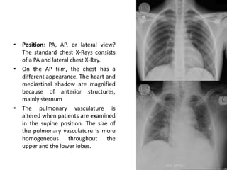
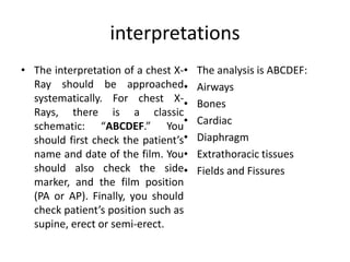
X ray chest
• There are 3 types of chest films;
• AnteroPosterior (AP)
• PosteroAnterior (PA)
• Lateral
• The ideal timing can be defined as the end of
inspiration, and the patient should hold his
breath at that time. Meanwhile, the X-ray tube
should be 180 cm away.
quality
• Assessing The Image Quality,
“RIPE” mnemonic is used;
Rotation, Inspiration, Position,
Exposure(Penetration).
• Rotation: The clavicles should
appear symmetrical and be seen
as equal length. The distance
between the thoracic spinal
process and clavicular heads
should be equal (Figure-1). If
there is a rotation, mediastinum
may look abnormal.
• Inspiration: On good inspiration, the
diaphragm should be seen at the level of
the 8th – 10th posterior rib or 5th – 6th
anterior rib.
• Position: PA, AP, or lateral view?
The standard chest X-Rays consists
of a PA and lateral chest X-Ray.
• On the AP film, the chest has a
different appearance. The heart and
mediastinal shadow are magnified
because of anterior structures,
mainly sternum
• The pulmonary vasculature is
altered when patients are examined
in the supine position. The size of
the pulmonary vasculature is more
homogeneous throughout the
upper and the lower lobes.
• Exposure / Penetration: Ideally,
you should be able to see the
heart, the blood vessels, and the
intervertebral spaces. Exposure
should be adequate if you are able
to see approximately T4 vertebra
and spinal process. If the film is
underexposed, you will not be able
to see them (Figure-6). If the film is
overexposed, details of bone
structures will be lost (Figure-7).
interpretations
• The interpretation of a chest X-
Ray should be approached
systematically. For chest X-
Rays, there is a classic
schematic: “ABCDEF.” You
should first check the patient’s
name and date of the film. You
should also check the side
marker, and the film position
(PA or AP). Finally, you should
check patient’s position such as
supine, erect or semi-erect.
• The analysis is ABCDEF:
• Airways
• Bones
• Cardiac
• Diaphragm
• Extrathoracic tissues
• Fields and Fissures
airway
bones
cardiac
diaphragm
Extrathorasic tissue (lungs)
Fluids and fissures
pathologies
Silhaete sign
Silhoutte can be applied to collapse
x ray chest.pptx
x ray chest.pptx
x ray chest.pptx
x ray chest.pptx
x ray chest.pptx
x ray chest.pptx
x ray chest.pptx
x ray chest.pptx

x ray chest.pptx

  • 1.
  • 2.
    • There are3 types of chest films; • AnteroPosterior (AP) • PosteroAnterior (PA) • Lateral • The ideal timing can be defined as the end of inspiration, and the patient should hold his breath at that time. Meanwhile, the X-ray tube should be 180 cm away.
  • 3.
    quality • Assessing TheImage Quality, “RIPE” mnemonic is used; Rotation, Inspiration, Position, Exposure(Penetration). • Rotation: The clavicles should appear symmetrical and be seen as equal length. The distance between the thoracic spinal process and clavicular heads should be equal (Figure-1). If there is a rotation, mediastinum may look abnormal.
  • 4.
    • Inspiration: Ongood inspiration, the diaphragm should be seen at the level of the 8th – 10th posterior rib or 5th – 6th anterior rib.
  • 5.
    • Position: PA,AP, or lateral view? The standard chest X-Rays consists of a PA and lateral chest X-Ray. • On the AP film, the chest has a different appearance. The heart and mediastinal shadow are magnified because of anterior structures, mainly sternum • The pulmonary vasculature is altered when patients are examined in the supine position. The size of the pulmonary vasculature is more homogeneous throughout the upper and the lower lobes.
  • 6.
    • Exposure /Penetration: Ideally, you should be able to see the heart, the blood vessels, and the intervertebral spaces. Exposure should be adequate if you are able to see approximately T4 vertebra and spinal process. If the film is underexposed, you will not be able to see them (Figure-6). If the film is overexposed, details of bone structures will be lost (Figure-7).
  • 7.
    interpretations • The interpretationof a chest X- Ray should be approached systematically. For chest X- Rays, there is a classic schematic: “ABCDEF.” You should first check the patient’s name and date of the film. You should also check the side marker, and the film position (PA or AP). Finally, you should check patient’s position such as supine, erect or semi-erect. • The analysis is ABCDEF: • Airways • Bones • Cardiac • Diaphragm • Extrathoracic tissues • Fields and Fissures
  • 8.
  • 9.
  • 10.
  • 11.
  • 12.
  • 13.
  • 14.
  • 23.
  • 25.
    Silhoutte can beapplied to collapse