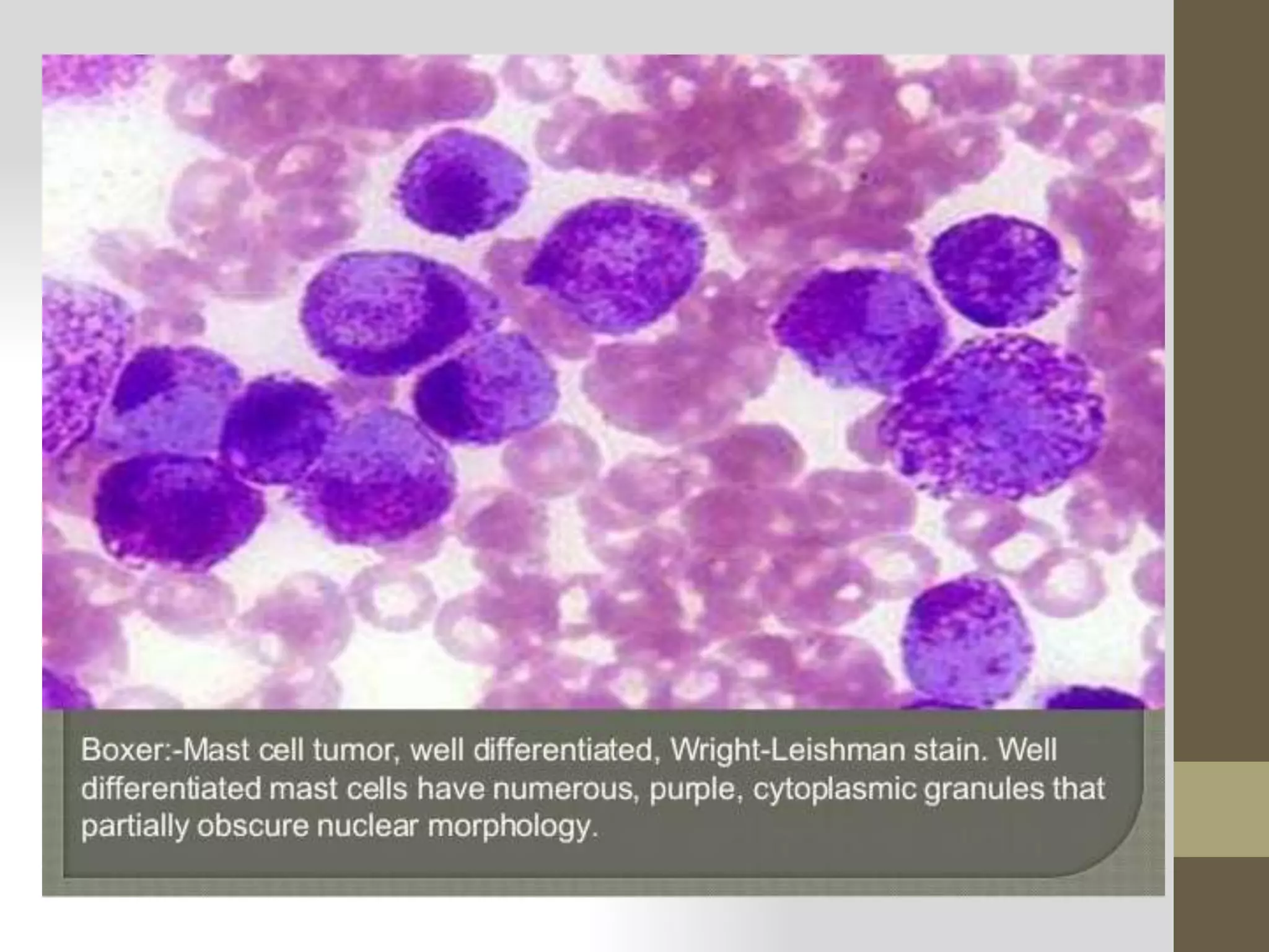
SlideShare a Scribd company logo
Preparation of blood smear
with different
staining method
Dr. Ankur Patel
Ankurvety001@gmail.com
Blood film ????
• A blood film or peripheral blood smear is a thin layer
of blood smeared on a microscope slide and then stained in such a
way to allow the various blood cells to be examined
microscopically.
Example of properly prepared thick and thin film blood smears
Aim of blood smear
• Blood films are usually examined to
investigate hematological problems (disorders of the blood)
and, occasionally, to look for parasites within the blood such
as malaria and filaria.
• Examination of thin blood films is important in the
investigation and management of anaemia, infections, and
other conditions which produce changes in the appearance
of blood cells and differential white cell count.
• A blood film report can provide rapidly and at low cost,
useful information about a patient’s condition.
The peripheral blood film (PBF) is of two types:
1. Thin blood film
2. Thick blood film
Three basic steps to make blood film:
1. Preparation of blood smear.
2. Fixation of blood smear.
3. Staining of blood smear.
Preparation of blood smear
1) THIN BLOOD FILM
Thin PBF can be prepared from anticoagulated blood
obtained by venepuncutre or from free flowing finger prick
blood by any of the following three techniques :
1. Slide method
2. Cover glass method
3. Spin method
Slide Method
Procedure
 Place a drop of blood in the centre of a clean glass
slide 1 to 2 cm from one end.
 Place another slide (spreader) with smooth edge at
an angle of 30-45⁰ near the drop of blood.
 Move the spreader backward so that it makes contact with
drop of blood.
 Then move the spreader forward rapidly over the slide.
 A thin peripheral blood film is thus prepared
 Dry it and stain it.
Cover Glass Method
Procedure
 Take a clean cover glass.
 Touch it on to the drop of a blood.
 Place it on another similar cover glass in crosswise direction
with side containing drop of blood facing down.
 Pull the cover glass quickly.
 Dry it and stain it.
 Mount it with a mountant, film side down on a clean glass
slide.
Spin Method
This is an automated method.
Procedure
 Place a drop of blood in the centre of a glass slide.
 Spin at a high speed in a special centrifuge, cytospin.
 Blood spreads uniformly.
 Dry it and stain it.
THICK BLOOD FILM
This is prepared for detecting blood parasites such as
malaria and microfilaria.
Procedure:
 Place a large drop of blood in the centre of a clean glass slide.
 Spread it in a circular area of 1.5 cm with the help of a
stick or end of another glass slide.
 Dry it
 Staing
The shape of blood film
Qualities of a Good Blood Film
i. It should not cover the entire surface of slide.
ii. It should have smooth and even appearance.
iii. It should be free from waves and holes.
iv.It should not have irregular tail.
Thethicknessofthespreadwhenpullingthesmear
isdeterminedby :
1. The angle of the spreader slide. (the greater the angle,
the thicker and shorter the smear).
2. Size of the blood drop.
3. Speed of spreading.
Notes:
1. If the haematocrit increased, the angel of the spreader
slide should be decreased.
2. If the haematocrit decreased, the angel of the
spreader slide should be increased.
large angle
low HCT
small angle
high HCT
Parts of a Thin Blood Film
A peripheral blood film consists of 3 parts :
1. Head i.e. the portion of blood film near the drop of blood.
2. Body i.e. the main part of the blood film.
3. Tail i.e. the tapering end of the blood film.
Commoncauseof a poorblood smear:
1. Drop of blood too large or too small
2. Spreader slide pushed across the slide in a jerky manner
3. Failure in keep the entire edge of the spreader slide against
the slide while making the smear
4. Failure in keep the spreader slide at a 30⁰ angel with the
slide
5. Failure to push the spreader slide completely across the
slide
6. Irregular spread with ridges and long tail: edges of spreader
dirty or chipped ; dusty slide
7. Holes in film – slide contaminated with fat or grease and air
bubbles
8. Cellular degenerative changes: delay in fixing inadequate
fixing time or methanol contaminated with water
A. Blood film with jagged tail made from a spreader with
achipped end.
B. Film which is too thick
C. Film which is too long, too wide, uneven thickness and
made on a greasy slide.
D. A well-made blood film.
Examples of unacceptablesmears
Biologic causes of a poor smear :
1. Cold agglutinin - RBCs will clump together.
Warm the blood at 37° C for 5 minutes, and then
remake the smear.
2. Lipemia - holes will appear in the smear.
There is nothing you can do to correct this.
3. Rouleaux - RBC’s will form into stacks resembling coins.
There is nothing you can do to correct this.
Fixation of blood smear
• To preserve the morphology of the cells, films must be
fixed as soon as possible after they have dried.
• It is important to prevent contact with water before
fixation is complete.
• Methyl alcohol (methanol) is the choice, although ethyl
alcohol ("absolute alcohol") can be used.
• Methylated spirit (95% ethanol) must not be used as it
contains water.
• To fix the films, place them in a covered staining jar or
tray containing the alcohol for 2-3 minutes. In humid
climates it might be necessary to replace the methanol 2-
3 times per day; the old portions can be used for storing
clean slides.
Different fixatives
Staining of blood smear
Variousstainsforperipheralbloodfilm:
 Romanowsky stains are universally employed for staining
of blood films. All Romanowsky combinations have two
essential ingredients i.e. methylene blue and eosin or
azure.
• Methylene blue is the basic dye and has affinity for acidic
component of the cell (i.e. nucleus) and eosin/azure is
the acidic dye and has affinity for basic component of cell
(i.e. cytoplasm).
• Most Romanowsky stains are prepared in methyl alcohol
so that they combine fixation and staining.
VariousstainsincludedunderRomanowskystainare
asunder:
1. Leishman stain
2. Giemsa stain
3. Wright stain
4. Field stain
5. Jenner stain
6. JSB stain
Leishman Stain:
Preparation
• Dissolve 0.2 g of powdered Leishman’s dye in 100 ml of
acetone-free methyl alcohol in a conical flask.
• Warm it to 50°C for half an hour with occasional shaking.
• Cool it and filter it.
Procedure for staining
• Pour Leishman’s stain dropwise (counting the drops) on the
slide and wait for 2 minutes. This allows fixation of the PBF in
methyl alcohol.
• Add double the quantity of buffered water dropwise over
the slide (i.e. double the number of drops).
• Mix by rocking for 8 minutes.
• Wash in water for 1 to 2 minutes.
• Dry in air and examine under oil immersion lens of the
microscope.
Metarubricyte (top left) and polychromatophilic erythrocyte (arrow) in
regenerative anemia. A monocyte (top right) also is present (Dog,
blood, Wright-Leishman stain).
Giemsa Stain
Preparation
• Stock solution of Giemsa stain is prepared by mixing 0.15 g of
Giemsa powder in 12.5 ml of glycerine and 12.5 ml of methyl
alcohol.
• Before use dissolve one volume of stock solution in nine volumes
of buffered water (dilution 1:9).
Procedure:
• Fill staining dish with staining solution
• Place thin film and thick films into the staining dish.
• Stain blood slides for 45 minutes
• Wash in water.
• Dry it and examine under oil immersion lens of the
microscope.
Caution:
Thick films need careful
rinsing!
(since they are not fixed
before staining)
rinsed
Mistake:
Thick films are not
properly! Blood is lost!
Trypanosoma org. In blood
Wright'sstain:
• Wright’s stain is used to differentiate nuclear and/or cytoplasmic
morphology of platelets, RBCs, WBCs, and parasites.
Procedure
• Thin blood films (only) – Dip Method
1. Dip air dried blood film in undiluted stain for 15 to 30 seconds
(double the staining time for bone marrow smears).
2. Decolorize the stained smears by immersion in distilled or deionized
water and air dry
3. Let air dry in a vertical position.
• Thin blood films (only) – Rack Method
1. Lay air dried slides on staining rack and flood with stain; stain for 10 to
15 seconds(double the staining time for bone marrow smears).
2.Add an equal volume of deionized/distilled water and stain for 10
seconds.
3.Rinse the slide by dipping in deionized/distilled water for 30 seconds.The
slide may also be rinsed by swishing or washing with deionized/distilled
water.
Thickbloodfilms(only):
1. Allow film to air dry thoroughly for several hours or overnight. Do
not dry films in an incubator or by heat, because this will fix the
blood and interfere with the lysing of the RBCs.
Note: If a rapid diagnosis of malaria is needed, thick films can
be made slightly thinner than usual, allowed to dry for 1 h, and
then stained.
2. Lake the thick film by immersing in distilled or deionized water for
10 min.
3. Allow the film to air dry thoroughly.
4. Fix air-dried film in absolute methanol for 30 seconds in a Coplin
jar containing absolute methanol.
5. Allow the film to air dry.
6. Dip air dried blood film in undiluted stain for 15 to 30 seconds
(double the staining time for bone marrow smears).
7. Decolorize the stained smears by immersion in distilled or
deionized water and air dry
8. Let air dry in a vertical position.
VenousbloodfromPekinduck(Wright'sstain)
Trypanosoma brucei_thin_wright-giemsa stain
Arrow indicatemalarial parasites
Fieldstain: (thin film)
Materials:
• Methanol (absolute)
• Field’s stain A und B
• Tube with water
• Staining dishes
• Filter paper
Procedure:
A. Fix thin film with methanol
for 1 min.
B. Dry microscopic slide on filter paper
C. Immerse slide in Field’s stain B (Eosin) for 5 seconds
D. Immediately wash with water
E. Immerse slide in Field‘s stain A (Methylene blue) for 10 sec
F.Immediately wash with water
G. Dry thin films
Materials:
• Methanol No!
• Field‘s stain A und B
• Tube with water
• Filter paper
Procedure:
A. Immerse thick film in Field‘s stain A (Methylene blue) for 3 sec
Do not forget:
• Thick films need to be
• haemolysed and are
• therefore not fixed with
• methanol
Fieldstain: (thick film)
B. Rinse immediately in tap water
C. Immerse thick film in Field’s stain B (Eosin) for 3 seconds
D. Then rinse immediately with tap water
E. Let the slide carefully dry
Jenner stain:
• The Jenner stain Solution is a mixture of several thiazin
dyes in a methanol solvent
• Ionic and noionic forces are involved in the binding of
these dyes
• The staining solution has anionic and cationic properties
• The negatively charged phosphoric acid groups of DNA
attract the purple polychromatic cationic dyes to the
nuclei
• The blue basophilic granules are stained by the
polychromatic cationic dyes
ImmersionStainingProtocol:
• Thoroughly dry blood or bone marrow smears
• Fix smears in absolute methanol for 15 seconds to 5 minutes
• Stain smears in Jenners Stain Solution for 2 minutes
• Stain in mixture of 50ml of Jenners Stain Solution, 75ml of
PH 6.6 Phosphate Buffer Solution and 175ml deionized
water for 5 minutes
• Rinse in standing deionized water for 1.5 minutes or rinse
briefly in running deionized water
• Air dry smears
• Examine smears under a microscope
HorizontalStainingProtocol:
• Place slide with thoroughly dried film in a horizontal
staining rack
• Flood smear with absolute methanol for 15-30 seconds and
then drain
• Flood smear with 1ml Jenners Stain Solution and let stand
for 3 minutes
• Add 1mL of pH 6.6 Phosphate Buffer solution and 1 ml
deionized water to smear and let stand for 45 seconds
• Rinse briefly with running deionized water
• Air dry and examine under a microscope
• Perform immuno chemical staining procedure according to
manufacturer.
J.S.B.Stain:
Materials and reagents required:
• Eosin yellow (water soluble)
• Methylene blue
• Potassium dichromate
• Di-sodium hydrogen phosphate (dihydrate)
• 1% sulphuric Acid.
• Round bottom flask (2 lit.)
• Healing mantle
• Distilled water
• Staining jars.
Staining technique:
• Prepare thin and thick smears from malaria cases on micro slides
• De-haemoglobinise the thick smear
• Fix the thin smear in methanol for few minutes
• Take 3 staining jars for J.S.B. I, J.S.B.II and tap water
• Dip the smears in J.S.B. II for few seconds and immediatedly wash
in water
• Drain the slides free of excess water
• Dip the smears in J.S.B.I for 30-40 seconds
• Wash well in water and dry
• Examine the smears under oil immersion
Staining of Thick Smear:
• It can be stained with any of the Romanowsky stains
• listed above except that before staining, the smear is
dehaemoglobinised by putting it in distilled water for 10
minute
Autostainers
• Currently, automatic staining machines are available
which enable a large batch of slides to be stained with a
uniform quality.
Jaswant Singh Battacharya (JSB) Stain for thick and thin films:
• This is the standard method used by the laboratories under
the National Malaria Eradication Programme in India
PrecautionsinStainingofperipheralbloodfilm
1) Dark blue blood film:
It can be due to overstaining, inadequate washing or
improper pH of the buffer. In this RBCs are blue, nuclear chromatin
is black,granules of the neutrophils are overstained and granules
of the eosinophils are blue or grey.
2) Light pink blood film:
In this RBCs are bright red, the nuclear chromatin is pale
blue and granules of the eosinophils are dark red. It can be due to
understaining, prolonged washing, mounting the film before
drying and improper pH of the buffer.
3) Precipitate on the blood film:
This could be due to inadequate filtration of the stain, dust
on the slide, drying during staining and inadequate washing.
TO ACIDIC SUITABLE TO BASIC
RETICULOCYTE COUNT
Prabin Shah BScMLT, MSc(Biochemistry)
RETICULOCYTES
🟔Reticulocytes are young , premature, non
nucleated red blood cells, contain reticular material
(RNA) that stain gray blue.
🟔Reticulum is present in newly released blood cells
for 1-2 days before the cell reach its full mature
state.
RETICULOCYTE STAINS
🟔Reticulocytes are visualized by supra-vital staining (such
as new methylene blue, Brilliant Cresyl Blue, Pure azure
blue) that precipitate the RNA and organelles, forming a
filamentous network of reticulum
🟔On Wright stain. the Reticulocyte appears
polychromatophilic or as a Macrocytic blue red cell.
PRINCIPLE
🟔 Whole blood is incubated with supra-vital
staining (new methylene blue). The vital stain
causes the ribosomal and residual RNA to
coprecipitate with the few remaining mitochondria
and ferritin masses in living young erythrocytes to
form dark-blue clusters and filaments (reticulum).
🟔 Smears of this mixture are then prepared and
examined. The number of reticulocytes in 1000
red blood cells is determined. This number is
divided by 10 to obtain the reticulocyte count in
percent.
SPECIMEN
 Whole blood that is anticoagulated with either EDTA
or heparin is suitable.
 Capillary blood drawn into heparinized tubes or
immediately mixed with stain may also be used.
 Red blood cells must still be living when the test is
performed therefore it is best to perform it promptly
after blood collection.
 Blood may be used up to 8 hours after collection.
 Stained smears retain their color for a prolonged
period of time.
REQUIREMENTS
1. Commercially prepared liquid new methylene blue
solution. It should be stored in a brown bottle. If
precipitate is a problem on the smear, the stain
should be filtered prior to use.
2. Microscope slides
3. Microscope
4. 10 x 75 mm test tubes
5. Pasteur pipets (with bulb if pipets are glass)
6. Capillary tubes
7. Miller ocular (if available)
PROCEDURE
Preparation of smears
1. Add 3-4 drops of new methylene blue solution to 3-4
drops of thoroughly mixed EDTA anticoagulated blood to
a glass 10 x 75 mm test tube.
2. Mix the contents by gently shaking and allow to incubate
at room temperature for a minimum of 10 minutes.
3. At the end of 10 minutes, gently mix the blood/stain
solution.
4. Using a capillary tube, place a drop of the mixture on
each of three slides near the frosted edge as you would
when making a peripheral smear.
5. Using the wedge smear technique, make acceptable
smears not too thick or thin.
6. Label the slides with patient name, ID# and date.
7. Allow to air dry. (Do not blow to hasten to drying.)
COUNTING PROCEDURE
 Place the first slide on the microscope stage and,
using the low power objective (10x), find an area in
the thin portion of the smear in which the red cells are
evenly distributed and are not touching each other.
 Carefully change to the oil immersion objective (100x)
and further located an area in which there are
approximately 100 red cells per oil immersion field.
 Do this by finding a field where the cells are evenly
distributed and mentally divide the field into 4
quadrants. Count the cells in 1 quadrant. If there are
about 25, you are in a good area. There will be a lot of
open space between the red cells.
2. Be sure to count all cells that contain a blue-staining
filament or at least 2 or more discrete blue aggregates
of reticulum in the erythrocyte.
3. Count 1000 red cells in consecutive oil immersion
fields. Record the number reticulocytes seen.
4. You may count 500 cells on two slides each. They
should agree within ± 15% of each other. If they do
not, repeat the reticulocyte count on the third smear.
5. Calculate the result as follow:
%Retix= Reticulocyte counted/10
MILLER DISCS METHOD
1. Use a 100x objective and a 10x ocular secured with a Miller
disc.
 The Miller disc imposes two squares (one 9 times the area of the
other) onto the field of view.
 Find a suitable area of the smear. A good area will show 3-10
RBCs in the smaller square of the Miller disc.
2. Count the reticulocytes within the entire large square
including those that are touching the lines on the left and
bottom of the ruled area. Count RBCs in the smaller square
whether they contain stained RNA or not. A retic in the
smaller square should be counted as an RBC and a retic.
Record RBC # counted and retic # counted separately.
3. Continue counting until a minimum of 111 RBCs have been
observed (usually 15-20 fields). This would correspond to
999 RBCs counted with the standard procedure.
The Miller disc may be placed in one of the ocular
lenses to aid in the counting of the reticulocytes.
%Reticulocyte= total reticulocyte in square A*100
total RBC in square B*9
REFERENCE VALUES
 RBCs life span ~ 100 days, ± 20 days
 Reticulocyte ~ 1 day in peripheral blood, Then
the B.M. replaces approximately 1 % of the adult
red blood cells every day.
 Normal value :
 0.5 to 1.5/100 red blood cells (or
, 0.5 to
1.5%)
 Absolute count :25 to 75 X 109/L
 Newborn (0-2 weeks):2.5-6.0%
 Normal Reticulocyte I
ndex :1-3%
RETICULOCYTE
REPORTING PATTERN
🟔Absolute Reticulocyte Count (ARC): is the actual
number of reticulocytes in 1L of whole blood. This is
calculated by multiplying the reticulocytes % by the
RBCs count and dividing by 100.
🟔Corrected Reticulocyte Count is calculated based
on a normal hematocrit of 45%.
🟔 Reticulocyte Production Index (RPI) = Corrected retic
count (%) / # Days (Maturation time)
CORRECTED RETICULOCYTE COUNT
 In states of anemia, the reticulocyte percentage is
not a true reflection of reticulocyte production. A
correction factor must be used so as not to
overestimate marrow production, because each
reticulocyte is released into whole blood containing
few RBCs - a low hematocrit (Hct) - thus relatively
increasing the percentage.
 The corrected reticulocyte count my be calculated
by the following formula:
RETICULOCYTE PRODUCTION INDEX(RPI)
 The RI is a measurement for reticulocytes when anemia is
present
 Estimating RBC production by using the corrected
reticulocyte count may yield erroneously high values in
patients when there is a premature release of younger
reticulocytes from the marrow (owing to increased
erythropoietin stimulation).
 The premature reticulocytes are called “stress or shift”
reticulocytes. These result when the reticulocytes of the
bone marrow pool are shifted to the circulation pool to
compensate for anemia.
 The younger stress reticulocytes present with more
filamentous reticulum. The mature reticulocyte may present
with granular dots representing reticulum. Normally,
reticulocytes lose their reticulum within 24 to 27 hours after
entering the peripheral circulation.
 The premature stress retics have increased reticulum
and require 2 to 2.5 days to lose their reticulum,
resulting in a longer peripheral blood maturation time.
 The peripheral blood smear should be reviewed
carefully for the presence of many polychromatophilic
macrocytes, thus indicating stress reticulocytes and
the need for correction for both the RBC count and the
presence of stress reticulocytes. The value obtained is
called the reticulocyte production index (RPI).
Maturation Time Hematocrit %
1 day 45
1.5 day 35
2 day 24
3 day 15
INTERPRETATION
 The Reticulocyte count is an important diagnostic
tool: The number of Reticulocytes is a good indicator of
bone marrow activity, because it represents recent
production. It is used to differentiate anemia's caused by
bone marrow failure from those caused by hemorrhage
or hemolysis.
 It used also to check the effectiveness of treatment in
prenicious anemia and folate and iron deficiency.
 To assess the recovery of bone marrow function in
aplastic anemia and to determine the effects of
radioactive substance on exposed workers.
 A low reticulocyte count may mean a need for a bone
marrow biopsy. This can tell if is a problem with how new
reticulocytes are made by the bone marrow.
 Reticulocytosis (Increased RBC Production)
⚫ Reticulocyte Index >3%, Reticulocyte Count >1.5%
1. Acute blood loss or hemorrhage
2. Post-Splenectomy
3. Acute Hemolytic Anemia (Microangiopathic
Anemia)
4. Hemoglobinopathy
 Sickle Cell Anemia
 Thalassemia major
5. Post-Anemia Treatment
 Folate Supplementation
 Iron Supplementation
 Vitamin B12 Supplementation
 Reticulocytopenia (Decreased RBC Production)
⚫ Reticulocyte Index <1%, Reticulocyte Count <0.5%
1. Aplastic Anemia
2. Bone Marrow infiltrate
3. Bone Marrow suppression or failure
1. Sepsis
2. Chemotherapy or radiotherapy
4. Disordered RBC maturation
1. Iron Deficiency Anemia
2. Vitamin B12 Deficiency
3. Folate Deficiency
4. Sideroblastic Anemia
5. Anemia of Chronic Disease
6. Hypothyroidism
5. Blood transfusion
6. Liver disease
FACTORS AFFECTING THE TEST
Reasons you may not be able to have the
test or why the results may not be helpful
include:
⚫ Taking medicines, such as levodopa,
corticotropin, azathioprine (Imuran),
chloramphenicol (Chloromycetin), dactinomycin
(Cosmegen), medicines to reduce a fever,
medicines to treat malaria, and methotrexate and
other cancer chemotherapy medicines.
⚫ Getting radiation therapy
⚫ Taking sulfonamide antibiotics (such as Bactrim or
Septra)
⚫ Being pregnant
⚫ Having a recent blood transfusion
ERROR SOURCES
1. A refractile appearance of erythrocytes should not be
confused with reticulocytes.
2. Filtration of the stain is necessary when precipitated
material is present which can resemble a reticulocyte.
3. Erythrocyte inclusions should not be mistaken for
Reticulocytes.
 Howell-Jolly bodies appear as one or sometime two, deep-purple
dense structures.
 Heinz bodies stain a light blue-green and are usually present at the
edge of the erythrocyte.
 Pappenheimer bodies are more often confused with reticulocytes
and are the most difficult to distinguish. These purple-staining iron
deposits generally appear as several granules in a small cluster. If
Pappenheimer bodies are suspected, stain with Wright-Giemsa to
verify their presence. Hemoglobin H inclusions will appear as multiple
small dots in every cell.
4. Falsely decreased reticulocyte counts can result
from under staining the blood with new methylene
blue. Be sure the stain/blood mixture incubates the
full 10 minutes.
5. High glucose levels can cause reticulocytes to stain
poorly.
6. There is high degree of inaccuracy in the manual
reticulocyte count owing to error (± 2%) in low
counts and ± 7% in high counts) and a lack of
reproducibility because of the inaccuracy of the
blood film. This inaccuracy has been overcome by
the use of automated instruments using flow
cytometry.
7. If no reticulocytes are observed after scanning at
least two slides, report “none seen”.

More Related Content

Similar to MergeResult_2023_06_19_02_48_32 (1).pptx

HEMATOLOGY PRACTICAL NOTES.pptx
HEMATOLOGY PRACTICAL NOTES.pptxHEMATOLOGY PRACTICAL NOTES.pptx
HEMATOLOGY PRACTICAL NOTES.pptx
AmosiRichard
 
pbs-fields staining-converted"""""""...pdf
pbs-fields staining-converted"""""""...pdfpbs-fields staining-converted"""""""...pdf
pbs-fields staining-converted"""""""...pdf
r8fdq7w2m9
 
Blood examination
Blood examinationBlood examination
Blood examination
Mathew Tut Moses Kol mose
 
Blood film for malaria
Blood film for malariaBlood film for malaria
Blood film for malaria
Ibrahim Hussein
 
bloodfilmformalaria-150420154949-conversion-gate01.pdf
bloodfilmformalaria-150420154949-conversion-gate01.pdfbloodfilmformalaria-150420154949-conversion-gate01.pdf
bloodfilmformalaria-150420154949-conversion-gate01.pdf
jnrkwesi
 
LABORATORY DIAGNOSIS OF malaria.pptx
LABORATORY DIAGNOSIS OF malaria.pptxLABORATORY DIAGNOSIS OF malaria.pptx
LABORATORY DIAGNOSIS OF malaria.pptx
Ananya147165
 
Peripheral Blood Smear Examination
Peripheral Blood Smear ExaminationPeripheral Blood Smear Examination
Peripheral Blood Smear Examination
Dr Snehal Kosale
 
romanowskystains-180521083448 (1).pptx
romanowskystains-180521083448 (1).pptxromanowskystains-180521083448 (1).pptx
romanowskystains-180521083448 (1).pptx
rohitshrivastava97
 
Peripheral blood smear [autosaved]
Peripheral blood smear [autosaved]Peripheral blood smear [autosaved]
Peripheral blood smear [autosaved]
Ankit Singh
 
Artefacts in hemat part 2
Artefacts in hemat part 2Artefacts in hemat part 2
Artefacts in hemat part 2
DrShrikant Sonune
 
Module 4-Speciemn collection.pptxghjfffghh
Module 4-Speciemn collection.pptxghjfffghhModule 4-Speciemn collection.pptxghjfffghh
Module 4-Speciemn collection.pptxghjfffghh
Yasinmohammed3571@gmail.com
 
Tissue processing 2012
Tissue processing 2012Tissue processing 2012
Tissue processing 2012Dhiraj Shukla
 
Smear preparation for differential leucocyte count
Smear preparation for differential leucocyte countSmear preparation for differential leucocyte count
Smear preparation for differential leucocyte count
Nidhi Jodhwani
 
Pbs Prepararion Using Wrights Stain
Pbs Prepararion Using Wrights StainPbs Prepararion Using Wrights Stain
Pbs Prepararion Using Wrights Stain
Tshering Namgyal Wangdi
 
Blood Smear preparation.pdf
Blood Smear preparation.pdfBlood Smear preparation.pdf
Blood Smear preparation.pdf
ItsMe468321
 
DLC practical ppt
DLC  practical pptDLC  practical ppt
DLC practical ppt
Reena Gollapalli
 

Similar to MergeResult_2023_06_19_02_48_32 (1).pptx (20)

HEMATOLOGY PRACTICAL NOTES.pptx
HEMATOLOGY PRACTICAL NOTES.pptxHEMATOLOGY PRACTICAL NOTES.pptx
HEMATOLOGY PRACTICAL NOTES.pptx
 
STAINING
STAINING STAINING
STAINING
 
pbs-fields staining-converted"""""""...pdf
pbs-fields staining-converted"""""""...pdfpbs-fields staining-converted"""""""...pdf
pbs-fields staining-converted"""""""...pdf
 
Blood examination
Blood examinationBlood examination
Blood examination
 
Blood film for malaria
Blood film for malariaBlood film for malaria
Blood film for malaria
 
bloodfilmformalaria-150420154949-conversion-gate01.pdf
bloodfilmformalaria-150420154949-conversion-gate01.pdfbloodfilmformalaria-150420154949-conversion-gate01.pdf
bloodfilmformalaria-150420154949-conversion-gate01.pdf
 
LABORATORY DIAGNOSIS OF malaria.pptx
LABORATORY DIAGNOSIS OF malaria.pptxLABORATORY DIAGNOSIS OF malaria.pptx
LABORATORY DIAGNOSIS OF malaria.pptx
 
stains
stainsstains
stains
 
Lecture 3.stains
Lecture 3.stainsLecture 3.stains
Lecture 3.stains
 
Peripheral Blood Smear Examination
Peripheral Blood Smear ExaminationPeripheral Blood Smear Examination
Peripheral Blood Smear Examination
 
romanowskystains-180521083448 (1).pptx
romanowskystains-180521083448 (1).pptxromanowskystains-180521083448 (1).pptx
romanowskystains-180521083448 (1).pptx
 
Romanowsky stains
Romanowsky stainsRomanowsky stains
Romanowsky stains
 
Peripheral blood smear [autosaved]
Peripheral blood smear [autosaved]Peripheral blood smear [autosaved]
Peripheral blood smear [autosaved]
 
Artefacts in hemat part 2
Artefacts in hemat part 2Artefacts in hemat part 2
Artefacts in hemat part 2
 
Module 4-Speciemn collection.pptxghjfffghh
Module 4-Speciemn collection.pptxghjfffghhModule 4-Speciemn collection.pptxghjfffghh
Module 4-Speciemn collection.pptxghjfffghh
 
Tissue processing 2012
Tissue processing 2012Tissue processing 2012
Tissue processing 2012
 
Smear preparation for differential leucocyte count
Smear preparation for differential leucocyte countSmear preparation for differential leucocyte count
Smear preparation for differential leucocyte count
 
Pbs Prepararion Using Wrights Stain
Pbs Prepararion Using Wrights StainPbs Prepararion Using Wrights Stain
Pbs Prepararion Using Wrights Stain
 
Blood Smear preparation.pdf
Blood Smear preparation.pdfBlood Smear preparation.pdf
Blood Smear preparation.pdf
 
DLC practical ppt
DLC  practical pptDLC  practical ppt
DLC practical ppt
 

More from vandana thakur

nursing class cellularadaptation and apoptosis.pptx
nursing class cellularadaptation and apoptosis.pptxnursing class cellularadaptation and apoptosis.pptx
nursing class cellularadaptation and apoptosis.pptx
vandana thakur
 
Nursing class lecture cell injury 2nd class.pptx
Nursing class lecture  cell injury 2nd class.pptxNursing class lecture  cell injury 2nd class.pptx
Nursing class lecture cell injury 2nd class.pptx
vandana thakur
 
poster on carcinosarcoma of gall bladder and liver.pptx
poster on carcinosarcoma of gall bladder and liver.pptxposter on carcinosarcoma of gall bladder and liver.pptx
poster on carcinosarcoma of gall bladder and liver.pptx
vandana thakur
 
lectureoncellinjury and adaptation-170214073507.pptx
lectureoncellinjury and adaptation-170214073507.pptxlectureoncellinjury and adaptation-170214073507.pptx
lectureoncellinjury and adaptation-170214073507.pptx
vandana thakur
 
chronic granulomatous lesion of colon along with carcinosarcomapptx
chronic granulomatous lesion of colon along with carcinosarcomapptxchronic granulomatous lesion of colon along with carcinosarcomapptx
chronic granulomatous lesion of colon along with carcinosarcomapptx
vandana thakur
 
acuteleukemiacomplt-161017163342 (1).pptx
acuteleukemiacomplt-161017163342 (1).pptxacuteleukemiacomplt-161017163342 (1).pptx
acuteleukemiacomplt-161017163342 (1).pptx
vandana thakur
 
blood tests.pptx
blood tests.pptxblood tests.pptx
blood tests.pptx
vandana thakur
 
3_2020_01_19!04_20_03_PM.pptx
3_2020_01_19!04_20_03_PM.pptx3_2020_01_19!04_20_03_PM.pptx
3_2020_01_19!04_20_03_PM.pptx
vandana thakur
 
ch7neoplasia-.pptx
ch7neoplasia-.pptxch7neoplasia-.pptx
ch7neoplasia-.pptx
vandana thakur
 
AIDS (1).pptx
AIDS (1).pptxAIDS (1).pptx
AIDS (1).pptx
vandana thakur
 
lectureoncellinjury-170214073507.pptx
lectureoncellinjury-170214073507.pptxlectureoncellinjury-170214073507.pptx
lectureoncellinjury-170214073507.pptx
vandana thakur
 
Nutritional anemias
Nutritional anemiasNutritional anemias
Nutritional anemias
vandana thakur
 

More from vandana thakur (13)

nursing class cellularadaptation and apoptosis.pptx
nursing class cellularadaptation and apoptosis.pptxnursing class cellularadaptation and apoptosis.pptx
nursing class cellularadaptation and apoptosis.pptx
 
Nursing class lecture cell injury 2nd class.pptx
Nursing class lecture  cell injury 2nd class.pptxNursing class lecture  cell injury 2nd class.pptx
Nursing class lecture cell injury 2nd class.pptx
 
poster on carcinosarcoma of gall bladder and liver.pptx
poster on carcinosarcoma of gall bladder and liver.pptxposter on carcinosarcoma of gall bladder and liver.pptx
poster on carcinosarcoma of gall bladder and liver.pptx
 
lectureoncellinjury and adaptation-170214073507.pptx
lectureoncellinjury and adaptation-170214073507.pptxlectureoncellinjury and adaptation-170214073507.pptx
lectureoncellinjury and adaptation-170214073507.pptx
 
chronic granulomatous lesion of colon along with carcinosarcomapptx
chronic granulomatous lesion of colon along with carcinosarcomapptxchronic granulomatous lesion of colon along with carcinosarcomapptx
chronic granulomatous lesion of colon along with carcinosarcomapptx
 
acuteleukemiacomplt-161017163342 (1).pptx
acuteleukemiacomplt-161017163342 (1).pptxacuteleukemiacomplt-161017163342 (1).pptx
acuteleukemiacomplt-161017163342 (1).pptx
 
blood tests.pptx
blood tests.pptxblood tests.pptx
blood tests.pptx
 
3_2020_01_19!04_20_03_PM.pptx
3_2020_01_19!04_20_03_PM.pptx3_2020_01_19!04_20_03_PM.pptx
3_2020_01_19!04_20_03_PM.pptx
 
ch7neoplasia-.pptx
ch7neoplasia-.pptxch7neoplasia-.pptx
ch7neoplasia-.pptx
 
CLASS 3 CI.pptx
CLASS 3 CI.pptxCLASS 3 CI.pptx
CLASS 3 CI.pptx
 
AIDS (1).pptx
AIDS (1).pptxAIDS (1).pptx
AIDS (1).pptx
 
lectureoncellinjury-170214073507.pptx
lectureoncellinjury-170214073507.pptxlectureoncellinjury-170214073507.pptx
lectureoncellinjury-170214073507.pptx
 
Nutritional anemias
Nutritional anemiasNutritional anemias
Nutritional anemias
 

Recently uploaded

5 Warning Signs Your BMW's Intelligent Battery Sensor Needs Attention
5 Warning Signs Your BMW's Intelligent Battery Sensor Needs Attention5 Warning Signs Your BMW's Intelligent Battery Sensor Needs Attention
5 Warning Signs Your BMW's Intelligent Battery Sensor Needs Attention
Bertini's German Motors
 
Antique Plastic Traders Company Profile
Antique Plastic Traders Company ProfileAntique Plastic Traders Company Profile
Antique Plastic Traders Company Profile
Antique Plastic Traders
 
gtyccccccccccccccccccccccccccccccccccccccccccccccccccccccc
gtycccccccccccccccccccccccccccccccccccccccccccccccccccccccgtyccccccccccccccccccccccccccccccccccccccccccccccccccccccc
gtyccccccccccccccccccccccccccccccccccccccccccccccccccccccc
4thzenzstar
 
What Does the PARKTRONIC Inoperative, See Owner's Manual Message Mean for You...
What Does the PARKTRONIC Inoperative, See Owner's Manual Message Mean for You...What Does the PARKTRONIC Inoperative, See Owner's Manual Message Mean for You...
What Does the PARKTRONIC Inoperative, See Owner's Manual Message Mean for You...
Autohaus Service and Sales
 
Tyre Industrymarket overview with examples of CEAT
Tyre Industrymarket overview with examples of CEATTyre Industrymarket overview with examples of CEAT
Tyre Industrymarket overview with examples of CEAT
kshamashah95
 
Statistics5,c.xz,c.;c.;d.c;d;ssssss.pptx
Statistics5,c.xz,c.;c.;d.c;d;ssssss.pptxStatistics5,c.xz,c.;c.;d.c;d;ssssss.pptx
Statistics5,c.xz,c.;c.;d.c;d;ssssss.pptx
coc7987515756
 
Empowering Limpopo Entrepreneurs Consulting SMEs.pptx
Empowering Limpopo Entrepreneurs  Consulting SMEs.pptxEmpowering Limpopo Entrepreneurs  Consulting SMEs.pptx
Empowering Limpopo Entrepreneurs Consulting SMEs.pptx
Precious Mvulane CA (SA),RA
 
What Exactly Is The Common Rail Direct Injection System & How Does It Work
What Exactly Is The Common Rail Direct Injection System & How Does It WorkWhat Exactly Is The Common Rail Direct Injection System & How Does It Work
What Exactly Is The Common Rail Direct Injection System & How Does It Work
Motor Cars International
 
Wondering if Your Mercedes EIS is at Fault Here’s How to Tell
Wondering if Your Mercedes EIS is at Fault Here’s How to TellWondering if Your Mercedes EIS is at Fault Here’s How to Tell
Wondering if Your Mercedes EIS is at Fault Here’s How to Tell
Vic Auto Collision & Repair
 
Core technology of Hyundai Motor Group's EV platform 'E-GMP'
Core technology of Hyundai Motor Group's EV platform 'E-GMP'Core technology of Hyundai Motor Group's EV platform 'E-GMP'
Core technology of Hyundai Motor Group's EV platform 'E-GMP'
Hyundai Motor Group
 
一比一原版(AIS毕业证)奥克兰商学院毕业证成绩单如何办理
一比一原版(AIS毕业证)奥克兰商学院毕业证成绩单如何办理一比一原版(AIS毕业证)奥克兰商学院毕业证成绩单如何办理
一比一原版(AIS毕业证)奥克兰商学院毕业证成绩单如何办理
eygkup
 
Why Isn't Your BMW X5's Comfort Access Functioning Properly Find Out Here
Why Isn't Your BMW X5's Comfort Access Functioning Properly Find Out HereWhy Isn't Your BMW X5's Comfort Access Functioning Properly Find Out Here
Why Isn't Your BMW X5's Comfort Access Functioning Properly Find Out Here
Masters European & Gapanese Auto Repair
 
Digital Fleet Management - Why Your Business Need It?
Digital Fleet Management - Why Your Business Need It?Digital Fleet Management - Why Your Business Need It?
Digital Fleet Management - Why Your Business Need It?
jennifermiller8137
 
What Causes 'Trans Failsafe Prog' to Trigger in BMW X5
What Causes 'Trans Failsafe Prog' to Trigger in BMW X5What Causes 'Trans Failsafe Prog' to Trigger in BMW X5
What Causes 'Trans Failsafe Prog' to Trigger in BMW X5
European Service Center
 
Why Is Your BMW X3 Hood Not Responding To Release Commands
Why Is Your BMW X3 Hood Not Responding To Release CommandsWhy Is Your BMW X3 Hood Not Responding To Release Commands
Why Is Your BMW X3 Hood Not Responding To Release Commands
Dart Auto
 
Ec460b lc Excavator Volvo Service Repair.pdf
Ec460b lc Excavator Volvo Service Repair.pdfEc460b lc Excavator Volvo Service Repair.pdf
Ec460b lc Excavator Volvo Service Repair.pdf
Excavator
 
Things to remember while upgrading the brakes of your car
Things to remember while upgrading the brakes of your carThings to remember while upgrading the brakes of your car
Things to remember while upgrading the brakes of your car
jennifermiller8137
 
Ec330B Lc Excavator Volvo Service Repair.pdf
Ec330B Lc Excavator Volvo Service Repair.pdfEc330B Lc Excavator Volvo Service Repair.pdf
Ec330B Lc Excavator Volvo Service Repair.pdf
Excavator
 
What do the symbols on vehicle dashboard mean?
What do the symbols on vehicle dashboard mean?What do the symbols on vehicle dashboard mean?
What do the symbols on vehicle dashboard mean?
Hyundai Motor Group
 
Bài tập - Tiếng anh 11 Global Success UNIT 1 - Bản HS.doc
Bài tập - Tiếng anh 11 Global Success UNIT 1 - Bản HS.docBài tập - Tiếng anh 11 Global Success UNIT 1 - Bản HS.doc
Bài tập - Tiếng anh 11 Global Success UNIT 1 - Bản HS.doc
daothibichhang1
 

Recently uploaded (20)

5 Warning Signs Your BMW's Intelligent Battery Sensor Needs Attention
5 Warning Signs Your BMW's Intelligent Battery Sensor Needs Attention5 Warning Signs Your BMW's Intelligent Battery Sensor Needs Attention
5 Warning Signs Your BMW's Intelligent Battery Sensor Needs Attention
 
Antique Plastic Traders Company Profile
Antique Plastic Traders Company ProfileAntique Plastic Traders Company Profile
Antique Plastic Traders Company Profile
 
gtyccccccccccccccccccccccccccccccccccccccccccccccccccccccc
gtycccccccccccccccccccccccccccccccccccccccccccccccccccccccgtyccccccccccccccccccccccccccccccccccccccccccccccccccccccc
gtyccccccccccccccccccccccccccccccccccccccccccccccccccccccc
 
What Does the PARKTRONIC Inoperative, See Owner's Manual Message Mean for You...
What Does the PARKTRONIC Inoperative, See Owner's Manual Message Mean for You...What Does the PARKTRONIC Inoperative, See Owner's Manual Message Mean for You...
What Does the PARKTRONIC Inoperative, See Owner's Manual Message Mean for You...
 
Tyre Industrymarket overview with examples of CEAT
Tyre Industrymarket overview with examples of CEATTyre Industrymarket overview with examples of CEAT
Tyre Industrymarket overview with examples of CEAT
 
Statistics5,c.xz,c.;c.;d.c;d;ssssss.pptx
Statistics5,c.xz,c.;c.;d.c;d;ssssss.pptxStatistics5,c.xz,c.;c.;d.c;d;ssssss.pptx
Statistics5,c.xz,c.;c.;d.c;d;ssssss.pptx
 
Empowering Limpopo Entrepreneurs Consulting SMEs.pptx
Empowering Limpopo Entrepreneurs  Consulting SMEs.pptxEmpowering Limpopo Entrepreneurs  Consulting SMEs.pptx
Empowering Limpopo Entrepreneurs Consulting SMEs.pptx
 
What Exactly Is The Common Rail Direct Injection System & How Does It Work
What Exactly Is The Common Rail Direct Injection System & How Does It WorkWhat Exactly Is The Common Rail Direct Injection System & How Does It Work
What Exactly Is The Common Rail Direct Injection System & How Does It Work
 
Wondering if Your Mercedes EIS is at Fault Here’s How to Tell
Wondering if Your Mercedes EIS is at Fault Here’s How to TellWondering if Your Mercedes EIS is at Fault Here’s How to Tell
Wondering if Your Mercedes EIS is at Fault Here’s How to Tell
 
Core technology of Hyundai Motor Group's EV platform 'E-GMP'
Core technology of Hyundai Motor Group's EV platform 'E-GMP'Core technology of Hyundai Motor Group's EV platform 'E-GMP'
Core technology of Hyundai Motor Group's EV platform 'E-GMP'
 
一比一原版(AIS毕业证)奥克兰商学院毕业证成绩单如何办理
一比一原版(AIS毕业证)奥克兰商学院毕业证成绩单如何办理一比一原版(AIS毕业证)奥克兰商学院毕业证成绩单如何办理
一比一原版(AIS毕业证)奥克兰商学院毕业证成绩单如何办理
 
Why Isn't Your BMW X5's Comfort Access Functioning Properly Find Out Here
Why Isn't Your BMW X5's Comfort Access Functioning Properly Find Out HereWhy Isn't Your BMW X5's Comfort Access Functioning Properly Find Out Here
Why Isn't Your BMW X5's Comfort Access Functioning Properly Find Out Here
 
Digital Fleet Management - Why Your Business Need It?
Digital Fleet Management - Why Your Business Need It?Digital Fleet Management - Why Your Business Need It?
Digital Fleet Management - Why Your Business Need It?
 
What Causes 'Trans Failsafe Prog' to Trigger in BMW X5
What Causes 'Trans Failsafe Prog' to Trigger in BMW X5What Causes 'Trans Failsafe Prog' to Trigger in BMW X5
What Causes 'Trans Failsafe Prog' to Trigger in BMW X5
 
Why Is Your BMW X3 Hood Not Responding To Release Commands
Why Is Your BMW X3 Hood Not Responding To Release CommandsWhy Is Your BMW X3 Hood Not Responding To Release Commands
Why Is Your BMW X3 Hood Not Responding To Release Commands
 
Ec460b lc Excavator Volvo Service Repair.pdf
Ec460b lc Excavator Volvo Service Repair.pdfEc460b lc Excavator Volvo Service Repair.pdf
Ec460b lc Excavator Volvo Service Repair.pdf
 
Things to remember while upgrading the brakes of your car
Things to remember while upgrading the brakes of your carThings to remember while upgrading the brakes of your car
Things to remember while upgrading the brakes of your car
 
Ec330B Lc Excavator Volvo Service Repair.pdf
Ec330B Lc Excavator Volvo Service Repair.pdfEc330B Lc Excavator Volvo Service Repair.pdf
Ec330B Lc Excavator Volvo Service Repair.pdf
 
What do the symbols on vehicle dashboard mean?
What do the symbols on vehicle dashboard mean?What do the symbols on vehicle dashboard mean?
What do the symbols on vehicle dashboard mean?
 
Bài tập - Tiếng anh 11 Global Success UNIT 1 - Bản HS.doc
Bài tập - Tiếng anh 11 Global Success UNIT 1 - Bản HS.docBài tập - Tiếng anh 11 Global Success UNIT 1 - Bản HS.doc
Bài tập - Tiếng anh 11 Global Success UNIT 1 - Bản HS.doc
 

MergeResult_2023_06_19_02_48_32 (1).pptx

  • 1. Preparation of blood smear with different staining method Dr. Ankur Patel Ankurvety001@gmail.com
  • 2. Blood film ???? • A blood film or peripheral blood smear is a thin layer of blood smeared on a microscope slide and then stained in such a way to allow the various blood cells to be examined microscopically. Example of properly prepared thick and thin film blood smears
  • 3. Aim of blood smear • Blood films are usually examined to investigate hematological problems (disorders of the blood) and, occasionally, to look for parasites within the blood such as malaria and filaria. • Examination of thin blood films is important in the investigation and management of anaemia, infections, and other conditions which produce changes in the appearance of blood cells and differential white cell count. • A blood film report can provide rapidly and at low cost, useful information about a patient’s condition.
  • 4. The peripheral blood film (PBF) is of two types: 1. Thin blood film 2. Thick blood film Three basic steps to make blood film: 1. Preparation of blood smear. 2. Fixation of blood smear. 3. Staining of blood smear.
  • 6. 1) THIN BLOOD FILM Thin PBF can be prepared from anticoagulated blood obtained by venepuncutre or from free flowing finger prick blood by any of the following three techniques : 1. Slide method 2. Cover glass method 3. Spin method
  • 7. Slide Method Procedure  Place a drop of blood in the centre of a clean glass slide 1 to 2 cm from one end.  Place another slide (spreader) with smooth edge at an angle of 30-45⁰ near the drop of blood.  Move the spreader backward so that it makes contact with drop of blood.  Then move the spreader forward rapidly over the slide.  A thin peripheral blood film is thus prepared  Dry it and stain it.
  • 8. Cover Glass Method Procedure  Take a clean cover glass.  Touch it on to the drop of a blood.  Place it on another similar cover glass in crosswise direction with side containing drop of blood facing down.  Pull the cover glass quickly.  Dry it and stain it.  Mount it with a mountant, film side down on a clean glass slide.
  • 9. Spin Method This is an automated method. Procedure  Place a drop of blood in the centre of a glass slide.  Spin at a high speed in a special centrifuge, cytospin.  Blood spreads uniformly.  Dry it and stain it.
  • 10. THICK BLOOD FILM This is prepared for detecting blood parasites such as malaria and microfilaria. Procedure:  Place a large drop of blood in the centre of a clean glass slide.  Spread it in a circular area of 1.5 cm with the help of a stick or end of another glass slide.  Dry it  Staing
  • 11.
  • 12. The shape of blood film
  • 13. Qualities of a Good Blood Film i. It should not cover the entire surface of slide. ii. It should have smooth and even appearance. iii. It should be free from waves and holes. iv.It should not have irregular tail.
  • 14. Thethicknessofthespreadwhenpullingthesmear isdeterminedby : 1. The angle of the spreader slide. (the greater the angle, the thicker and shorter the smear). 2. Size of the blood drop. 3. Speed of spreading. Notes: 1. If the haematocrit increased, the angel of the spreader slide should be decreased. 2. If the haematocrit decreased, the angel of the spreader slide should be increased.
  • 15. large angle low HCT small angle high HCT
  • 16. Parts of a Thin Blood Film A peripheral blood film consists of 3 parts : 1. Head i.e. the portion of blood film near the drop of blood. 2. Body i.e. the main part of the blood film. 3. Tail i.e. the tapering end of the blood film.
  • 17. Commoncauseof a poorblood smear: 1. Drop of blood too large or too small 2. Spreader slide pushed across the slide in a jerky manner 3. Failure in keep the entire edge of the spreader slide against the slide while making the smear 4. Failure in keep the spreader slide at a 30⁰ angel with the slide 5. Failure to push the spreader slide completely across the slide 6. Irregular spread with ridges and long tail: edges of spreader dirty or chipped ; dusty slide 7. Holes in film – slide contaminated with fat or grease and air bubbles 8. Cellular degenerative changes: delay in fixing inadequate fixing time or methanol contaminated with water
  • 18. A. Blood film with jagged tail made from a spreader with achipped end. B. Film which is too thick C. Film which is too long, too wide, uneven thickness and made on a greasy slide. D. A well-made blood film.
  • 20. Biologic causes of a poor smear : 1. Cold agglutinin - RBCs will clump together. Warm the blood at 37° C for 5 minutes, and then remake the smear. 2. Lipemia - holes will appear in the smear. There is nothing you can do to correct this. 3. Rouleaux - RBC’s will form into stacks resembling coins. There is nothing you can do to correct this.
  • 22. • To preserve the morphology of the cells, films must be fixed as soon as possible after they have dried. • It is important to prevent contact with water before fixation is complete. • Methyl alcohol (methanol) is the choice, although ethyl alcohol ("absolute alcohol") can be used. • Methylated spirit (95% ethanol) must not be used as it contains water. • To fix the films, place them in a covered staining jar or tray containing the alcohol for 2-3 minutes. In humid climates it might be necessary to replace the methanol 2- 3 times per day; the old portions can be used for storing clean slides.
  • 25. Variousstainsforperipheralbloodfilm:  Romanowsky stains are universally employed for staining of blood films. All Romanowsky combinations have two essential ingredients i.e. methylene blue and eosin or azure. • Methylene blue is the basic dye and has affinity for acidic component of the cell (i.e. nucleus) and eosin/azure is the acidic dye and has affinity for basic component of cell (i.e. cytoplasm). • Most Romanowsky stains are prepared in methyl alcohol so that they combine fixation and staining.
  • 26. VariousstainsincludedunderRomanowskystainare asunder: 1. Leishman stain 2. Giemsa stain 3. Wright stain 4. Field stain 5. Jenner stain 6. JSB stain
  • 27. Leishman Stain: Preparation • Dissolve 0.2 g of powdered Leishman’s dye in 100 ml of acetone-free methyl alcohol in a conical flask. • Warm it to 50°C for half an hour with occasional shaking. • Cool it and filter it. Procedure for staining • Pour Leishman’s stain dropwise (counting the drops) on the slide and wait for 2 minutes. This allows fixation of the PBF in methyl alcohol. • Add double the quantity of buffered water dropwise over the slide (i.e. double the number of drops). • Mix by rocking for 8 minutes. • Wash in water for 1 to 2 minutes. • Dry in air and examine under oil immersion lens of the microscope.
  • 28.
  • 29. Metarubricyte (top left) and polychromatophilic erythrocyte (arrow) in regenerative anemia. A monocyte (top right) also is present (Dog, blood, Wright-Leishman stain).
  • 30. Giemsa Stain Preparation • Stock solution of Giemsa stain is prepared by mixing 0.15 g of Giemsa powder in 12.5 ml of glycerine and 12.5 ml of methyl alcohol. • Before use dissolve one volume of stock solution in nine volumes of buffered water (dilution 1:9).
  • 31. Procedure: • Fill staining dish with staining solution • Place thin film and thick films into the staining dish. • Stain blood slides for 45 minutes • Wash in water. • Dry it and examine under oil immersion lens of the microscope.
  • 32. Caution: Thick films need careful rinsing! (since they are not fixed before staining) rinsed Mistake: Thick films are not properly! Blood is lost!
  • 33.
  • 34.
  • 35.
  • 37.
  • 38. Wright'sstain: • Wright’s stain is used to differentiate nuclear and/or cytoplasmic morphology of platelets, RBCs, WBCs, and parasites. Procedure • Thin blood films (only) – Dip Method 1. Dip air dried blood film in undiluted stain for 15 to 30 seconds (double the staining time for bone marrow smears). 2. Decolorize the stained smears by immersion in distilled or deionized water and air dry 3. Let air dry in a vertical position. • Thin blood films (only) – Rack Method 1. Lay air dried slides on staining rack and flood with stain; stain for 10 to 15 seconds(double the staining time for bone marrow smears). 2.Add an equal volume of deionized/distilled water and stain for 10 seconds. 3.Rinse the slide by dipping in deionized/distilled water for 30 seconds.The slide may also be rinsed by swishing or washing with deionized/distilled water.
  • 39. Thickbloodfilms(only): 1. Allow film to air dry thoroughly for several hours or overnight. Do not dry films in an incubator or by heat, because this will fix the blood and interfere with the lysing of the RBCs. Note: If a rapid diagnosis of malaria is needed, thick films can be made slightly thinner than usual, allowed to dry for 1 h, and then stained. 2. Lake the thick film by immersing in distilled or deionized water for 10 min. 3. Allow the film to air dry thoroughly. 4. Fix air-dried film in absolute methanol for 30 seconds in a Coplin jar containing absolute methanol. 5. Allow the film to air dry. 6. Dip air dried blood film in undiluted stain for 15 to 30 seconds (double the staining time for bone marrow smears). 7. Decolorize the stained smears by immersion in distilled or deionized water and air dry 8. Let air dry in a vertical position.
  • 43. Fieldstain: (thin film) Materials: • Methanol (absolute) • Field’s stain A und B • Tube with water • Staining dishes • Filter paper Procedure: A. Fix thin film with methanol for 1 min.
  • 44. B. Dry microscopic slide on filter paper C. Immerse slide in Field’s stain B (Eosin) for 5 seconds D. Immediately wash with water E. Immerse slide in Field‘s stain A (Methylene blue) for 10 sec F.Immediately wash with water G. Dry thin films
  • 45. Materials: • Methanol No! • Field‘s stain A und B • Tube with water • Filter paper Procedure: A. Immerse thick film in Field‘s stain A (Methylene blue) for 3 sec Do not forget: • Thick films need to be • haemolysed and are • therefore not fixed with • methanol Fieldstain: (thick film)
  • 46. B. Rinse immediately in tap water C. Immerse thick film in Field’s stain B (Eosin) for 3 seconds D. Then rinse immediately with tap water E. Let the slide carefully dry
  • 47. Jenner stain: • The Jenner stain Solution is a mixture of several thiazin dyes in a methanol solvent • Ionic and noionic forces are involved in the binding of these dyes • The staining solution has anionic and cationic properties • The negatively charged phosphoric acid groups of DNA attract the purple polychromatic cationic dyes to the nuclei • The blue basophilic granules are stained by the polychromatic cationic dyes
  • 48. ImmersionStainingProtocol: • Thoroughly dry blood or bone marrow smears • Fix smears in absolute methanol for 15 seconds to 5 minutes • Stain smears in Jenners Stain Solution for 2 minutes • Stain in mixture of 50ml of Jenners Stain Solution, 75ml of PH 6.6 Phosphate Buffer Solution and 175ml deionized water for 5 minutes • Rinse in standing deionized water for 1.5 minutes or rinse briefly in running deionized water • Air dry smears • Examine smears under a microscope
  • 49. HorizontalStainingProtocol: • Place slide with thoroughly dried film in a horizontal staining rack • Flood smear with absolute methanol for 15-30 seconds and then drain • Flood smear with 1ml Jenners Stain Solution and let stand for 3 minutes • Add 1mL of pH 6.6 Phosphate Buffer solution and 1 ml deionized water to smear and let stand for 45 seconds • Rinse briefly with running deionized water • Air dry and examine under a microscope • Perform immuno chemical staining procedure according to manufacturer.
  • 50. J.S.B.Stain: Materials and reagents required: • Eosin yellow (water soluble) • Methylene blue • Potassium dichromate • Di-sodium hydrogen phosphate (dihydrate) • 1% sulphuric Acid. • Round bottom flask (2 lit.) • Healing mantle • Distilled water • Staining jars.
  • 51. Staining technique: • Prepare thin and thick smears from malaria cases on micro slides • De-haemoglobinise the thick smear • Fix the thin smear in methanol for few minutes • Take 3 staining jars for J.S.B. I, J.S.B.II and tap water • Dip the smears in J.S.B. II for few seconds and immediatedly wash in water • Drain the slides free of excess water • Dip the smears in J.S.B.I for 30-40 seconds • Wash well in water and dry • Examine the smears under oil immersion
  • 52. Staining of Thick Smear: • It can be stained with any of the Romanowsky stains • listed above except that before staining, the smear is dehaemoglobinised by putting it in distilled water for 10 minute Autostainers • Currently, automatic staining machines are available which enable a large batch of slides to be stained with a uniform quality.
  • 53. Jaswant Singh Battacharya (JSB) Stain for thick and thin films: • This is the standard method used by the laboratories under the National Malaria Eradication Programme in India
  • 54. PrecautionsinStainingofperipheralbloodfilm 1) Dark blue blood film: It can be due to overstaining, inadequate washing or improper pH of the buffer. In this RBCs are blue, nuclear chromatin is black,granules of the neutrophils are overstained and granules of the eosinophils are blue or grey. 2) Light pink blood film: In this RBCs are bright red, the nuclear chromatin is pale blue and granules of the eosinophils are dark red. It can be due to understaining, prolonged washing, mounting the film before drying and improper pH of the buffer. 3) Precipitate on the blood film: This could be due to inadequate filtration of the stain, dust on the slide, drying during staining and inadequate washing.
  • 55. TO ACIDIC SUITABLE TO BASIC
  • 56.
  • 57. RETICULOCYTE COUNT Prabin Shah BScMLT, MSc(Biochemistry)
  • 58. RETICULOCYTES 🟔Reticulocytes are young , premature, non nucleated red blood cells, contain reticular material (RNA) that stain gray blue. 🟔Reticulum is present in newly released blood cells for 1-2 days before the cell reach its full mature state.
  • 59. RETICULOCYTE STAINS 🟔Reticulocytes are visualized by supra-vital staining (such as new methylene blue, Brilliant Cresyl Blue, Pure azure blue) that precipitate the RNA and organelles, forming a filamentous network of reticulum 🟔On Wright stain. the Reticulocyte appears polychromatophilic or as a Macrocytic blue red cell.
  • 60. PRINCIPLE 🟔 Whole blood is incubated with supra-vital staining (new methylene blue). The vital stain causes the ribosomal and residual RNA to coprecipitate with the few remaining mitochondria and ferritin masses in living young erythrocytes to form dark-blue clusters and filaments (reticulum). 🟔 Smears of this mixture are then prepared and examined. The number of reticulocytes in 1000 red blood cells is determined. This number is divided by 10 to obtain the reticulocyte count in percent.
  • 61. SPECIMEN  Whole blood that is anticoagulated with either EDTA or heparin is suitable.  Capillary blood drawn into heparinized tubes or immediately mixed with stain may also be used.  Red blood cells must still be living when the test is performed therefore it is best to perform it promptly after blood collection.  Blood may be used up to 8 hours after collection.  Stained smears retain their color for a prolonged period of time.
  • 62. REQUIREMENTS 1. Commercially prepared liquid new methylene blue solution. It should be stored in a brown bottle. If precipitate is a problem on the smear, the stain should be filtered prior to use. 2. Microscope slides 3. Microscope 4. 10 x 75 mm test tubes 5. Pasteur pipets (with bulb if pipets are glass) 6. Capillary tubes 7. Miller ocular (if available)
  • 63. PROCEDURE Preparation of smears 1. Add 3-4 drops of new methylene blue solution to 3-4 drops of thoroughly mixed EDTA anticoagulated blood to a glass 10 x 75 mm test tube. 2. Mix the contents by gently shaking and allow to incubate at room temperature for a minimum of 10 minutes. 3. At the end of 10 minutes, gently mix the blood/stain solution. 4. Using a capillary tube, place a drop of the mixture on each of three slides near the frosted edge as you would when making a peripheral smear. 5. Using the wedge smear technique, make acceptable smears not too thick or thin. 6. Label the slides with patient name, ID# and date. 7. Allow to air dry. (Do not blow to hasten to drying.)
  • 64. COUNTING PROCEDURE  Place the first slide on the microscope stage and, using the low power objective (10x), find an area in the thin portion of the smear in which the red cells are evenly distributed and are not touching each other.  Carefully change to the oil immersion objective (100x) and further located an area in which there are approximately 100 red cells per oil immersion field.  Do this by finding a field where the cells are evenly distributed and mentally divide the field into 4 quadrants. Count the cells in 1 quadrant. If there are about 25, you are in a good area. There will be a lot of open space between the red cells.
  • 65. 2. Be sure to count all cells that contain a blue-staining filament or at least 2 or more discrete blue aggregates of reticulum in the erythrocyte. 3. Count 1000 red cells in consecutive oil immersion fields. Record the number reticulocytes seen. 4. You may count 500 cells on two slides each. They should agree within ± 15% of each other. If they do not, repeat the reticulocyte count on the third smear. 5. Calculate the result as follow: %Retix= Reticulocyte counted/10
  • 66. MILLER DISCS METHOD 1. Use a 100x objective and a 10x ocular secured with a Miller disc.  The Miller disc imposes two squares (one 9 times the area of the other) onto the field of view.  Find a suitable area of the smear. A good area will show 3-10 RBCs in the smaller square of the Miller disc. 2. Count the reticulocytes within the entire large square including those that are touching the lines on the left and bottom of the ruled area. Count RBCs in the smaller square whether they contain stained RNA or not. A retic in the smaller square should be counted as an RBC and a retic. Record RBC # counted and retic # counted separately. 3. Continue counting until a minimum of 111 RBCs have been observed (usually 15-20 fields). This would correspond to 999 RBCs counted with the standard procedure.
  • 67. The Miller disc may be placed in one of the ocular lenses to aid in the counting of the reticulocytes. %Reticulocyte= total reticulocyte in square A*100 total RBC in square B*9
  • 68. REFERENCE VALUES  RBCs life span ~ 100 days, ± 20 days  Reticulocyte ~ 1 day in peripheral blood, Then the B.M. replaces approximately 1 % of the adult red blood cells every day.  Normal value :  0.5 to 1.5/100 red blood cells (or , 0.5 to 1.5%)  Absolute count :25 to 75 X 109/L  Newborn (0-2 weeks):2.5-6.0%  Normal Reticulocyte I ndex :1-3%
  • 70. REPORTING PATTERN 🟔Absolute Reticulocyte Count (ARC): is the actual number of reticulocytes in 1L of whole blood. This is calculated by multiplying the reticulocytes % by the RBCs count and dividing by 100. 🟔Corrected Reticulocyte Count is calculated based on a normal hematocrit of 45%. 🟔 Reticulocyte Production Index (RPI) = Corrected retic count (%) / # Days (Maturation time)
  • 71. CORRECTED RETICULOCYTE COUNT  In states of anemia, the reticulocyte percentage is not a true reflection of reticulocyte production. A correction factor must be used so as not to overestimate marrow production, because each reticulocyte is released into whole blood containing few RBCs - a low hematocrit (Hct) - thus relatively increasing the percentage.  The corrected reticulocyte count my be calculated by the following formula:
  • 72. RETICULOCYTE PRODUCTION INDEX(RPI)  The RI is a measurement for reticulocytes when anemia is present  Estimating RBC production by using the corrected reticulocyte count may yield erroneously high values in patients when there is a premature release of younger reticulocytes from the marrow (owing to increased erythropoietin stimulation).  The premature reticulocytes are called “stress or shift” reticulocytes. These result when the reticulocytes of the bone marrow pool are shifted to the circulation pool to compensate for anemia.  The younger stress reticulocytes present with more filamentous reticulum. The mature reticulocyte may present with granular dots representing reticulum. Normally, reticulocytes lose their reticulum within 24 to 27 hours after entering the peripheral circulation.
  • 73.  The premature stress retics have increased reticulum and require 2 to 2.5 days to lose their reticulum, resulting in a longer peripheral blood maturation time.  The peripheral blood smear should be reviewed carefully for the presence of many polychromatophilic macrocytes, thus indicating stress reticulocytes and the need for correction for both the RBC count and the presence of stress reticulocytes. The value obtained is called the reticulocyte production index (RPI).
  • 74. Maturation Time Hematocrit % 1 day 45 1.5 day 35 2 day 24 3 day 15
  • 75. INTERPRETATION  The Reticulocyte count is an important diagnostic tool: The number of Reticulocytes is a good indicator of bone marrow activity, because it represents recent production. It is used to differentiate anemia's caused by bone marrow failure from those caused by hemorrhage or hemolysis.  It used also to check the effectiveness of treatment in prenicious anemia and folate and iron deficiency.  To assess the recovery of bone marrow function in aplastic anemia and to determine the effects of radioactive substance on exposed workers.  A low reticulocyte count may mean a need for a bone marrow biopsy. This can tell if is a problem with how new reticulocytes are made by the bone marrow.
  • 76.  Reticulocytosis (Increased RBC Production) ⚫ Reticulocyte Index >3%, Reticulocyte Count >1.5% 1. Acute blood loss or hemorrhage 2. Post-Splenectomy 3. Acute Hemolytic Anemia (Microangiopathic Anemia) 4. Hemoglobinopathy  Sickle Cell Anemia  Thalassemia major 5. Post-Anemia Treatment  Folate Supplementation  Iron Supplementation  Vitamin B12 Supplementation
  • 77.  Reticulocytopenia (Decreased RBC Production) ⚫ Reticulocyte Index <1%, Reticulocyte Count <0.5% 1. Aplastic Anemia 2. Bone Marrow infiltrate 3. Bone Marrow suppression or failure 1. Sepsis 2. Chemotherapy or radiotherapy 4. Disordered RBC maturation 1. Iron Deficiency Anemia 2. Vitamin B12 Deficiency 3. Folate Deficiency 4. Sideroblastic Anemia 5. Anemia of Chronic Disease 6. Hypothyroidism 5. Blood transfusion 6. Liver disease
  • 78. FACTORS AFFECTING THE TEST Reasons you may not be able to have the test or why the results may not be helpful include: ⚫ Taking medicines, such as levodopa, corticotropin, azathioprine (Imuran), chloramphenicol (Chloromycetin), dactinomycin (Cosmegen), medicines to reduce a fever, medicines to treat malaria, and methotrexate and other cancer chemotherapy medicines. ⚫ Getting radiation therapy ⚫ Taking sulfonamide antibiotics (such as Bactrim or Septra) ⚫ Being pregnant ⚫ Having a recent blood transfusion
  • 79. ERROR SOURCES 1. A refractile appearance of erythrocytes should not be confused with reticulocytes. 2. Filtration of the stain is necessary when precipitated material is present which can resemble a reticulocyte. 3. Erythrocyte inclusions should not be mistaken for Reticulocytes.  Howell-Jolly bodies appear as one or sometime two, deep-purple dense structures.  Heinz bodies stain a light blue-green and are usually present at the edge of the erythrocyte.  Pappenheimer bodies are more often confused with reticulocytes and are the most difficult to distinguish. These purple-staining iron deposits generally appear as several granules in a small cluster. If Pappenheimer bodies are suspected, stain with Wright-Giemsa to verify their presence. Hemoglobin H inclusions will appear as multiple small dots in every cell.
  • 80. 4. Falsely decreased reticulocyte counts can result from under staining the blood with new methylene blue. Be sure the stain/blood mixture incubates the full 10 minutes. 5. High glucose levels can cause reticulocytes to stain poorly. 6. There is high degree of inaccuracy in the manual reticulocyte count owing to error (± 2%) in low counts and ± 7% in high counts) and a lack of reproducibility because of the inaccuracy of the blood film. This inaccuracy has been overcome by the use of automated instruments using flow cytometry. 7. If no reticulocytes are observed after scanning at least two slides, report “none seen”.