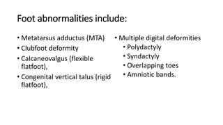
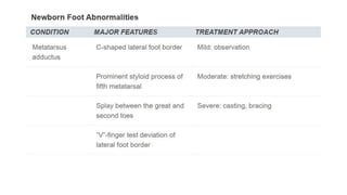
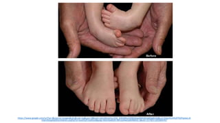
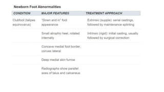
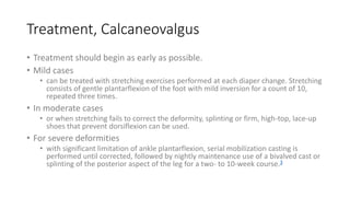
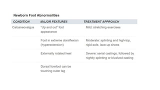
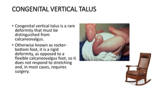
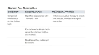
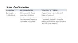
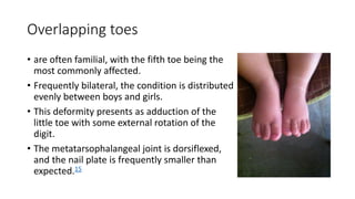
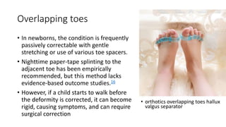
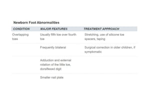
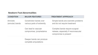
1. The Newborn Foot
By ABDULLAH ALANAZI
Family medicine
Twitter@ASKanzi
Askalanzi@gmail.com
2. Objectives
• Abstract
• Examination Techniques
• Common Foot Abnormalities
• At the end of each topic there is a table summarizing the important points
• Digital Deformities
3. Abstract
• An examination of the feet is an essential component of an evaluation of a
newborn. A thorough examination can be performed quickly. Despite its small
size, the newborn foot is a complex structure. Most deformities can be
diagnosed easily with physical examination alone, using few diagnostic studies.
A thorough examination includes assessment of vascular, dermatologic, and
neurologic status of the lower extremities, and observation, palpation, and
evaluation of joint range of motion in both feet. Common newborn foot
abnormalities include metatarsus adductus, clubfoot deformity, calcaneovalgus
(flexible flatfoot), congenital vertical talus (rigid flatfoot), and multiple digital
deformities—polydactyly, syndactyly, overlapping toes, and amniotic bands.
Most treatments include conservative measures, such as observation,
stretching, and splinting, which can be performed easily in the family medicine
setting. Cases that require surgical correction should be referred to a
subspecialist with expertise in correcting lower extremity deformities in
children. When surgery is indicated, procedures generally are postponed for six
to nine months so that the child will better tolerate anesthesia.
4. Examination Techniques
• Despite its small size, the newborn
foot is complex, consisting of 26 to 28
bones.
• The foot can be divided into three
anatomic regions (Figure 1):
• the hindfoot or rearfoot (talus and
calcaneus);
• the midfoot (navicular bone, cuboid
bone, and three cuneiform bones); and
• the forefoot (metatarsals and
phalanges). Differences between a
newborn foot and an adult foot are
summarized in Table 1.
5. Examination Techniques
• Simultaneous observation of both feet can reveal many deformities.
• The skin should be examined for unusual creases or folds that can be
formed by various foot deviations. Certain areas of the skin might be
abnormally taut, indicating extra tension on the skin, while the skin
on the opposite side of the foot might reveal loose, excessive
skinfolds.
6. Examination Techniques
• During the next part of the examination, various foot and ankle joints
are moved through their respective ranges of motion. The joints
should be assessed for flexibility or rigidity, unusual positions, lack of
motion, and asymmetry.
• Finally, the vascular examination consists of assessment of capillary
refill and skin color, because pulses are difficult to palpate.
Fortunately, the majority of newborns exhibit excellent lower
extremity vascular supply, unless it is compromised by an extrinsic
factor, such as an intrauterine amniotic band.
9. METATARSUS ADDUCTUS (MTA)
• One of the most common foot deformities, occurring
in one to two cases per 1,000 live births.1
• It is defined as a transverse plane deformity in
Lisfranc's (tarsometatarsal) joints in which the
metatarsals are deviated medially (Figure 2).
• medial deviation of all metatarsals with normal
relationship between talus and calcaneus.
10. On inspection (MTA)
• The toes angle abruptly toward the
midline, creating a C-shaped lateral
foot border with a prominent styloid
process of the fifth
metatarsal.2 (Figures 3a and 3b).
• A splay can appear between the
great and second toes.
• Skin examination frequently reveals a
deep skin cleft at the medial midfoot.
• Bilateral metatarsus adductus. (A) medial deviation of the forefoot. (B) Note rounded
lateral foot border(C-shaped border).
11. “V”-finger test
• A simple test that can raise the clinician's
suspicions of MTA
• In this test, the heel of the foot is placed in the
“V” formed by the index and middle fingers,
and the lateral aspect of the foot is observed
from a plantar side for medial or lateral
deviation from the middle finger.
• Medial deviation from the middle finger at the
styloid process indicates MTA.3
12. Treatment (MTA)
• It is based on the severity of the condition and is controversial.
• While some authors1 advocate only observation without active intervention
for mild cases, others would intervene early in all severe cases or by two
months of age, if the condition is not resolved.4
• Other authors3 recommend treatment as soon as possible, especially in
moderate to severe cases.
• Based on the natural course of the condition, a more conservative approach
seems reasonable.
• Of MTA cases identified at birth, 85 to 90 percent resolve by one year of
age.1,3
• Another prospective study5 confirmed these findings—87 percent of MTA
cases had resolved by six years of age, with only about 4 percent remaining at
age 16.
13. Treatment (MTA)
• Mild
• (flexible, passively correctable) MTA requires only parental reassurance.
• Moderate
• (semi-flexible, reducible) MTA can be treated with stretching exercises at every
diaper change.
• First, the heel is stabilized within the notch between the thumb and index finger.
Then, the forefoot is slightly pulled distally, held between the thumb and index finger
of the other hand, and gently pushed into a corrected position.1
• For the majority of MTA cases, the prognosis is quite good.
• In severe cases,
• excessive compensation at the level of the mediotarsal joint can lead to the
development of bunions, hammertoes, and other disorders.6 Therefore, severe
(rigid) MTA can be referred for serial casting and bracing. Evidence-based
comparisons of splinting or casting versus manipulation alone are not yet available.4
14.
15. CLUBFOOT
• Clubfoot, or talipes equinovarus,
• A congenital deformity that typically
has four main components:
• inversion and adduction of the
forefoot;
• inversion of the heel and hindfoot;
• equinus (limitation of extension) of
the ankle and subtalar joint;
• internal rotation of the leg.
16. On inspection Clubfoot
• the “down and in” appearance of the foot,
which somewhat resembles that of MTA, is
obvious (Figure 5).3
• The foot appears smaller, with a flexible, softer
heel because of the hypoplastic calcaneus.
• The medial border of the foot is concave with a
deep medial skin furrow, and the lateral border
is highly convex.
• The heel is usually small and is internally
rotated, making the soles of the feet face each
other in cases of bilateral deformities.
17. • On testing, there is pronounced
tightness of the Achilles tendon
with very little dorsiflexion, which
differentiates clubfoot from
MTA (Figure 6).
• Radiographs of clubfeet usually
reveal roughly parallel axes of the
talus and calcaneus (Figure7).
18. Treatment
• Clubfoot can be classified into extrinsic (supple) type, which is essentially a
severe positional or soft tissue deformity; and intrinsic (rigid) type, where
manual reduction is impossible. The type of clubfoot determines the
specific therapy. Extrinsic clubfoot can be treated by serial casting, while
intrinsic clubfoot eventually may require surgery. Plaster casting should be
attempted on virtually all clubfeet, supple or rigid, as soon as practical.
Casts initially are changed at semiweekly to weekly intervals and are
continued until the deformity responds and is corrected fully. Persistent
cast treatments by experienced clinicians have been reported to be
successful in most patients.9 However, if a plateau is reached in treatment,
surgery by a specialist in pediatric foot deformities should be considered,
usually when the child is between six and nine months of age.10 The goal is
to obtain a stable, “platform-like” position of the foot for future
ambulation.
21. CALCANEOVALGUS
• The axis of calcaneovalgus deformity is in the
tibiotalar joint, where the foot is positioned in
extreme hyperextension, with its dorsum
frequently touching the distal leg.
• Females are affected more often than males,
and this deformity can be unilateral or
bilateral.3
• It occurs in about 5 percent of all
newborns5 and is associated with external
rotation of the calcaneus, an overstretched
Achilles tendon, and tight anterior leg
musculature, all of which warrant treatment.11
22. On inspection, calcaneovalgus
• the foot has an “up and out”
appearance, with the dorsal
forefoot practically touching
the anterior aspect of the ankle
and lower leg (Figure 8).
• The ankle generally can be
plantarflexed to only 90
degrees or less. Radiographs
can confirm clinical diagnosis
• Metatarsus adductus foot (left) compared
with calcaneovalgus foot (right).
23. Treatment, Calcaneovalgus
• Calcaneovalgus is a positional deformity that is highly amenable to
treatment.
• According to some authors,10,12 it has an excellent natural history and can
spontaneously resolve on its own.
• Others3 advocate a more aggressive approach because of the possibility of
future complications, such as
• permanent muscle imbalance, peroneal tendon dislocation, and delayed
ambulation.
• Generally, the more severe the limitation of ankle plantar flexion, the more
treatment is warranted.
24. Treatment, Calcaneovalgus
• Treatment should begin as early as possible.
• Mild cases
• can be treated with stretching exercises performed at each diaper change. Stretching
consists of gentle plantarflexion of the foot with mild inversion for a count of 10,
repeated three times.
• In moderate cases
• or when stretching fails to correct the deformity, splinting or firm, high-top, lace-up
shoes that prevent dorsiflexion can be used.
• For severe deformities
• with significant limitation of ankle plantarflexion, serial mobilization casting is
performed until corrected, followed by nightly maintenance use of a bivalved cast or
splinting of the posterior aspect of the leg for a two- to 10-week course.3
25.
26. CONGENITAL VERTICAL TALUS
• Congenital vertical talus is a rare
deformity that must be
distinguished from
calcaneovalgus.
• Otherwise known as rocker-
bottom foot, it is a rigid
deformity, as opposed to a
flexible calcaneovalgus foot, so it
does not respond to stretching
and, in most cases, requires
surgery.
29. POLYDACTYLY
• The primary cause of polydactyly is thought to be genetic. Polydactyly
usually involves border digits, especially the fifth. Although some
cases involve only a distal phalanx, other cases are much more
complex, involving the entire digit, with duplication of nails, tendons,
and vascular structures.
• Treatment of complex cases involving bone requires removal of
duplicated structures, whereas cases involving soft tissue can be
treated with only ligation sutures applied in the nursery.
• Surgery generally is performed at six to nine months of age, before
the child is ambulatory but able to tolerate general anesthesia.10,15
30.
31.
32. SYNDACTYLY
• Syndactyly (webbed toes or
fingers) occurs in approximately
one in 2,000 to .15 There are
various levels of syndactylization,
from partial to complete.
• The most frequent site is between
the second and third toes.
• Syndactyly is thought to be
genetic, with an autosomal
dominant pattern of inheritance.
33. SYNDACTYLY
• Simple syndactyly is more of a cosmetic problem than a functional
one, and rarely requires treatment.10
• A radiographic evaluation is not indicated unless radical treatment is
being contemplated.
• If the parents desire surgery, it is advisable to wait until the child is
old enough to take part in the decision and participate in
postoperative care.
• The most common complication of the surgery is skin-flap slough,
leading to a recurrence of the problem.
34.
35. Overlapping toes
• are often familial, with the fifth toe being the
most commonly affected.
• Frequently bilateral, the condition is distributed
evenly between boys and girls.
• This deformity presents as adduction of the
little toe with some external rotation of the
digit.
• The metatarsophalangeal joint is dorsiflexed,
and the nail plate is frequently smaller than
expected.15
36. Overlapping toes
• In newborns, the condition is frequently
passively correctable with gentle
stretching or use of various toe spacers.
• Nighttime paper-tape splinting to the
adjacent toe has been empirically
recommended, but this method lacks
evidence-based outcome studies.16
• However, if a child starts to walk before
the deformity is corrected, it can become
rigid, causing symptoms, and can require
surgical correction
• orthotics overlapping toes hallux
valgus separator
37.
38. AMNIOTIC (ANNULAR) BANDS
• Amniotic bands commonly involve toes and fingers. They are produced by
thin bands of amniotic membrane wrapping around various parts of the
extremity in utero.15 Simple bands create swelling of the distal part of the
toe with concomitant lymphedema, and deeper bands can produce
complete amputations.17
• Contractions and resultant bulbous ends of the involved digits are seen
easily during examination. In addition to lymphedema, it is essential to rule
out vascular compromise, which is manifested by pale, cool skin with
delayed capillary refill.
• Simple bands are mainly cosmetic problems and do not require treatment,
but complex bands, especially ones that produce neurovascular
compromise, should be surgically released.17,18