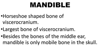
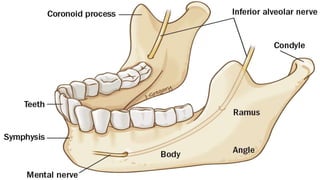
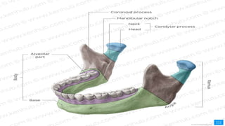
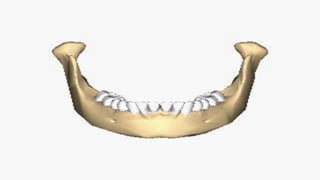
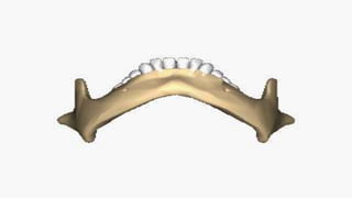
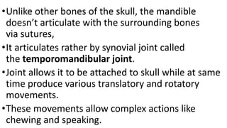
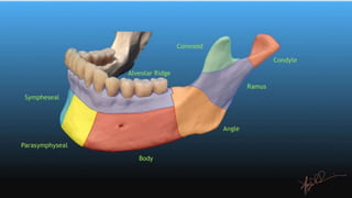
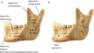
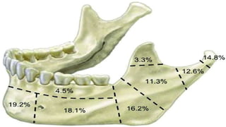
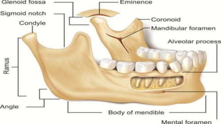
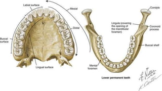
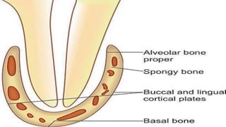
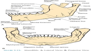
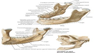
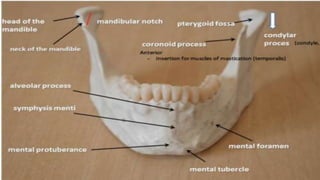
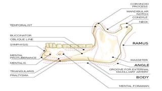
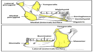
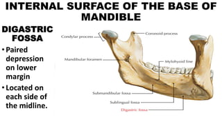
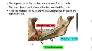
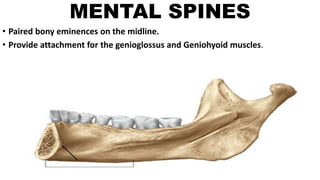
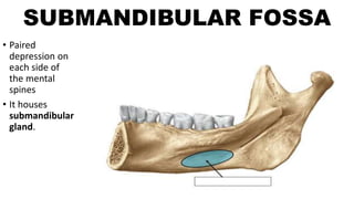
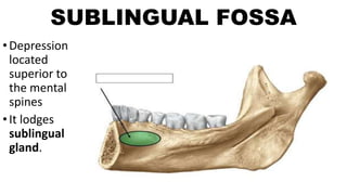
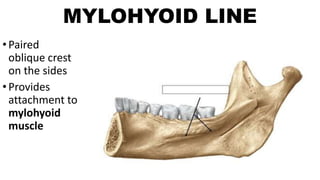
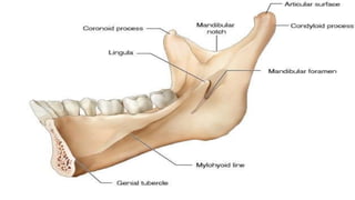
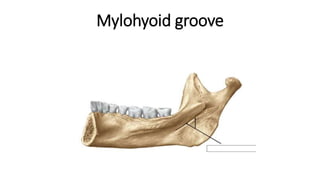
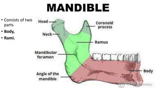
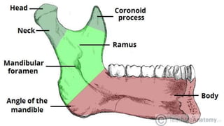
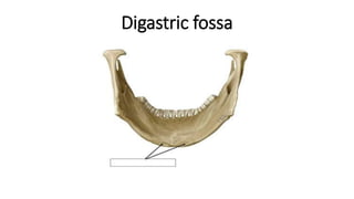
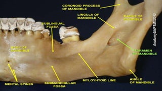
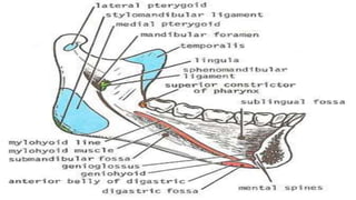
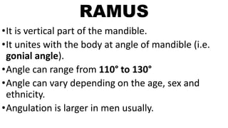
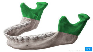
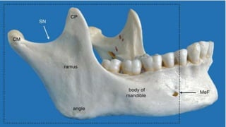
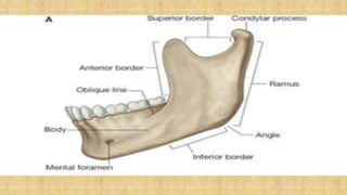
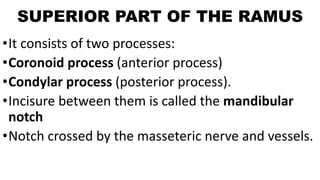
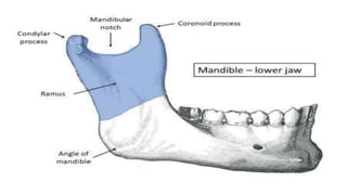
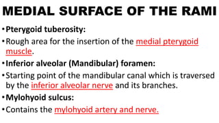
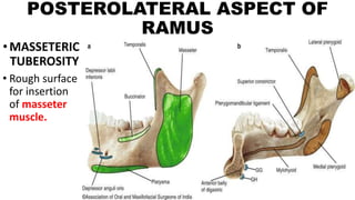
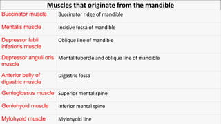
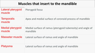
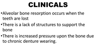
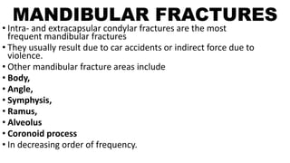
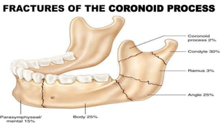
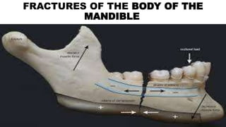
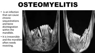
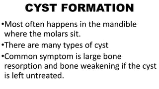
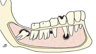
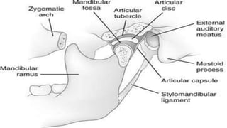
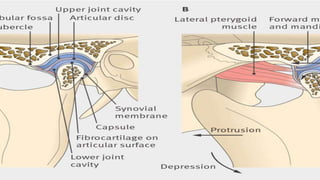
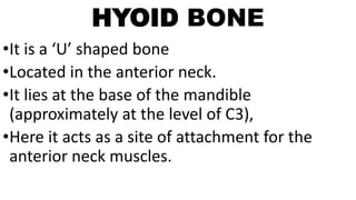
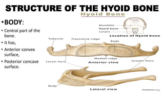
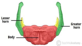
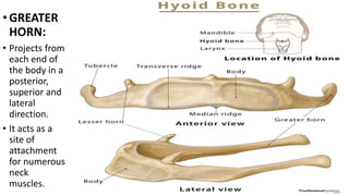
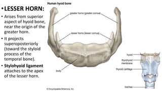
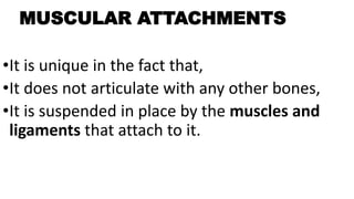
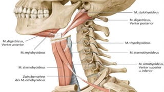
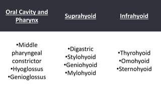
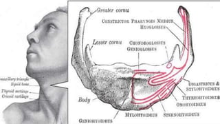
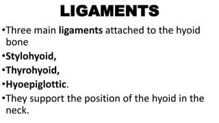
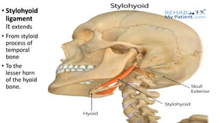
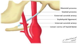
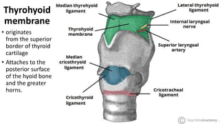
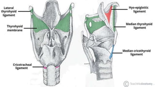
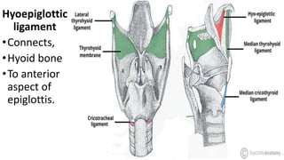
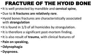
2. MANDIBLE
•Horseshoe shaped bone of
viscerocranium.
•Largest bone of viscerocranium.
•Besides the bones of the middle ear,
mandible is only mobile bone in the skull.
3.
4.
5.
6.
7. •Unlike other bones of the skull, the mandible
doesn’t articulate with the surrounding bones
via sutures,
•It articulates rather by synovial joint called
the temporomandibular joint.
•Joint allows it to be attached to skull while at same
time produce various translatory and rotatory
movements.
•These movements allow complex actions like
chewing and speaking.
13. ALVEOLAR PART OF BODY OF MANDIBLE
•It is the upper portion of the body.
•It consists of two bony lamellae:
•Thick buccal lamella,
•Thin lingual lamella.
•They are parallel to each other,
•They form shallow trench on the upper surface of the alveolar
part.
•Lamellae are connected by interalveolar septa,
•These septae cut the trench into sockets which house the
mandibular teeth.
14.
15.
16.
17.
18.
19. BASE OF BODY OF MANDIBLE
•It is inferior part of body
•It has several anatomical landmarks.
•Mandibular symphysis:
•Fibrous tissue in the midline,
•It ossifies by the first year of life.
•It unites the left and right halves of the mandible in
order to form a single, symmetrical bone.
42. RAMUS
•It is vertical part of the mandible.
•It unites with the body at angle of mandible (i.e.
gonial angle).
•Angle can range from 110° to 130°
•Angle can vary depending on the age, sex and
ethnicity.
•Angulation is larger in men usually.
43.
44.
45.
46. SUPERIOR PART OF THE RAMUS
•It consists of two processes:
•Coronoid process (anterior process)
•Condylar process (posterior process).
•Incisure between them is called the mandibular
notch
•Notch crossed by the masseteric nerve and vessels.
47.
48. MEDIAL SURFACE OF THE RAMI
•Pterygoid tuberosity:
•Rough area for the insertion of the medial pterygoid
muscle.
•Inferior alveolar (Mandibular) foramen:
•Starting point of the mandibular canal which is traversed
by the inferior alveolar nerve and its branches.
•Mylohyoid sulcus:
•Contains the mylohyoid artery and nerve.
51. Muscles that originate from the mandible
Buccinator muscle Buccinator ridge of mandible
Mentalis muscle Incisive fossa of mandible
Depressor labii
inferioris muscle
Oblique line of mandible
Depressor anguli oris
muscle
Mental tubercle and oblique line of mandible
Anterior belly of
digastric muscle
Digastric fossa
Genioglossus muscle Superior mental spine
Geniohyoid muscle Inferior mental spine
Mylohyoid muscle Mylohyoid line
52. Muscles that insert to the mandible
Lateral pterygoid
muscle
Pterygoid fossa
Temporalis
muscle
Apex and medial surface of coronoid process of mandible
Medial pterygoid
muscle
Medial surface of ramus (pterygoid tuberosity) and angle of
mandible
Masseter muscle Lateral surface of ramus and angle of mandible
Platysma Lateral surface of ramus and angle of mandible
53. CLINICALS
•Alveolar bone resorption occurs when the
teeth are lost
•There is a lack of structures to support the
bone
•There is increased pressure upon the bone due
to chronic denture wearing.
54. MANDIBULAR FRACTURES
• Intra- and extracapsular condylar fractures are the most
frequent mandibular fractures
• They usually result due to car accidents or indirect force due to
violence.
• Other mandibular fracture areas include
• Body,
• Angle,
• Symphysis,
• Ramus,
• Alveolus
• Coronoid process
• In decreasing order of frequency.
55. MANDIBULAR FRACTURES
•A mandibular fracture rarely occurs in isolation.
•Fracture on one side is frequently associated with a
fracture on the contralateral side.
•Therefore, if one fracture is observed, another
should be searched for.
•E.g.: Fractured neck of the mandible is often
observed in conjunction with a fracture of the
contralateral mandibular body.
60. TOOTH APLASIA
•It is common in,
•Third molars,
•Premolars
•Lateral incisors.
•This can lead to gaps in the teeth and
an uneven alveolar ridge.
61.
62. OSTEORADIONECROSIS
• It is a
disorder
that occurs
due
to cancer
treatment
• Bone
disintegrates
because of
radiation.
63. OSTEOMYELITIS
• is an infection
that can cause
chronic
sequestrations
and bone
disintegration
within the
mandible.
• It is irreversible
and the mandible
often needs
resecting.
64. CYST FORMATION
•Most often happens in the mandible
where the molars sit.
•There are many types of cyst
•Common symptom is large bone
resorption and bone weakening if the cyst
is left untreated.
65.
66.
67.
68. HYOID BONE
•It is a ‘U’ shaped bone
•Located in the anterior neck.
•It lies at the base of the mandible
(approximately at the level of C3),
•Here it acts as a site of attachment for the
anterior neck muscles.
69.
70. STRUCTURE OF THE HYOID BONE
•BODY:
• Central part of the
bone.
• It has,
• Anterior convex
surface,
• Posterior concave
surface.
71.
72. •GREATER
HORN:
• Projects from
each end of
the body in a
posterior,
superior and
lateral
direction.
• It acts as a
site of
attachment
for numerous
neck
muscles.
73. •LESSER HORN:
• Arises from superior
aspect of hyoid bone,
near the origin of the
greater horn.
• It projects
superoposteriorly
(toward the styloid
process of the
temporal bone).
• Stylohyoid ligament
attaches to the apex
of the lesser horn.
74.
75. MUSCULAR ATTACHMENTS
•It is unique in the fact that,
•It does not articulate with any other bones,
•It is suspended in place by the muscles and
ligaments that attach to it.
79. LIGAMENTS
•Three main ligaments attached to the hyoid
bone
•Stylohyoid,
•Thyrohyoid,
•Hyoepiglottic.
•They support the position of the hyoid in the
neck.
85. FRACTURE OF THE HYOID BONE
•It is well protected by mandible and cervical spine,
•Due to it fractures are relatively rare.
•Hyoid bones fractures are characteristically associated
with strangulation
•It is found in 1/3 of all homicides by strangulation.
•It is therefore a significant post-mortem finding.
•It is also result of trauma, with clinical features of
•Pain on speaking,
•Odynophagia
•Dyspnoea.