
Recommended
More Related Content
What's hot
What's hot (20)
Similar to The Cell Cycle (Mitosis & Meiosis) by Shiven Trambadia
Similar to The Cell Cycle (Mitosis & Meiosis) by Shiven Trambadia (20)
Recently uploaded
Recently uploaded (20)
The Cell Cycle (Mitosis & Meiosis) by Shiven Trambadia
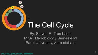
- 1. The_Cell_Cycle_Shiven_Trambadia The Cell Cycle By, Shiven R. Trambadia M.Sc. Microbiology Semester-1 Parul University, Ahmedabad. 1
- 2. The_Cell_Cycle_Shiven_Trambadia The Cell Cycle ● The cell cycle is an ordered series of events. It is the sequence of events by which a cell duplicates its genome and eventually divides into two daughter cells. ● The cell cycle has two main phases - interphase and M-phase. ● The period of actual division, corresponding to the visible mitosis, is called M phase (mitosis phase). ● The Interphase is the time during which the cell is preparing for division by undergoing both cell growth and DNA replication in an orderly manner. 2
- 3. The_Cell_Cycle_Shiven_Trambadia ● The interphase is further subdivided into; ○ G1 phase (Gap 1, the period between the end of M phase and the start of DNA replication); ○ S phase (Synthesis, the period during which DNA synthesis occurs); and ○ G2 phase (Gap 2, the gap period following DNA replication and preceding the initiation of the M phase) ● Cells that do not divide enter into G0 state. Most cells in our body are in G0 state. 3
- 4. The_Cell_Cycle_Shiven_Trambadia Mitosis ● Mitosis was firstly introduced by Walther Flemming in 1882. ● Mitosis partitions newly replicated chromosomes equally into two daughter cells. ● Mitosis leads to one round of DNA replication followed by one round of Chromosome segregation and giving rise to two genetically identical daughter cells. 4
- 5. The_Cell_Cycle_Shiven_Trambadia Stages of M-phase ● The M-phase starts with karyokinesis (nuclear division), forming two daughter nuclei and generally ends up with cytokinesis (cytoplasm division). ● Conventionally mitosis is divided into 4 substages: prophase, metaphase, anaphase and telophase. ● Cells show different physical characteristics when reaching in each of these phases. 5
- 6. The_Cell_Cycle_Shiven_Trambadia Microscopic image of Mitosis 6
- 7. The_Cell_Cycle_Shiven_Trambadia Prophase ● The diffuse chromatin in interphase, now slowly starts to condense during the beginning of prophase. ● Chromosomes are duplicated during the previous S-phase and consists of two sister chromatids, which are held together at a constricted region called, Centromere. ● At centromere, sister chromatids are held together by a membrane of the SMC (Structural Maintenance of Chromosomes), included in the family of proteins, called cohesin. ● At the end of prophase, cytoplasmic microtubules disassemble and starts the formation of mitotic spindle. 7
- 8. The_Cell_Cycle_Shiven_Trambadia Prophase ● At the end of prophase nucleolus disappears along with the nuclear envelope. ● Most of the cells mark the end of the prophase when nuclear envelope disrupts. But, nuclear envelope disruption is not universally observed feature of mitosis. Eg; Yeast cells don’t loose nuclear envelope during cell division. 8
- 9. The_Cell_Cycle_Shiven_Trambadia Metaphase ● Metaphase is an very dynamic part of the cell cycle. ● Spindle fibres rapidly assemble and disassemble as they grow out of the Microtubule Organizing Center (MTOC), seeking out attachment sites at chromosome. ● For attachment of spindle fibres, each chromosome has a special structure called kinetochore. ● There are two kinetochores located at the centromere, on each chromosome facing in opposite directions. ● Chromosomes are now lined up to the Equator of the cell. 9
- 10. The_Cell_Cycle_Shiven_Trambadia Anaphase ● The kinetochore microtubules pull the two kinetochores towards the opposite poles, at anaphase. ● The cohesins holding together the two sister chromatids, are now digested by another protein, Separase. ● Until anaphase the separase is kept in active form by another protein, Securin. ● After separation, each chromatid, which are now called as chromosome, are pulled towards the polls they face, opposite to each other. 10
- 11. The_Cell_Cycle_Shiven_Trambadia Telophase ● Separated daughter chromosomes arrive at the poles and the kinetochore microtubules disappears, at Telophase. ● Still polar microtubule elongates more, and a new nuclear envelope re-forms around each set of daughter chromosomes. ● Condensed chromatin expands again and the nucleoli reappears, marking the end of the mitosis. 11
- 12. The_Cell_Cycle_Shiven_Trambadia Cytokinesis ● Division of cytoplasm after karyokinesis is referred as cytokinesis. ● Cytokinesis begins in anaphase and ends in telophase. ● Mechanism of cytokinesis in animals and plants cell vary, in former it is completed by cleavage and in latter by cell plate formation. 12
- 13. The_Cell_Cycle_Shiven_Trambadia Meiosis ● Farmer and Moore, explained that a specialized cell division in which number of chromosomes is reduced to half is ● This reduction in chromosome number is achieved by one round of DNA replication, followed by two rounds of chromosome segregation with no intervening rounds of DNA replication. ● Meiosis is divided into two parts, meiosis I and meiosis II. At the end of meiotic process, there are four daughter cells, each having half the total number of chromosomes as parent cell. 13
- 15. The_Cell_Cycle_Shiven_Trambadia Meiosis 1 ● Prophase 1 of meiosis 1 is divided into five sub-stages: Leptotene, Zygotene, Pachytene, Diplotene and Diakinesis. ● Leptotene: although each chromosome has replicated and consists of two sister chromatids, these chromatids are unusually closely apposed, and each chromosome therefore appears to be single. ● In leptotene to zygotene transition, the tips of the chromosomes move until most end up in a limited region near each other, this arrangement is called a Bouquet stage. ● It facilitates homologous chromosome pairing and synapsis. 15
- 16. The_Cell_Cycle_Shiven_Trambadia Zygotene ● As soon as the intimate pairing, between the two homologs initiates it marks the starting of Zygotene. ● Synapsis often starts when the homologous end of the two chromosomes are brought together on the nuclear envelope and proceeds inward in a zipper fashion from both ends. ● Each pair of homologous chromosomes forms as a result of synapsis is called a bivalent. 16
- 17. The_Cell_Cycle_Shiven_Trambadia Pachytene ● As soon as the synapsis is completed all along the chromosomes, the cells are said to have entered the pachytene stage. ● The paired chromosomes now get shorten and thicken. ● At this stage , large recombination nodules appear at the intervals on the synaptonemal complexes. ● Crossing over is a reciprocal exchange of equal and corresponding segment between non-sister chromatids in a pair of homologous chromosome. ● It leads to the recombination of genes present on two homologous chromosomes. 17
- 18. The_Cell_Cycle_Shiven_Trambadia Diplotene ● Desynapsis begins in the diplotene stage of meiotic prophase 1. ● Synaptonemal complex dissolves, allowing the two homologous chromosomes in a bivalent to pull away from each other to some extent. ● But, each bivalent remains attached by one or more chiasmata. 18
- 19. The_Cell_Cycle_Shiven_Trambadia Diakinesis ● Diplotene merges imperceptibly into diakinesis, the stage of transition to metaphase, as RNA synthesis ceases and the chromosomes condense, thicken and become detached from the nuclear envelope. ● Now each bivalent consists of four separate chromatids, with each pair of sister chromatids linked at their centromeres. ● Now cell enters Metaphase 1. 19
- 21. The_Cell_Cycle_Shiven_Trambadia Metaphase 1 ● Breakdown of nuclear membrane and attachment of kinetochore microtubules to bivalent, marks the initiation of Metaphase 1. ● Homologous pairs move together along the metaphase plate. ● Independent assortment is based on the physical orientation of chromosomes of each bivalent along the metaphase plate, with respect to that of other bivalents along the same equatorial line. 21
- 22. The_Cell_Cycle_Shiven_Trambadia Metaphase 1 to Anaphase 1 22
- 23. The_Cell_Cycle_Shiven_Trambadia Anaphase 1 ● Homologous chromosomes are separated in Anaphase 1. ● Sister chromatids stay together. ● Special meiosis specific cohesin, Rec8 inhibits the separation of sister chromatids. ● Homologous chromosomes separate and move to opposite poles. The meiotic division is hence, called the reductional division, as it reduces the chromosome number to half the diploid number in each daughter cell. 23
- 24. The_Cell_Cycle_Shiven_Trambadia Telophase 1 ● As soon as the chromosomes arrive at the poles meiosis 1 is said to have been ended effectively. ● Now each daughter cell has half the number of chromosomes left, with a pair of chromatids. ● New nuclear membrane surrounds each haploid set. ● Chromosomes are uncoiled back to chromatin. ● Cytokinesis take place, generating two daughter cells. 24
- 25. The_Cell_Cycle_Shiven_Trambadia Meiosis 2 ● Time gap between Meiosis 1 and Meiosis 2 is called interkinesis. ● It is usually short or may not occur at all, at this stage no DNA replication occurs. ● Meiosis 2 is similar to that of Mitosis. ● But, no S phase occurs during Meiosis 2. ● Now the chromatids of each chromosome are not identical due to recombination taken place in prophase 1 of meiosis 1. ● Meiosis 2 separates the sister chromatids producing two daughter cells each with a haploid chromosome number. 25