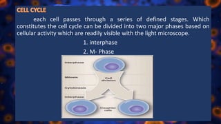
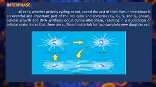
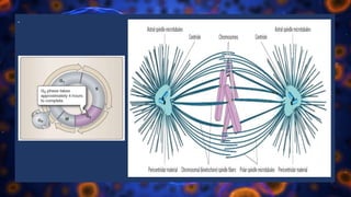
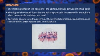
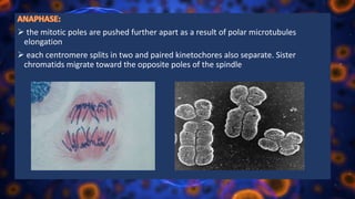
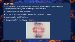
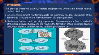
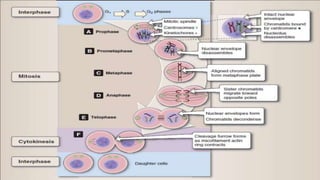
The cell cycle, or cell-division cycle, is the series of events that take place in a cell that cause it to divide into two daughter cells. These events include the duplication of its DNA (DNA replication) and some of its organelles, and subsequently the partitioning of its cytoplasm and other components into two daughter cells in a process called cell division.
here u will find every detail of cell cycle.
for more details ,visit @biOlOgy BINGE-insight learning
Cell cycle is the most fundamental and important process by which eukaryotic cells duplicate and divide. This slide will talk about the method to analysis the cell cycle.
The cell cycle, or cell-division cycle, is the series of events that take place in a cell leading to duplication of its DNA (DNA replication) and division of cytoplasm and organelles to produce two daughter cells.
This slide describes the various stages of the Eukaryotic cell cycle. The diagrams included here explains the various changes that take place during the mitotic division of a eukaryotic cell.
The cell cycle, or cell-division cycle, is the series of events that take place in a cell leading to its division and duplication of its DNA to produce two daughter cells. In bacteria, which lack a cell nucleus, the cell cycle is divided into the B, C, and D periods
OVERVIEW OF CELL CYCLE
Explained in brief phases of cell cycle . Given a explanation of each phase in detail, also explained the significance of meiosis in brief.
Multicellular organisms develop from a single cell known as zygote by the process of mitosis. Asexual reproduction in some organisms like amoeba and vegetative reproduction in plants takes place by mitosis. This type of cell division involves many steps and it does not alter the genetic material.
describe cell cycle and cell cycle control system for downloading the presentation , more presentations , infographics and blogs visit :
studyscienceblog.wordpress.com
This presentation include the process of cell division. It hope it will helpful for all the medical students. Cell division is the series of events of equally dividing of one single mother cell into two identical daughter cell. Cell cycle and cell division terms are alternately used. Cell division is an important part of the all living processes.
At the time of cell division, RNA replication is a natural process.
The cell cycle, or cell-division cycle, is the series of events that take place in a cell that cause it to divide into two daughter cells.
These events include the duplication of its DNA (DNA replication) and some of its organelles, and subsequently the partitioning of its cytoplasm and other components into two daughter cells in a process called cell division.
There are two types of cell division
A) Mitosis and Binary fission – (Asexual reproduction) and B) Meiosis – (Sexual reproduction)
In prokaryotic cell, the cell division occurs via a process termed as Binary fission.
• In eukaryotic cell, the cell cycle can be divided in two periods i.e Interphase and Mitosis.
• During Interphase, the cell grows and DNA is replicated.
During Mitotic phase, the replicated DNA and cytoplasmic contents are separated, and cell divides.
The duration of cycle varies from hours to years. A typical human cell cycle has duration of 24 hours.
Some cells, such as skin cells, are constantly going through cell cycle, while other cells may divide rarely.
Some cells don’t grow and divide once they mature for ex. Neuron
Eukaryotic cell have a more complex cell cycle than prokaryotic cell.
Professional air quality monitoring systems provide immediate, on-site data for analysis, compliance, and decision-making.
Monitor common gases, weather parameters, particulates.
Slide 1: Title Slide
Extrachromosomal Inheritance
Slide 2: Introduction to Extrachromosomal Inheritance
Definition: Extrachromosomal inheritance refers to the transmission of genetic material that is not found within the nucleus.
Key Components: Involves genes located in mitochondria, chloroplasts, and plasmids.
Slide 3: Mitochondrial Inheritance
Mitochondria: Organelles responsible for energy production.
Mitochondrial DNA (mtDNA): Circular DNA molecule found in mitochondria.
Inheritance Pattern: Maternally inherited, meaning it is passed from mothers to all their offspring.
Diseases: Examples include Leber’s hereditary optic neuropathy (LHON) and mitochondrial myopathy.
Slide 4: Chloroplast Inheritance
Chloroplasts: Organelles responsible for photosynthesis in plants.
Chloroplast DNA (cpDNA): Circular DNA molecule found in chloroplasts.
Inheritance Pattern: Often maternally inherited in most plants, but can vary in some species.
Examples: Variegation in plants, where leaf color patterns are determined by chloroplast DNA.
Slide 5: Plasmid Inheritance
Plasmids: Small, circular DNA molecules found in bacteria and some eukaryotes.
Features: Can carry antibiotic resistance genes and can be transferred between cells through processes like conjugation.
Significance: Important in biotechnology for gene cloning and genetic engineering.
Slide 6: Mechanisms of Extrachromosomal Inheritance
Non-Mendelian Patterns: Do not follow Mendel’s laws of inheritance.
Cytoplasmic Segregation: During cell division, organelles like mitochondria and chloroplasts are randomly distributed to daughter cells.
Heteroplasmy: Presence of more than one type of organellar genome within a cell, leading to variation in expression.
Slide 7: Examples of Extrachromosomal Inheritance
Four O’clock Plant (Mirabilis jalapa): Shows variegated leaves due to different cpDNA in leaf cells.
Petite Mutants in Yeast: Result from mutations in mitochondrial DNA affecting respiration.
Slide 8: Importance of Extrachromosomal Inheritance
Evolution: Provides insight into the evolution of eukaryotic cells.
Medicine: Understanding mitochondrial inheritance helps in diagnosing and treating mitochondrial diseases.
Agriculture: Chloroplast inheritance can be used in plant breeding and genetic modification.
Slide 9: Recent Research and Advances
Gene Editing: Techniques like CRISPR-Cas9 are being used to edit mitochondrial and chloroplast DNA.
Therapies: Development of mitochondrial replacement therapy (MRT) for preventing mitochondrial diseases.
Slide 10: Conclusion
Summary: Extrachromosomal inheritance involves the transmission of genetic material outside the nucleus and plays a crucial role in genetics, medicine, and biotechnology.
Future Directions: Continued research and technological advancements hold promise for new treatments and applications.
Slide 11: Questions and Discussion
Invite Audience: Open the floor for any questions or further discussion on the topic.
Salas, V. (2024) "John of St. Thomas (Poinsot) on the Science of Sacred Theol...Studia Poinsotiana
I Introduction
II Subalternation and Theology
III Theology and Dogmatic Declarations
IV The Mixed Principles of Theology
V Virtual Revelation: The Unity of Theology
VI Theology as a Natural Science
VII Theology’s Certitude
VIII Conclusion
Notes
Bibliography
All the contents are fully attributable to the author, Doctor Victor Salas. Should you wish to get this text republished, get in touch with the author or the editorial committee of the Studia Poinsotiana. Insofar as possible, we will be happy to broker your contact.
Observation of Io’s Resurfacing via Plume Deposition Using Ground-based Adapt...Sérgio Sacani
Since volcanic activity was first discovered on Io from Voyager images in 1979, changes
on Io’s surface have been monitored from both spacecraft and ground-based telescopes.
Here, we present the highest spatial resolution images of Io ever obtained from a groundbased telescope. These images, acquired by the SHARK-VIS instrument on the Large
Binocular Telescope, show evidence of a major resurfacing event on Io’s trailing hemisphere. When compared to the most recent spacecraft images, the SHARK-VIS images
show that a plume deposit from a powerful eruption at Pillan Patera has covered part
of the long-lived Pele plume deposit. Although this type of resurfacing event may be common on Io, few have been detected due to the rarity of spacecraft visits and the previously low spatial resolution available from Earth-based telescopes. The SHARK-VIS instrument ushers in a new era of high resolution imaging of Io’s surface using adaptive
optics at visible wavelengths.
Seminar of U.V. Spectroscopy by SAMIR PANDASAMIR PANDA
Spectroscopy is a branch of science dealing the study of interaction of electromagnetic radiation with matter.
Ultraviolet-visible spectroscopy refers to absorption spectroscopy or reflect spectroscopy in the UV-VIS spectral region.
Ultraviolet-visible spectroscopy is an analytical method that can measure the amount of light received by the analyte.
Deep Behavioral Phenotyping in Systems Neuroscience for Functional Atlasing a...Ana Luísa Pinho
Functional Magnetic Resonance Imaging (fMRI) provides means to characterize brain activations in response to behavior. However, cognitive neuroscience has been limited to group-level effects referring to the performance of specific tasks. To obtain the functional profile of elementary cognitive mechanisms, the combination of brain responses to many tasks is required. Yet, to date, both structural atlases and parcellation-based activations do not fully account for cognitive function and still present several limitations. Further, they do not adapt overall to individual characteristics. In this talk, I will give an account of deep-behavioral phenotyping strategies, namely data-driven methods in large task-fMRI datasets, to optimize functional brain-data collection and improve inference of effects-of-interest related to mental processes. Key to this approach is the employment of fast multi-functional paradigms rich on features that can be well parametrized and, consequently, facilitate the creation of psycho-physiological constructs to be modelled with imaging data. Particular emphasis will be given to music stimuli when studying high-order cognitive mechanisms, due to their ecological nature and quality to enable complex behavior compounded by discrete entities. I will also discuss how deep-behavioral phenotyping and individualized models applied to neuroimaging data can better account for the subject-specific organization of domain-general cognitive systems in the human brain. Finally, the accumulation of functional brain signatures brings the possibility to clarify relationships among tasks and create a univocal link between brain systems and mental functions through: (1) the development of ontologies proposing an organization of cognitive processes; and (2) brain-network taxonomies describing functional specialization. To this end, tools to improve commensurability in cognitive science are necessary, such as public repositories, ontology-based platforms and automated meta-analysis tools. I will thus discuss some brain-atlasing resources currently under development, and their applicability in cognitive as well as clinical neuroscience.
Comparing Evolved Extractive Text Summary Scores of Bidirectional Encoder Rep...University of Maribor
Slides from:
11th International Conference on Electrical, Electronics and Computer Engineering (IcETRAN), Niš, 3-6 June 2024
Track: Artificial Intelligence
https://www.etran.rs/2024/en/home-english/
(May 29th, 2024) Advancements in Intravital Microscopy- Insights for Preclini...Scintica Instrumentation
Intravital microscopy (IVM) is a powerful tool utilized to study cellular behavior over time and space in vivo. Much of our understanding of cell biology has been accomplished using various in vitro and ex vivo methods; however, these studies do not necessarily reflect the natural dynamics of biological processes. Unlike traditional cell culture or fixed tissue imaging, IVM allows for the ultra-fast high-resolution imaging of cellular processes over time and space and were studied in its natural environment. Real-time visualization of biological processes in the context of an intact organism helps maintain physiological relevance and provide insights into the progression of disease, response to treatments or developmental processes.
In this webinar we give an overview of advanced applications of the IVM system in preclinical research. IVIM technology is a provider of all-in-one intravital microscopy systems and solutions optimized for in vivo imaging of live animal models at sub-micron resolution. The system’s unique features and user-friendly software enables researchers to probe fast dynamic biological processes such as immune cell tracking, cell-cell interaction as well as vascularization and tumor metastasis with exceptional detail. This webinar will also give an overview of IVM being utilized in drug development, offering a view into the intricate interaction between drugs/nanoparticles and tissues in vivo and allows for the evaluation of therapeutic intervention in a variety of tissues and organs. This interdisciplinary collaboration continues to drive the advancements of novel therapeutic strategies.
Nutraceutical market, scope and growth: Herbal drug technologyLokesh Patil
As consumer awareness of health and wellness rises, the nutraceutical market—which includes goods like functional meals, drinks, and dietary supplements that provide health advantages beyond basic nutrition—is growing significantly. As healthcare expenses rise, the population ages, and people want natural and preventative health solutions more and more, this industry is increasing quickly. Further driving market expansion are product formulation innovations and the use of cutting-edge technology for customized nutrition. With its worldwide reach, the nutraceutical industry is expected to keep growing and provide significant chances for research and investment in a number of categories, including vitamins, minerals, probiotics, and herbal supplements.
Richard's aventures in two entangled wonderlandsRichard Gill
Since the loophole-free Bell experiments of 2020 and the Nobel prizes in physics of 2022, critics of Bell's work have retreated to the fortress of super-determinism. Now, super-determinism is a derogatory word - it just means "determinism". Palmer, Hance and Hossenfelder argue that quantum mechanics and determinism are not incompatible, using a sophisticated mathematical construction based on a subtle thinning of allowed states and measurements in quantum mechanics, such that what is left appears to make Bell's argument fail, without altering the empirical predictions of quantum mechanics. I think however that it is a smoke screen, and the slogan "lost in math" comes to my mind. I will discuss some other recent disproofs of Bell's theorem using the language of causality based on causal graphs. Causal thinking is also central to law and justice. I will mention surprising connections to my work on serial killer nurse cases, in particular the Dutch case of Lucia de Berk and the current UK case of Lucy Letby.
2. Multicellular organisms are composed of a variety of specialized cells organized
into a cellular community. When an organism requires additional cells, either for
growth or to replace those normally lost, new ones must be produced by cell division,
or proliferation. Somatic cells are generated by the division of existing cells in an
orderly sequence of events. They duplicate their contents and then divide to produce
two identical daughter cells. This sequence of duplication is known as the cell cycle
and is the essential mechanism of eukaryotic reproduction. Cell division occurs
throughout the life of the organism, although different cell types divide more or less
often than others. Cells display remarkable variation in their proliferative capacity,
depending on the cell type and the age of the individual.
the new cell originate only from other living cells, the process by which this
occurs is called cell division.
3. each cell passes through a series of defined stages. Which
constitutes the cell cycle can be divided into two major phases based on
cellular activity which are readily visible with the light microscope.
1. interphase
2. M- Phase
4. all cells, whether actively cycling or not, spend the vast of their lives in interphase is
an eventful and important part of the cell cycle and comprises G0, G1, S, and G2 phases,
cellular growth and DNA synthesis occur during interphase, resulting in a duplication of
cellular materials so that there are sufficient materials for two complete new daughter cell.
5. named for the gap that follows mitosis and the next round of DNA synthesis
G1 phase is generally both a growth phase & preparation time for DNA synthesis of S
phase
RNA and protein also synthesized in this phase in addition organelles and
intracellular structure are duplicated and the cells grows during this phase
the length of G1 phase is variable among cell types. Very rapidly cells, such as
growing embryonic cell
these cells in G1 phase that are not committed to DNA synthesis are in a specialized
resting state called G0 phase. Some inactive or quiescent cells in G0 phase may
reenter the active phase of cell cycle upon proper stimulation.
the restriction point is critical for cell cycle regulation is located with in G1 phase and
if passed with commit a cell to continuing into DNA synthesis within S phase
6. occur the DNA replication
46 chromosomes in a human cells is copied to form a sister chromatid
ATP- depended unwinding of chromatin structure by DNA helicase exposes the binding
sites of for DNA polymerase that will catalyze the synthesis of new DNA in the 5’ to 3’
direction
7. multiple replication forks are activated on each chromosome & entire genome is
duplicated within the time span of S phase
after this chromosome synthesis this strands are condensed into tightly coiled
heterochromatin
time of preparation for the nuclear division of mitosis
this safety gap allows the cell to ensure that DNA synthesis is complete before
proceeding to nuclear division in mitosis
8. G2 is a check point where intracellular regulatory molecules asses nuclear integrity
mitosis or nuclear division is a continuous process and can be divided into five phase
based on progress made to a specific point in the overall nuclear division
that occur in 1 hour in mitosis
after completion division resulting in the formation of two sepate daughter cells
from the on parent cell
10. in prophase the nuclear envelop remains intact while the chromatin that was
duplicated during S phase condenses into defined chromosomal structures called
chromatids.
chromosomes of mitotic cells contain two chromatids connected to each other at a
centromere
specialized protein complexes, called KINETOCHORES, form and associate with each
chromatid.
mitotic spindle microtubules will attach each kinetochore as chromosomes are moved
apart later in mitosis
the microtubules of the cytoplasm disassemble and then reorganize on the surface of
the nucleus to form the mitotic spindle. Two centriole pairs push away from each other
by growing bandles of microtubules forming the mitotic spindle. The nucleolus, the
organelle within the nucleus where ribosomes are made, disassembles in prophase.
11. chromosomal microtubules attach to kinetochores of chromosomes
chromosomes are moved to spindle equator
13. chromatids aligned at the equator of the spindle, halfway between the two poles
the aligned chromatids form the metaphase plate cells be arrested in metaphase
when microtubule inhibitors are used
karyotype analyses used to determine the over all chromosome composition and
structure most often require cells in metaphase.
14. the mitotic poles are pushed further apart as a result of polar microtubules
elongation
each centromere splits in two and paired kinetochores also separate. Sister
chromatids migrate toward the opposite poles of the spindle
15. the last phase of nuclear division, telophase is characterized by kinetochore
microtubule disambly and mitotic spindle dissociation
chromosomes become dispersed
nuclear envelope assembles around chromosomes clusters
golgi complex and ER reforms
daughter cells formed by cytokinesis
16. in order to create two distinct, separate daughter cells. Cytoplasmic division follows
nuclear division
an actin microfilament ring forms to crate the machinery needed. Contraction of this
actin based structure results in the formation of a cleavage furrow.
the furrow deepens until opposing edges meet. Plasma membrane fuse on each side
of the deep cleavage furrow and the result is the formation of two separate daughter
cells, each identical to the other and to the original parent cell
17.
18. • REFERENCE:
Gerald Karp - Cell and Molecular Biology_ Concepts and Experiments-Wiley (2010), 1ST edition
188-195.
(Lippincott’s Illustrated Reviews) Nalini Chandar, Susan Viselli - Cell and Molecular Biology
(Lippincott’s Illustrated Reviews) -Lippincott Williams & Wilkins (2010), 6th edition, 569-588.