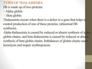
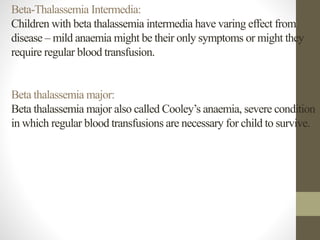
Nursing college
Thalassemia (Case Study)
Prepare by:MohammedHassan
Case Study
Child was diagnosed with the thalassemia major 5 yrs. back and is
undergoing blood transfusion every month since then. Patient also
gives the history of splenectomy. Family history revealed presence of
thalassemia in one of his family members. Personal history was non-
contributory. Patient was poorly built and nourished with short stature.
Extraoral examination of the patient revealed frontal bossing,
maxillary protrusion, saddle nose and frontal bossing giving an
appearance of ‘chip-munk facies’or rodent facies’ Patient exhibited
asthenic body with weakness of scapular girdle muscles. Patient’s
finger nails and skin extremities exhibited yellowish tinge and sclera
showed icterus There was a presence surgical scar in the left
hypochondrium which was matched with history of splenectomy.
Thalassemia is a hereditary disease of the blood, caused by an
imbalance in the production of the hemoglobin molecule, which is
the central protein found in red blood cells, which carries oxygen
from the lungs to the rest of the body. Hemoglobin consists of two
types of protein chains, two alpha chains and two beta chains.
TYPES OF THALASSEMIA
Hb is made up of two proteins:
–Alpha globin
– Beta globin
Thalassemia occurs when there is a defect in a gene that helps in
control production of one of these proteins. (abnormal Hb
synthesis).
Alpha thalassemia is caused by reduced or absent synthesis of alpha
globin chains, and beta thalassemia is caused by reduced or absent
synthesis of beta globin chains. Imbalances of globin chains cause
hemolysis and impair erythropoiesis.
Alpha Thalassemia:
*Alpha thalassemia occurs when one or more of the four genes
needed for making the alpha globin chain of haemoglobin are
variant or missing.
* Moderate to severe anaemia results when more than two genes are
affected.
* The most severe form of alpha thalassemia is known as alpha
thalassemia major
Beta thalassemia:
* Beta thalassemia occurs when one or both of the two genes needed
making the polypeptide globin chain of haemoglobin is defective.
* The severity of illness depends on whether one or both genes are
affected and the nature of abnormality.
* If both genes are affected, anaemia can range from moderate to
severe.
Beta-thalassemia is grouped into three categories
1. Minor (trait),
2. Intermedia and
3. Major (cooley's anemia).
Beta-thalassemia Minor (trait):
* If one gene is affected, person is carrier and has mild anemia. This
condition is called beta thalassemia trait or beta thalassemia minor.
* Beta thalassemia minor often goes undiagnosed because kids with
the condition have no real symptoms other than mild anemia and small
red blood cells.
Beta-Thalassemia Intermedia:
Children with beta thalassemia intermedia have varing effect from
disease – mild anaemia might be their only symptoms or might they
require regular blood transfusion.
Beta thalassemia major:
Beta thalassemia major also called Cooley’s anaemia, severe condition
in which regular blood transfusions are necessary for child to survive.
CLINICAL MANIFESTATIONS
* alpha-thalassemia may have mild anemia & are typically
asymptomatic.
* Children born with beta-thalassemia major (Cooley anemia) are
normal at birth, but develop severe anemia during first year of life.
* Children also have pain, failure to thrive, frequent infections,
diarrhea, splenomegaly, hepatomegaly, jaundice from RBC hemolysis,
and bone marrow hyperplasia.
* Other symptoms are: – Fatigue, Weakness, Pale or yellowish skin,
Abdominal swelling, Dark urine, Bone deformities in face and
Shortness of breath
DIAGNOSTIC EVALUATION
* History collection.
* Physical examination (can reveal spleenomegaly).
* Blood test including the findings:
– RBC’s will appear small & abnormal in shape when looked under
microscope
– CBC reveals anemia.
–Atest called Hb electrophoresis shows the presence of an abnormal
form of Hb and elevated levels of HbF and HbA2; limited amount of
HbA
–Atest called mutational analysis can help detect alpha thalassemia.
*Amniocentesis (fetal diagnosis for a specific type of thalassemia).
* Molecular diagnostic tests can determine whether a mutation is
present after 8 weeks of gestation.
MEDICALMANAGEMENT:
Treatment for thalassemia depends on which type have and how
severe it is.
* Treatments for mild thalassemia
- Signs and symptoms are usually mild with thalassemia minor and
little, if any, treatment is needed.
- Occasionally, may need a blood transfusion, particularly after
surgery, after having a baby or to help manage thalassemia
complications.
* Treatments for moderate to severe thalassemia may include:
- Frequent and regular blood transfusions of packed RBCs to
maintain Hb levels above 10 g/dL.
- Washed, packed RBCs are usually used to minimize the possibility
of transfusion reactions. If unavailable, leukofiltered cells can be
substituted.
- The frequency and amount of transfusions depend on the size of
the child, usually 10 to 15 mLpacked RBC per kg body weight
every 2 to 3 weeks.
- Iron chelation therapy with deferoxamine (Desferal) reduces the
toxic adverse effects of excess iron; increases iron excretion through
urine and feces.
- I.V. infusion of 100 to 150 mg/kg per day given in hospital during
blood transfusion or for child with high ferritin level and poor
compliance with home chelation therapy.
* SURGICAL MANAGEMENT:
- Bone marrow transplantation
- Spleenectomy may be done to decrease transfusion requirements
(because RBC’s may be sequestered in spleen , if iron supplements
used during blood transfusion)
NURSING MANAGEMENT:
•Assessment:
– History
– Physical examination
–Assess client’s skin, heart, spleen, liver, pancreas, lymph nodes,
endocrine, skeletal, etc.
– Measure growth and development parameters.
• Nursing diagnosis:
* Ineffective Tissue Perfusion related to abnormal Hb
* Chronic Pain related to progression of disease in bone
*Activity Intolerance related to bone pain, cardiac dysfunction, and
anemia
* Risk for Infection related to progressive anemia and splenectomy
* Deficient Knowledge related to iron chelation therapy
* Disturbed Body Image related to endocrine and skeletal
abnormalities
* Ineffective Family Coping related to poor prognosis
* Impaired nutritional pattern less than body requirement related to
inadequate nutritional intake and anorexia.
• Intervention:
–Assess the client’s general conditions.
– Monitor vital signs.
– Monitor intake output.
– Manage fluid overload.
– Monitor regular blood transfusion to keep Hb at 10.5 gm/dl.
– 24 hours urine collection after chelating therapy to estimate
amount of iron excreted.
– Reassure the orange color of urine is normal (can show
dehydration).
thalassemia.pptx

thalassemia.pptx

  • 1.
    Nursing college Thalassemia (CaseStudy) Prepare by:MohammedHassan
  • 2.
    Case Study Child wasdiagnosed with the thalassemia major 5 yrs. back and is undergoing blood transfusion every month since then. Patient also gives the history of splenectomy. Family history revealed presence of thalassemia in one of his family members. Personal history was non- contributory. Patient was poorly built and nourished with short stature. Extraoral examination of the patient revealed frontal bossing, maxillary protrusion, saddle nose and frontal bossing giving an appearance of ‘chip-munk facies’or rodent facies’ Patient exhibited asthenic body with weakness of scapular girdle muscles. Patient’s finger nails and skin extremities exhibited yellowish tinge and sclera showed icterus There was a presence surgical scar in the left hypochondrium which was matched with history of splenectomy.
  • 4.
    Thalassemia is ahereditary disease of the blood, caused by an imbalance in the production of the hemoglobin molecule, which is the central protein found in red blood cells, which carries oxygen from the lungs to the rest of the body. Hemoglobin consists of two types of protein chains, two alpha chains and two beta chains.
  • 7.
    TYPES OF THALASSEMIA Hbis made up of two proteins: –Alpha globin – Beta globin Thalassemia occurs when there is a defect in a gene that helps in control production of one of these proteins. (abnormal Hb synthesis). Alpha thalassemia is caused by reduced or absent synthesis of alpha globin chains, and beta thalassemia is caused by reduced or absent synthesis of beta globin chains. Imbalances of globin chains cause hemolysis and impair erythropoiesis.
  • 8.
    Alpha Thalassemia: *Alpha thalassemiaoccurs when one or more of the four genes needed for making the alpha globin chain of haemoglobin are variant or missing. * Moderate to severe anaemia results when more than two genes are affected. * The most severe form of alpha thalassemia is known as alpha thalassemia major
  • 9.
    Beta thalassemia: * Betathalassemia occurs when one or both of the two genes needed making the polypeptide globin chain of haemoglobin is defective. * The severity of illness depends on whether one or both genes are affected and the nature of abnormality. * If both genes are affected, anaemia can range from moderate to severe. Beta-thalassemia is grouped into three categories 1. Minor (trait), 2. Intermedia and 3. Major (cooley's anemia).
  • 10.
    Beta-thalassemia Minor (trait): *If one gene is affected, person is carrier and has mild anemia. This condition is called beta thalassemia trait or beta thalassemia minor. * Beta thalassemia minor often goes undiagnosed because kids with the condition have no real symptoms other than mild anemia and small red blood cells.
  • 11.
    Beta-Thalassemia Intermedia: Children withbeta thalassemia intermedia have varing effect from disease – mild anaemia might be their only symptoms or might they require regular blood transfusion. Beta thalassemia major: Beta thalassemia major also called Cooley’s anaemia, severe condition in which regular blood transfusions are necessary for child to survive.
  • 12.
    CLINICAL MANIFESTATIONS * alpha-thalassemiamay have mild anemia & are typically asymptomatic. * Children born with beta-thalassemia major (Cooley anemia) are normal at birth, but develop severe anemia during first year of life. * Children also have pain, failure to thrive, frequent infections, diarrhea, splenomegaly, hepatomegaly, jaundice from RBC hemolysis, and bone marrow hyperplasia. * Other symptoms are: – Fatigue, Weakness, Pale or yellowish skin, Abdominal swelling, Dark urine, Bone deformities in face and Shortness of breath
  • 14.
    DIAGNOSTIC EVALUATION * Historycollection. * Physical examination (can reveal spleenomegaly). * Blood test including the findings: – RBC’s will appear small & abnormal in shape when looked under microscope – CBC reveals anemia. –Atest called Hb electrophoresis shows the presence of an abnormal form of Hb and elevated levels of HbF and HbA2; limited amount of HbA –Atest called mutational analysis can help detect alpha thalassemia. *Amniocentesis (fetal diagnosis for a specific type of thalassemia). * Molecular diagnostic tests can determine whether a mutation is present after 8 weeks of gestation.
  • 15.
    MEDICALMANAGEMENT: Treatment for thalassemiadepends on which type have and how severe it is. * Treatments for mild thalassemia - Signs and symptoms are usually mild with thalassemia minor and little, if any, treatment is needed. - Occasionally, may need a blood transfusion, particularly after surgery, after having a baby or to help manage thalassemia complications.
  • 16.
    * Treatments formoderate to severe thalassemia may include: - Frequent and regular blood transfusions of packed RBCs to maintain Hb levels above 10 g/dL. - Washed, packed RBCs are usually used to minimize the possibility of transfusion reactions. If unavailable, leukofiltered cells can be substituted. - The frequency and amount of transfusions depend on the size of the child, usually 10 to 15 mLpacked RBC per kg body weight every 2 to 3 weeks. - Iron chelation therapy with deferoxamine (Desferal) reduces the toxic adverse effects of excess iron; increases iron excretion through urine and feces.
  • 17.
    - I.V. infusionof 100 to 150 mg/kg per day given in hospital during blood transfusion or for child with high ferritin level and poor compliance with home chelation therapy. * SURGICAL MANAGEMENT: - Bone marrow transplantation - Spleenectomy may be done to decrease transfusion requirements (because RBC’s may be sequestered in spleen , if iron supplements used during blood transfusion)
  • 20.
    NURSING MANAGEMENT: •Assessment: – History –Physical examination –Assess client’s skin, heart, spleen, liver, pancreas, lymph nodes, endocrine, skeletal, etc. – Measure growth and development parameters.
  • 21.
    • Nursing diagnosis: *Ineffective Tissue Perfusion related to abnormal Hb * Chronic Pain related to progression of disease in bone *Activity Intolerance related to bone pain, cardiac dysfunction, and anemia * Risk for Infection related to progressive anemia and splenectomy * Deficient Knowledge related to iron chelation therapy * Disturbed Body Image related to endocrine and skeletal abnormalities * Ineffective Family Coping related to poor prognosis * Impaired nutritional pattern less than body requirement related to inadequate nutritional intake and anorexia.
  • 22.
    • Intervention: –Assess theclient’s general conditions. – Monitor vital signs. – Monitor intake output. – Manage fluid overload. – Monitor regular blood transfusion to keep Hb at 10.5 gm/dl. – 24 hours urine collection after chelating therapy to estimate amount of iron excreted. – Reassure the orange color of urine is normal (can show dehydration).