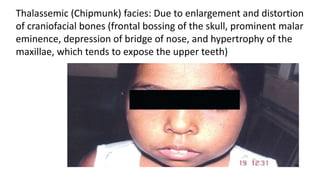
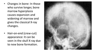
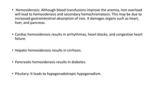
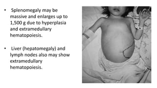
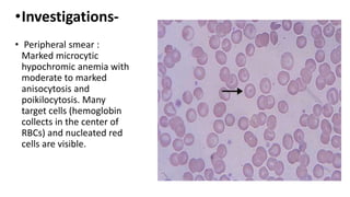
Thalassemia
Sonali Thakur
19104
Thalassemia syndrome is a
heterogeneous group of inherited
disorders which result from reduced or
absence of synthesis of one of the globin
chains (either α or β globin chain).
Classified into two main types -
depending on the defective globin chain- α or β
thalassemia.
α-Thalassemia Syndromes-
• Each cell has four genes coding for α-globin, two on each
chromosome.
• Each of the four α-globin genes normally contributes 25% of
the total α-globin chains.
• Severity of α-thalassemia depends on the number of α-
globin genes deleted or affected. Deleted genes may vary
from 1 to 4.
• Silent carrier state develops with deletion of one α-chain gene.
• α-thalassemia trait: It is due to deletion of two α-genes.
• HbH disease: It is due to deletion of three α-genes.
• Hb Bart’s hydrops fetalis: It develops if all four genes are absent.
It is incompatible with life and the infants are either stillborn or die
shortly after birth. They are pale, edematous, and have large liver
and spleen.
b-Thalassemia Major
• β-thalassemia major (Mediterranean or Cooley’s anemia) is
the homozygous form of β-thalassemia characterized by
absent or reduced synthesis of β-chain.
• Anemia is produced due to diminished synthesis of HbA,
ineffective erythropoiesis, and extravascular hemolysis.
Consequences of ineffective erythropoiesis:
• marked erythroid hyperplasia: Severe hemolytic anemia
stimulates erythropoietin (EPO) production by kidney leading
to marrow erythroid hyperplasia.
• Changes in the bone: Thalassemic facies and hair-on-end
appearance of skull X-ray
• Extramedullary hematopoiesis.
Clinical Features-
• Severe anemia: Infants with thalassemia major are well at birth but
develop moderate-to-severe anemia 6-9 months after birth, when
hemoglobin synthesis switches from HbF to HbA.
• Retardation of growth and development: Untreated/untransfused
children fail to thrive (growth retardation) and die early within 4–5
years of age from the effects of anemia. They are susceptible to
recurrent bacterial infections.
Thalassemic (Chipmunk) facies: Due to enlargement and distortion
of craniofacial bones (frontal bossing of the skull, prominent malar
eminence, depression of bridge of nose, and hypertrophy of the
maxillae, which tends to expose the upper teeth)
• Changes in bone: In those
who survive longer, bone
marrow hyperplasia
causes expansion and
widening of marrow and
gives the classical X-ray
changes.
• Hair-on-end (crew-cut)
appearance: It can be
seen in the skull X-ray due
to new bone formation.
• Hemosiderosis: Although blood transfusions improve the anemia, iron overload
will lead to hemosiderosis and secondary hemochromatosis. This may be due to
increased gastrointestinal absorption of iron. It damages organs such as heart,
liver, and pancreas.
• Cardiac hemosiderosis results in arrhythmias, heart blocks, and congestive heart
failure.
• Hepatic hemosiderosis results in cirrhosis.
• Pancreatic hemosiderosis results in diabetes.
• Pituitary: It leads to hypogonadotropic hypogonadism.
• Splenomegaly may be
massive and enlarges up to
1,500 g due to hyperplasia
and extramedullary
hematopoiesis.
• Liver (hepatomegaly) and
lymph nodes also may show
extramedullary
hematopoiesis.
•Investigations-
• Peripheral smear :
Marked microcytic
hypochromic anemia with
moderate to marked
anisocytosis and
poikilocytosis. Many
target cells (hemoglobin
collects in the center of
RBCs) and nucleated red
cells are visible.
• Osmotic fragility test shows increased resistance to
hemolysis.
• Skull radiograph shows a “hair-on-end”/crew-cut
appearance.
• HbF level is increased (30–92%) on hemoglobin
electrophoresis.
• There is markedly reduced or absent HbA.
• Aims of treatment:- To suppress ineffective erythropoiesis, prevent
deformities of bone, and allow normal activity and development.
Blood transfusions may be required every 4–6 weeks.
• Hypertransfusion to maintain Hb level between 10 and 12 g/dL is
probably adequate. It decreases the effect of chronic anemia and
prevents abnormal growth and development.
• Supertransfusion wherein the Hb level is maintained at 12 g/dL is
designed to completely suppress hematopoiesis.
Management
• Treatment of iron overload: The iron-chelating agent,
desferrioxamine (administered parenterally), is indicated if serum
ferritin >1,500 µg/L.
Ascorbic acid 200 mg daily along with desferrioxamine increases the
urinary excretion of iron in response to desferrioxamine.
Deferiprone and deferasirox are oral iron chelators.
.
• Splenectomy is indicated in children with massive symptomatic
splenomegaly and those with progressively increasing
requirement of blood transfusion or hypersplenism
• Bone marrow transplantation is done in young patients.
• Management of associated complications: eg.- congestive
heart failure and endocrinopathies
β-Thalassemia Minor (Trait)-
• β-thalassemia minor is more common than β-thalassemia major.
• It is a common carrier (heterozygous) state and is usually
asymptomatic.
• Anemia is mild or absent.
• Peripheral blood smear shows severe microcytic and hypochromic red
cells with target cells. It may be confused with iron deficiency.
• Serum ferritin and the iron stores are normal.
• Hb electrophoresis usually shows a raised HbA2 (3.5–7.5%) and often a
raised HbF.
• Iron should not be given to these patients unless there is associated iron
deficiency.
• Genetic counseling is recommended to prevent transmission of carrier
state from both parents.
• Prenatal diagnosis by chorionic villi biopsy at 11 weeks is done.
β-Thalassemia Intermedia-
• It is a clinical entity in which patients have a clinical spectrum
intermediate between thalassemia trait and thalassemia major.
• Patients are anemic and generally have mild-to-moderate anemia (Hb
7–9 g/dL).
• It is not transfusion dependent.
• Mild splenomegaly, bone deformities, gallstones, and chronic leg
ulcers may be seen.
• Folic acid supplementation should be given.
THANK YOU

DOC-20231023-WA0000.pptx

  • 1.
  • 2.
    Thalassemia syndrome isa heterogeneous group of inherited disorders which result from reduced or absence of synthesis of one of the globin chains (either α or β globin chain).
  • 3.
    Classified into twomain types - depending on the defective globin chain- α or β thalassemia.
  • 4.
    α-Thalassemia Syndromes- • Eachcell has four genes coding for α-globin, two on each chromosome. • Each of the four α-globin genes normally contributes 25% of the total α-globin chains. • Severity of α-thalassemia depends on the number of α- globin genes deleted or affected. Deleted genes may vary from 1 to 4.
  • 5.
    • Silent carrierstate develops with deletion of one α-chain gene. • α-thalassemia trait: It is due to deletion of two α-genes. • HbH disease: It is due to deletion of three α-genes. • Hb Bart’s hydrops fetalis: It develops if all four genes are absent. It is incompatible with life and the infants are either stillborn or die shortly after birth. They are pale, edematous, and have large liver and spleen.
  • 6.
    b-Thalassemia Major • β-thalassemiamajor (Mediterranean or Cooley’s anemia) is the homozygous form of β-thalassemia characterized by absent or reduced synthesis of β-chain. • Anemia is produced due to diminished synthesis of HbA, ineffective erythropoiesis, and extravascular hemolysis.
  • 7.
    Consequences of ineffectiveerythropoiesis: • marked erythroid hyperplasia: Severe hemolytic anemia stimulates erythropoietin (EPO) production by kidney leading to marrow erythroid hyperplasia. • Changes in the bone: Thalassemic facies and hair-on-end appearance of skull X-ray • Extramedullary hematopoiesis.
  • 8.
    Clinical Features- • Severeanemia: Infants with thalassemia major are well at birth but develop moderate-to-severe anemia 6-9 months after birth, when hemoglobin synthesis switches from HbF to HbA. • Retardation of growth and development: Untreated/untransfused children fail to thrive (growth retardation) and die early within 4–5 years of age from the effects of anemia. They are susceptible to recurrent bacterial infections.
  • 9.
    Thalassemic (Chipmunk) facies:Due to enlargement and distortion of craniofacial bones (frontal bossing of the skull, prominent malar eminence, depression of bridge of nose, and hypertrophy of the maxillae, which tends to expose the upper teeth)
  • 10.
    • Changes inbone: In those who survive longer, bone marrow hyperplasia causes expansion and widening of marrow and gives the classical X-ray changes. • Hair-on-end (crew-cut) appearance: It can be seen in the skull X-ray due to new bone formation.
  • 11.
    • Hemosiderosis: Althoughblood transfusions improve the anemia, iron overload will lead to hemosiderosis and secondary hemochromatosis. This may be due to increased gastrointestinal absorption of iron. It damages organs such as heart, liver, and pancreas. • Cardiac hemosiderosis results in arrhythmias, heart blocks, and congestive heart failure. • Hepatic hemosiderosis results in cirrhosis. • Pancreatic hemosiderosis results in diabetes. • Pituitary: It leads to hypogonadotropic hypogonadism.
  • 12.
    • Splenomegaly maybe massive and enlarges up to 1,500 g due to hyperplasia and extramedullary hematopoiesis. • Liver (hepatomegaly) and lymph nodes also may show extramedullary hematopoiesis.
  • 13.
    •Investigations- • Peripheral smear: Marked microcytic hypochromic anemia with moderate to marked anisocytosis and poikilocytosis. Many target cells (hemoglobin collects in the center of RBCs) and nucleated red cells are visible.
  • 14.
    • Osmotic fragilitytest shows increased resistance to hemolysis. • Skull radiograph shows a “hair-on-end”/crew-cut appearance. • HbF level is increased (30–92%) on hemoglobin electrophoresis. • There is markedly reduced or absent HbA.
  • 15.
    • Aims oftreatment:- To suppress ineffective erythropoiesis, prevent deformities of bone, and allow normal activity and development. Blood transfusions may be required every 4–6 weeks. • Hypertransfusion to maintain Hb level between 10 and 12 g/dL is probably adequate. It decreases the effect of chronic anemia and prevents abnormal growth and development. • Supertransfusion wherein the Hb level is maintained at 12 g/dL is designed to completely suppress hematopoiesis. Management
  • 16.
    • Treatment ofiron overload: The iron-chelating agent, desferrioxamine (administered parenterally), is indicated if serum ferritin >1,500 µg/L. Ascorbic acid 200 mg daily along with desferrioxamine increases the urinary excretion of iron in response to desferrioxamine. Deferiprone and deferasirox are oral iron chelators. .
  • 17.
    • Splenectomy isindicated in children with massive symptomatic splenomegaly and those with progressively increasing requirement of blood transfusion or hypersplenism • Bone marrow transplantation is done in young patients. • Management of associated complications: eg.- congestive heart failure and endocrinopathies
  • 18.
    β-Thalassemia Minor (Trait)- •β-thalassemia minor is more common than β-thalassemia major. • It is a common carrier (heterozygous) state and is usually asymptomatic. • Anemia is mild or absent. • Peripheral blood smear shows severe microcytic and hypochromic red cells with target cells. It may be confused with iron deficiency. • Serum ferritin and the iron stores are normal.
  • 19.
    • Hb electrophoresisusually shows a raised HbA2 (3.5–7.5%) and often a raised HbF. • Iron should not be given to these patients unless there is associated iron deficiency. • Genetic counseling is recommended to prevent transmission of carrier state from both parents. • Prenatal diagnosis by chorionic villi biopsy at 11 weeks is done.
  • 20.
    β-Thalassemia Intermedia- • Itis a clinical entity in which patients have a clinical spectrum intermediate between thalassemia trait and thalassemia major. • Patients are anemic and generally have mild-to-moderate anemia (Hb 7–9 g/dL). • It is not transfusion dependent. • Mild splenomegaly, bone deformities, gallstones, and chronic leg ulcers may be seen. • Folic acid supplementation should be given.
  • 21.