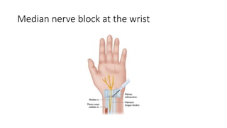
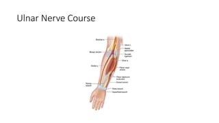
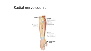
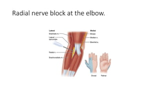
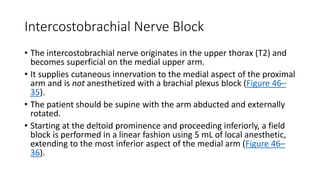
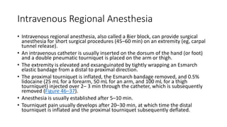
Blocks of the Terminal
Nerves
• Terminal nerves may be anesthetized anywhere along their course,
but the elbow and the wrist are the two most favored sites.
Median Nerve Block
• The median nerve is derived from the lateral and medial cords of the
brachial plexus.
• It enters the arm and runs just medial to the brachial artery near the
insertion of the biceps tendon.
• Just distal to this point, it gives off numerous motor branches to the
wrist and finger flexors and follows the interosseous membrane to the
wrist.
• At the level of the proximal wrist flexion crease, it lies directly behind
the palmaris longus tendon in the carpal tunnel.
Median nerve course.
• To block the median nerve at the elbow, the brachial artery is
identified in the antecubital crease just medial to the biceps insertion.
• A short 22-gauge insulated needle is inserted just medial to the artery
and directed toward the medial epicondyle until wrist flexion or
thumb opposition is elicited (Figure 46–24); 3–5 mL of local
anesthetic is then injected.
Median nerve block at the elbow
• To block the median nerve at the wrist, the palmaris longus tendon is
first identified by asking the patient to flex the wrist against
resistance.
• A short 22-gauge needle is inserted just medial and deep to the
palmaris longus tendon, and 3–5 mL of local anesthetic is injected
(Figure 46–26).
• With ultrasound, the median nerve may be identified at the level of
the mid-forearm between the muscle bellies of the flexor digitorum
profundus, flexor digitorum superficialis, and flexor pollicis longus
(transducer faces perpendicular to the trajectory of the nerves).
Median nerve block at the wrist
Ulnar Nerve Block
• The ulnar nerve is the continuation of the medial cord of the brachial
plexus and maintains a position medial to the axillary and brachial
arteries in the upper arm (Figure 46–27).
• At the distal third of the humerus, the nerve moves more medially
and passes under the arcuate ligament of the medial epicondyle.
• The nerve is frequently palpable just proximal to the medial
epicondyle.
• In the mid-forearm, the nerve lies between the flexor digitorum
profundus and the flexor carpi ulnaris.
• At the wrist, it is lateral to the flexor carpi ulnaris tendon and medial
to the ulnar artery.
Ulnar Nerve Course
• To block the ulnar nerve at the level of the elbow, an insulated 22-
gauge needle is inserted approximately one fingerbreadth proximal to
the arcuate ligament (Figure 46–28), and advanced until fourth/fifth
digit flexion or thumb adduction is elicited; 3–5 mL of local anesthetic
is then injected.
• To block the ulnar nerve at the wrist, the ulnar artery pulse is
palpated just lateral to the flexor carpi ulnaris tendon.
• The needle is inserted just medial to the artery (Figure 46–29) and 3–
5 mL of local anesthetic is injected.
• If ultrasound is used, the ulnar nerve may be identified just medial to
the ulnar artery.
Ulnar nerve block at the elbow with region of
anesthesia illustrated on the hand
Ulnar nerve block at the wrist
Radial Nerve Block
• The radial nerve—the terminal branch of the posterior cord of the brachial
plexus—courses posterior to the humerus, innervating the triceps muscle,
and enters the spiral groove of the humerus before it moves laterally at the
elbow (Figure 46–30).
• Terminal sensory branches include the lateral cutaneous nerve of the arm
and the posterior cutaneous nerve of the forearm.
• After exiting the spiral groove as it approaches the lateral epicondyle, the
radial nerve separates into superficial and deep branches.
• The deep branch remains close to the periosteum and innervates the
postaxial extensor group of the forearm.
• The superficial branch becomes superficial and follows the radial artery to
innervate the radial aspects of the dorsal wrist and the dorsal aspect of the
lateral three digits and half of the fourth.
Radial nerve course.
• To block the radial nerve at the elbow, the biceps tendon is identified
in the antecubital fossa.
• A short 22-gauge insulated needle is inserted just lateral to the
tendon and directed toward the lateral epicondyle (Figure 46–31)
until wrist or finger extension is elicited; 5 mL of local anesthetic is
then injected.
• With ultrasound, the radial nerve can be identified in cross-section
just proximal to the antecubital fossa between the biceps and
brachioradialis muscles.
Radial nerve block at the elbow.
• At the wrist, the superficial branch of the radial nerve lies just lateral
to the radial artery, which can be easily palpated lateral to the flexor
carpi radialis tendon (Figure 46–32).
• Using a short 22-gauge needle, 3–5 mL local anesthetic is injected
lateral to the artery.
• Ultrasound may be used at the level of the wrist or mid-forearm to
identify the radial nerve just lateral to the radial artery.
Radial nerve block at the wrist
• Musculocutaneous Nerve Block
• A musculocutaneous nerve block is essential to complete the
anesthesia for the forearm and wrist and is commonly included when
performing the axillary block.
• The musculocutaneous nerve is the terminal branch of the lateral
cord and the most proximal of the major nerves to emerge from the
brachial plexus (Figure 46–33).
• This nerve innervates the biceps and brachialis muscles and distally
terminates as the lateral antebrachial cutaneous nerve, supplying
sensory input to the lateral aspect of the forearm and wrist.
Musculocutaneous nerve course
• To target the musculocutaneous nerve following an axillary block, the needle is
redirected superior and proximal to the artery (see Figure 46–21), the
coracobrachialis muscle is pierced, and 5–10 mL of local anesthetic is injected,
with or without elicitation of elbow flexion. (Simple infiltration may be used,
although the success rate using this technique is questionable.)
• Ultrasound may be used to confirm the location of the musculocutaneous nerve
in the coracobrachialis muscle or between this muscle and the biceps (see Figure
46–22).
• Alternatively, the block can be performed at the elbow as the nerve courses
superficially at the interepicondylar line.
• The insertion of the biceps tendon is identified, and a short 22-guage needle is
inserted 1–2 cm laterally; 5–10 mL of local anesthetic is then injected as a field
block.
Digital Nerve Blocks
• Digital nerve blocks are used for minor operations on the fingers and to
supplement incomplete brachial plexus and terminal nerve blocks.
• Sensory innervation of each finger is provided by four small digital nerves
that enter each digit at its base in each of the four corners (Figure 46–34).
• A small-gauge needle is inserted at the medial and lateral aspects of the
base of the selected digit, and 2–3 mL of local anesthetic is inserted
without epinephrine.
• Addition of a vasoconstrictor (epinephrine) has been claimed to seriously
compromise blood flow to the digit; however, there are no case reports
involving lidocaine or other modern local anesthetics to confirm this claim.
Intercostobrachial Nerve Block
• The intercostobrachial nerve originates in the upper thorax (T2) and
becomes superficial on the medial upper arm.
• It supplies cutaneous innervation to the medial aspect of the proximal
arm and is not anesthetized with a brachial plexus block (Figure 46–
35).
• The patient should be supine with the arm abducted and externally
rotated.
• Starting at the deltoid prominence and proceeding inferiorly, a field
block is performed in a linear fashion using 5 mL of local anesthetic,
extending to the most inferior aspect of the medial arm (Figure 46–
36).
Intravenous Regional Anesthesia
• Intravenous regional anesthesia, also called a Bier block, can provide surgical
anesthesia for short surgical procedures (45–60 min) on an extremity (eg, carpal
tunnel release).
• An intravenous catheter is usually inserted on the dorsum of the hand (or foot)
and a double pneumatic tourniquet is placed on the arm or thigh.
• The extremity is elevated and exsanguinated by tightly wrapping an Esmarch
elastic bandage from a distal to proximal direction.
• The proximal tourniquet is inflated, the Esmarch bandage removed, and 0.5%
lidocaine (25 mL for a forearm, 50 mL for an arm, and 100 mL for a thigh
tourniquet) injected over 2– 3 min through the catheter, which is subsequently
removed (Figure 46–37).
• Anesthesia is usually established after 5–10 min.
• Tourniquet pain usually develops after 20–30 min, at which time the distal
tourniquet is inflated and the proximal tourniquet subsequently deflated.
• Patients usually tolerate the distal tourniquet for an additional 15–20
min because it is inflated over an anesthetized area.
• Even for surgical procedures of a very short duration, the tourniquet
must be left inflated for a total of at least 15–20 min to avoid a rapid
intravenous systemic bolus of local anesthetic resulting in toxicity.
• Slow deflation is also recommended to provide an additional margin
of safety.

terminal nerve block.pptx

  • 1.
    Blocks of theTerminal Nerves
  • 2.
    • Terminal nervesmay be anesthetized anywhere along their course, but the elbow and the wrist are the two most favored sites.
  • 3.
    Median Nerve Block •The median nerve is derived from the lateral and medial cords of the brachial plexus. • It enters the arm and runs just medial to the brachial artery near the insertion of the biceps tendon. • Just distal to this point, it gives off numerous motor branches to the wrist and finger flexors and follows the interosseous membrane to the wrist. • At the level of the proximal wrist flexion crease, it lies directly behind the palmaris longus tendon in the carpal tunnel.
  • 4.
  • 5.
    • To blockthe median nerve at the elbow, the brachial artery is identified in the antecubital crease just medial to the biceps insertion. • A short 22-gauge insulated needle is inserted just medial to the artery and directed toward the medial epicondyle until wrist flexion or thumb opposition is elicited (Figure 46–24); 3–5 mL of local anesthetic is then injected.
  • 6.
    Median nerve blockat the elbow
  • 7.
    • To blockthe median nerve at the wrist, the palmaris longus tendon is first identified by asking the patient to flex the wrist against resistance. • A short 22-gauge needle is inserted just medial and deep to the palmaris longus tendon, and 3–5 mL of local anesthetic is injected (Figure 46–26). • With ultrasound, the median nerve may be identified at the level of the mid-forearm between the muscle bellies of the flexor digitorum profundus, flexor digitorum superficialis, and flexor pollicis longus (transducer faces perpendicular to the trajectory of the nerves).
  • 8.
    Median nerve blockat the wrist
  • 9.
    Ulnar Nerve Block •The ulnar nerve is the continuation of the medial cord of the brachial plexus and maintains a position medial to the axillary and brachial arteries in the upper arm (Figure 46–27). • At the distal third of the humerus, the nerve moves more medially and passes under the arcuate ligament of the medial epicondyle. • The nerve is frequently palpable just proximal to the medial epicondyle. • In the mid-forearm, the nerve lies between the flexor digitorum profundus and the flexor carpi ulnaris. • At the wrist, it is lateral to the flexor carpi ulnaris tendon and medial to the ulnar artery.
  • 10.
  • 11.
    • To blockthe ulnar nerve at the level of the elbow, an insulated 22- gauge needle is inserted approximately one fingerbreadth proximal to the arcuate ligament (Figure 46–28), and advanced until fourth/fifth digit flexion or thumb adduction is elicited; 3–5 mL of local anesthetic is then injected. • To block the ulnar nerve at the wrist, the ulnar artery pulse is palpated just lateral to the flexor carpi ulnaris tendon. • The needle is inserted just medial to the artery (Figure 46–29) and 3– 5 mL of local anesthetic is injected. • If ultrasound is used, the ulnar nerve may be identified just medial to the ulnar artery.
  • 12.
    Ulnar nerve blockat the elbow with region of anesthesia illustrated on the hand
  • 13.
    Ulnar nerve blockat the wrist
  • 14.
    Radial Nerve Block •The radial nerve—the terminal branch of the posterior cord of the brachial plexus—courses posterior to the humerus, innervating the triceps muscle, and enters the spiral groove of the humerus before it moves laterally at the elbow (Figure 46–30). • Terminal sensory branches include the lateral cutaneous nerve of the arm and the posterior cutaneous nerve of the forearm. • After exiting the spiral groove as it approaches the lateral epicondyle, the radial nerve separates into superficial and deep branches. • The deep branch remains close to the periosteum and innervates the postaxial extensor group of the forearm. • The superficial branch becomes superficial and follows the radial artery to innervate the radial aspects of the dorsal wrist and the dorsal aspect of the lateral three digits and half of the fourth.
  • 15.
  • 16.
    • To blockthe radial nerve at the elbow, the biceps tendon is identified in the antecubital fossa. • A short 22-gauge insulated needle is inserted just lateral to the tendon and directed toward the lateral epicondyle (Figure 46–31) until wrist or finger extension is elicited; 5 mL of local anesthetic is then injected. • With ultrasound, the radial nerve can be identified in cross-section just proximal to the antecubital fossa between the biceps and brachioradialis muscles.
  • 17.
    Radial nerve blockat the elbow.
  • 18.
    • At thewrist, the superficial branch of the radial nerve lies just lateral to the radial artery, which can be easily palpated lateral to the flexor carpi radialis tendon (Figure 46–32). • Using a short 22-gauge needle, 3–5 mL local anesthetic is injected lateral to the artery. • Ultrasound may be used at the level of the wrist or mid-forearm to identify the radial nerve just lateral to the radial artery.
  • 19.
    Radial nerve blockat the wrist
  • 20.
    • Musculocutaneous NerveBlock • A musculocutaneous nerve block is essential to complete the anesthesia for the forearm and wrist and is commonly included when performing the axillary block. • The musculocutaneous nerve is the terminal branch of the lateral cord and the most proximal of the major nerves to emerge from the brachial plexus (Figure 46–33). • This nerve innervates the biceps and brachialis muscles and distally terminates as the lateral antebrachial cutaneous nerve, supplying sensory input to the lateral aspect of the forearm and wrist.
  • 21.
  • 22.
    • To targetthe musculocutaneous nerve following an axillary block, the needle is redirected superior and proximal to the artery (see Figure 46–21), the coracobrachialis muscle is pierced, and 5–10 mL of local anesthetic is injected, with or without elicitation of elbow flexion. (Simple infiltration may be used, although the success rate using this technique is questionable.) • Ultrasound may be used to confirm the location of the musculocutaneous nerve in the coracobrachialis muscle or between this muscle and the biceps (see Figure 46–22). • Alternatively, the block can be performed at the elbow as the nerve courses superficially at the interepicondylar line. • The insertion of the biceps tendon is identified, and a short 22-guage needle is inserted 1–2 cm laterally; 5–10 mL of local anesthetic is then injected as a field block.
  • 23.
    Digital Nerve Blocks •Digital nerve blocks are used for minor operations on the fingers and to supplement incomplete brachial plexus and terminal nerve blocks. • Sensory innervation of each finger is provided by four small digital nerves that enter each digit at its base in each of the four corners (Figure 46–34). • A small-gauge needle is inserted at the medial and lateral aspects of the base of the selected digit, and 2–3 mL of local anesthetic is inserted without epinephrine. • Addition of a vasoconstrictor (epinephrine) has been claimed to seriously compromise blood flow to the digit; however, there are no case reports involving lidocaine or other modern local anesthetics to confirm this claim.
  • 24.
    Intercostobrachial Nerve Block •The intercostobrachial nerve originates in the upper thorax (T2) and becomes superficial on the medial upper arm. • It supplies cutaneous innervation to the medial aspect of the proximal arm and is not anesthetized with a brachial plexus block (Figure 46– 35). • The patient should be supine with the arm abducted and externally rotated. • Starting at the deltoid prominence and proceeding inferiorly, a field block is performed in a linear fashion using 5 mL of local anesthetic, extending to the most inferior aspect of the medial arm (Figure 46– 36).
  • 25.
    Intravenous Regional Anesthesia •Intravenous regional anesthesia, also called a Bier block, can provide surgical anesthesia for short surgical procedures (45–60 min) on an extremity (eg, carpal tunnel release). • An intravenous catheter is usually inserted on the dorsum of the hand (or foot) and a double pneumatic tourniquet is placed on the arm or thigh. • The extremity is elevated and exsanguinated by tightly wrapping an Esmarch elastic bandage from a distal to proximal direction. • The proximal tourniquet is inflated, the Esmarch bandage removed, and 0.5% lidocaine (25 mL for a forearm, 50 mL for an arm, and 100 mL for a thigh tourniquet) injected over 2– 3 min through the catheter, which is subsequently removed (Figure 46–37). • Anesthesia is usually established after 5–10 min. • Tourniquet pain usually develops after 20–30 min, at which time the distal tourniquet is inflated and the proximal tourniquet subsequently deflated.
  • 26.
    • Patients usuallytolerate the distal tourniquet for an additional 15–20 min because it is inflated over an anesthetized area. • Even for surgical procedures of a very short duration, the tourniquet must be left inflated for a total of at least 15–20 min to avoid a rapid intravenous systemic bolus of local anesthetic resulting in toxicity. • Slow deflation is also recommended to provide an additional margin of safety.