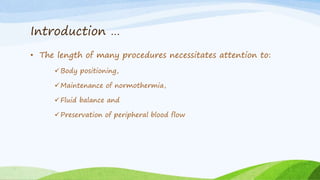
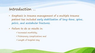
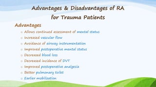
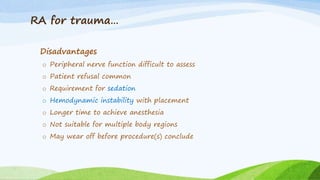
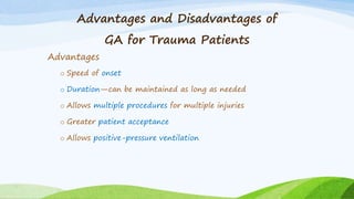
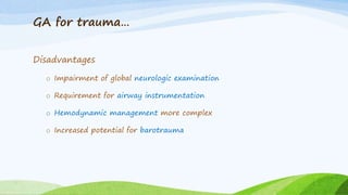
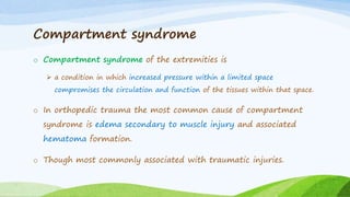
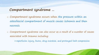
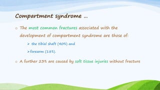
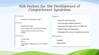
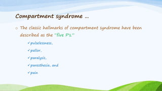
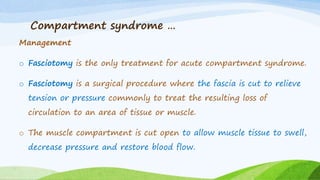
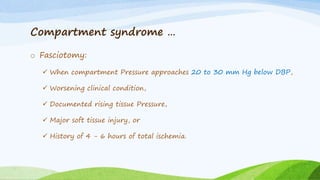
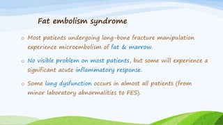
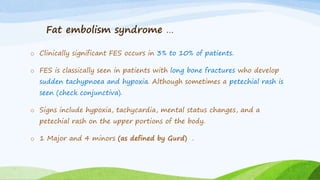
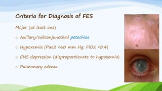
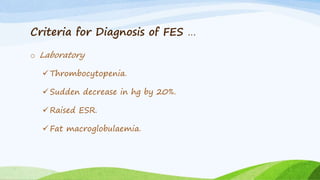
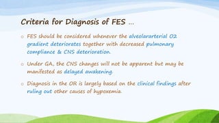
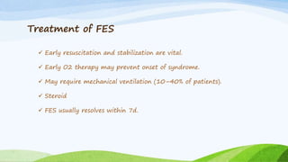
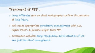
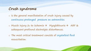
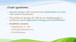
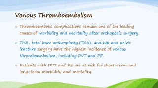
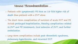
Orthopedic Trauma
considerations
Abraham Tarekegn
Lecturer, Department of Anesthesia
CMHS, University of Gondar
Outline
 Introduction
 Comparison of GA vs RA for orthopedic trauma
 Fat embolism syndrome
 Compartment syndrome
 crush syndrome
 VTE
Objectives
o Upon completion of this session you will be able to:
Describe special anesthetic issues related to orthopedic
trauma.
Utilize appropriate anesthesia options for OT.
Analyze advantages of one anesthetic technique over
the other.
Introduction
• Musculoskeletal injuries are the most frequent indication for
surgery in most trauma centers.
• Because many procedures can be appropriately managed
under RA, familiarity with these techniques is essential.
Introduction …
• The length of many procedures necessitates attention to:
Body positioning,
Maintenance of normothermia,
Fluid balance and
Preservation of peripheral blood flow
Introduction …
• Emphasis in trauma management of a multiple trauma
patient has included early stabilization of long-bone, spine,
pelvic, and acetabular fractures.
• Failure to do so results in:
 Increased morbidity,
 Pulmonary complications and
 Length of hospital stay
Advantages & Disadvantages of RA
for Trauma Patients
Advantages
o Allows continued assessment of mental status
o Increased vascular flow
o Avoidance of airway instrumentation
o Improved postoperative mental status
o Decreased blood loss
o Decreased incidence of DVT
o Improved postoperative analgesia
o Better pulmonary toilet
o Earlier mobilization
RA for trauma…
Disadvantages
o Peripheral nerve function difficult to assess
o Patient refusal common
o Requirement for sedation
o Hemodynamic instability with placement
o Longer time to achieve anesthesia
o Not suitable for multiple body regions
o May wear off before procedure(s) conclude
Advantages and Disadvantages of
GA for Trauma Patients
Advantages
o Speed of onset
o Duration—can be maintained as long as needed
o Allows multiple procedures for multiple injuries
o Greater patient acceptance
o Allows positive-pressure ventilation
GA for trauma…
Disadvantages
o Impairment of global neurologic examination
o Requirement for airway instrumentation
o Hemodynamic management more complex
o Increased potential for barotrauma
Compartment syndrome
o Compartment syndrome of the extremities is
 a condition in which increased pressure within a limited space
compromises the circulation and function of the tissues within that space.
o In orthopedic trauma the most common cause of compartment
syndrome is edema secondary to muscle injury and associated
hematoma formation.
o Though most commonly associated with traumatic injuries.
Compartment syndrome …
o Compartment syndrome occurs when the pressure within an
osteofascial compartment of muscle causes ischemia and then
necrosis.
o Compartment syndrome can also occur as a result of a number of causes
associated with trauma including:
 reperfusion injury, burns, drug overdose, and prolonged limb compression
Compartment syndrome …
o The most common fractures associated with the
development of compartment syndrome are those of:
 the tibial shaft (40%) and
forearm (18%).
o A further 23% are caused by soft tissue injuries without fracture
Risk Factors for The Development of
Compartment Syndrome
Compartment syndrome …
o The classic hallmarks of compartment syndrome have been
described as the “five P's.”
pulselessness,
pallor,
paralysis,
paresthesia, and
pain
Compartment syndrome …
Management
o Fasciotomy is the only treatment for acute compartment syndrome.
o Fasciotomy is a surgical procedure where the fascia is cut to relieve
tension or pressure commonly to treat the resulting loss of
circulation to an area of tissue or muscle.
o The muscle compartment is cut open to allow muscle tissue to swell,
decrease pressure and restore blood flow.
Compartment syndrome …
o Fasciotomy:
 When compartment Pressure approaches 20 to 30 mm Hg below DBP,
 Worsening clinical condition,
 Documented rising tissue Pressure,
 Major soft tissue injury, or
 History of 4 - 6 hours of total ischemia.
Fat embolism syndrome
o Most patients undergoing long-bone fracture manipulation
experience microembolism of fat & marrow.
o No visible problem on most patients, but some will experience a
significant acute inflammatory response.
o Some lung dysfunction occurs in almost all patients (from
minor laboratory abnormalities to FES).
Fat embolism syndrome …
o Clinically significant FES occurs in 3% to 10% of patients.
o FES is classically seen in patients with long bone fractures who develop
sudden tachypnoea and hypoxia. Although sometimes a petechial rash is
seen (check conjunctiva).
o Signs include hypoxia, tachycardia, mental status changes, and a
petechial rash on the upper portions of the body.
o 1 Major and 4 minors (as defined by Gurd) .
Criteria for Diagnosis of FES
Major (at least one)
o Axillary/subconjunctival petechiae
o Hypoxemia (Pao2 <60 mm Hg; FIO2 <0.4)
o CNS depression (disproportionate to hypoxemia)
o Pulmonary edema
Criteria for Diagnosis of FES …
Minor (at least four)
o Tachycardia (>110 beats/min)
o Hyperthermia
o Retinal fat emboli
o Urinary fat globules
o Decreased platelets/hematocrit (unexplained)
o Increased erythrocyte sedimentation rate
o Fat globules in sputum
Criteria for Diagnosis of FES …
o Laboratory
Thrombocytopenia.
Sudden decrease in hg by 20%.
Raised ESR.
Fat macroglobulaemia.
Criteria for Diagnosis of FES …
o FES should be considered whenever the alveolararterial O2
gradient deteriorates together with decreased pulmonary
compliance & CNS deterioration.
o Under GA, the CNS changes will not be apparent but may be
manifested as delayed awakening.
o Diagnosis in the OR is largely based on the clinical findings after
ruling out other causes of hypoxemia.
Treatment of FES
 Early resuscitation and stabilization are vital.
 Early O2 therapy may prevent onset of syndrome.
 May require mechanical ventilation (10–40% of patients).
 Steroid
 FES usually resolves within 7d.
Treatment of FES …
o Lung infiltrates seen on chest radiography confirm the presence
of lung injury.
o This needs appropriate ventilatory management with O2,
higher PEEP, & possible longer term MV.
o Treatment includes: early recognition, administration of O2,
and judicious fluid management.
Crush syndrome
o Is the general manifestation of crush injury caused by
continuous prolonged pressure on extremities.
o Muscle injury 2o to ischemia  Myoglobinuria  ARF &
subsequent profound electrolyte disturbances.
o The most critical treatment consists of crystalloid fluid
resuscitation.
Crush syndrome …
o Osmotic diuresis with mannitol and alkalinization of urine
with sodium bicarbonate.
o The preferred therapy for ARF 2o to rhabdomyolysis is
continuous renal replacement therapy and hemofiltration.
o Anesthetic concerns
 Myoglobinuria  ARF
 Electrolyte disturbance (K)
 Fluid disturbance
Venous Thromboembolism
o Thromboembolic complications remain one of the leading
causes of morbidity and mortality after orthopedic surgery.
o THA, total knee arthroplasty (TKA), and hip and pelvic
fracture surgery have the highest incidence of venous
thromboembolism, including DVT and PE.
o Patients with DVT and PE are at risk for short-term and
long-term morbidity and mortality.
Venous Thromboembolism …
o Patients with symptomatic PE have an 18-fold higher risk of
death than patients with a DVT alone.
o The short-term complications of survivors of acute DVT and PE
include prolonged hospitalization, bleeding complications related
to DVT and PE treatments, local extension of DVT, and further
embolization.
o Long-term complications include post-thrombotic syndrome,
pulmonary hypertension, and recurrent DVT.
Venous Thromboembolism …
Risk factors:
o advanced age greater than 60 years,
o obesity,
o prolonged immobility or bed rest more than 4 days,
o prior history of thromboembolism,
o cancer,
o pre-existing hypercoaguable state
o major surgery.
o procedures lasting more than 30 min,
o use of a tourniquet,
o lower extremity fracture
What to do for VTE
What to do for VTE …
Management
o Pharmacological prophylaxis and the routine use of mechanical
devices such as intermittent pneumatic compression (IPC) have
been shown to decrease the incidence of DVT and PE.
o While mechanical thromboprophylaxis should be considered for
every patient, the use of pharmacological anticoagulants must be
balanced against the risk of major bleeding.
Management …
o For patients at increased risk for DVT but having “normal” bleeding
risk,
mechanical prophylaxis
low-dose subcutaneous unfractionated heparin (LUFH),
warfarin, or
low-molecular-weight heparin (LMWH)
o Patients at significantly increased risk of bleeding may be managed
with mechanical prophylaxis alone until bleeding risk decreases.
Thank You!!!

Orthopedic trauma.pptx

  • 1.
    Orthopedic Trauma considerations Abraham Tarekegn Lecturer,Department of Anesthesia CMHS, University of Gondar
  • 2.
    Outline  Introduction  Comparisonof GA vs RA for orthopedic trauma  Fat embolism syndrome  Compartment syndrome  crush syndrome  VTE
  • 3.
    Objectives o Upon completionof this session you will be able to: Describe special anesthetic issues related to orthopedic trauma. Utilize appropriate anesthesia options for OT. Analyze advantages of one anesthetic technique over the other.
  • 4.
    Introduction • Musculoskeletal injuriesare the most frequent indication for surgery in most trauma centers. • Because many procedures can be appropriately managed under RA, familiarity with these techniques is essential.
  • 5.
    Introduction … • Thelength of many procedures necessitates attention to: Body positioning, Maintenance of normothermia, Fluid balance and Preservation of peripheral blood flow
  • 6.
    Introduction … • Emphasisin trauma management of a multiple trauma patient has included early stabilization of long-bone, spine, pelvic, and acetabular fractures. • Failure to do so results in:  Increased morbidity,  Pulmonary complications and  Length of hospital stay
  • 7.
    Advantages & Disadvantagesof RA for Trauma Patients Advantages o Allows continued assessment of mental status o Increased vascular flow o Avoidance of airway instrumentation o Improved postoperative mental status o Decreased blood loss o Decreased incidence of DVT o Improved postoperative analgesia o Better pulmonary toilet o Earlier mobilization
  • 8.
    RA for trauma… Disadvantages oPeripheral nerve function difficult to assess o Patient refusal common o Requirement for sedation o Hemodynamic instability with placement o Longer time to achieve anesthesia o Not suitable for multiple body regions o May wear off before procedure(s) conclude
  • 9.
    Advantages and Disadvantagesof GA for Trauma Patients Advantages o Speed of onset o Duration—can be maintained as long as needed o Allows multiple procedures for multiple injuries o Greater patient acceptance o Allows positive-pressure ventilation
  • 10.
    GA for trauma… Disadvantages oImpairment of global neurologic examination o Requirement for airway instrumentation o Hemodynamic management more complex o Increased potential for barotrauma
  • 11.
    Compartment syndrome o Compartmentsyndrome of the extremities is  a condition in which increased pressure within a limited space compromises the circulation and function of the tissues within that space. o In orthopedic trauma the most common cause of compartment syndrome is edema secondary to muscle injury and associated hematoma formation. o Though most commonly associated with traumatic injuries.
  • 12.
    Compartment syndrome … oCompartment syndrome occurs when the pressure within an osteofascial compartment of muscle causes ischemia and then necrosis. o Compartment syndrome can also occur as a result of a number of causes associated with trauma including:  reperfusion injury, burns, drug overdose, and prolonged limb compression
  • 13.
    Compartment syndrome … oThe most common fractures associated with the development of compartment syndrome are those of:  the tibial shaft (40%) and forearm (18%). o A further 23% are caused by soft tissue injuries without fracture
  • 14.
    Risk Factors forThe Development of Compartment Syndrome
  • 15.
    Compartment syndrome … oThe classic hallmarks of compartment syndrome have been described as the “five P's.” pulselessness, pallor, paralysis, paresthesia, and pain
  • 16.
    Compartment syndrome … Management oFasciotomy is the only treatment for acute compartment syndrome. o Fasciotomy is a surgical procedure where the fascia is cut to relieve tension or pressure commonly to treat the resulting loss of circulation to an area of tissue or muscle. o The muscle compartment is cut open to allow muscle tissue to swell, decrease pressure and restore blood flow.
  • 17.
    Compartment syndrome … oFasciotomy:  When compartment Pressure approaches 20 to 30 mm Hg below DBP,  Worsening clinical condition,  Documented rising tissue Pressure,  Major soft tissue injury, or  History of 4 - 6 hours of total ischemia.
  • 18.
    Fat embolism syndrome oMost patients undergoing long-bone fracture manipulation experience microembolism of fat & marrow. o No visible problem on most patients, but some will experience a significant acute inflammatory response. o Some lung dysfunction occurs in almost all patients (from minor laboratory abnormalities to FES).
  • 19.
    Fat embolism syndrome… o Clinically significant FES occurs in 3% to 10% of patients. o FES is classically seen in patients with long bone fractures who develop sudden tachypnoea and hypoxia. Although sometimes a petechial rash is seen (check conjunctiva). o Signs include hypoxia, tachycardia, mental status changes, and a petechial rash on the upper portions of the body. o 1 Major and 4 minors (as defined by Gurd) .
  • 20.
    Criteria for Diagnosisof FES Major (at least one) o Axillary/subconjunctival petechiae o Hypoxemia (Pao2 <60 mm Hg; FIO2 <0.4) o CNS depression (disproportionate to hypoxemia) o Pulmonary edema
  • 21.
    Criteria for Diagnosisof FES … Minor (at least four) o Tachycardia (>110 beats/min) o Hyperthermia o Retinal fat emboli o Urinary fat globules o Decreased platelets/hematocrit (unexplained) o Increased erythrocyte sedimentation rate o Fat globules in sputum
  • 22.
    Criteria for Diagnosisof FES … o Laboratory Thrombocytopenia. Sudden decrease in hg by 20%. Raised ESR. Fat macroglobulaemia.
  • 23.
    Criteria for Diagnosisof FES … o FES should be considered whenever the alveolararterial O2 gradient deteriorates together with decreased pulmonary compliance & CNS deterioration. o Under GA, the CNS changes will not be apparent but may be manifested as delayed awakening. o Diagnosis in the OR is largely based on the clinical findings after ruling out other causes of hypoxemia.
  • 24.
    Treatment of FES Early resuscitation and stabilization are vital.  Early O2 therapy may prevent onset of syndrome.  May require mechanical ventilation (10–40% of patients).  Steroid  FES usually resolves within 7d.
  • 25.
    Treatment of FES… o Lung infiltrates seen on chest radiography confirm the presence of lung injury. o This needs appropriate ventilatory management with O2, higher PEEP, & possible longer term MV. o Treatment includes: early recognition, administration of O2, and judicious fluid management.
  • 26.
    Crush syndrome o Isthe general manifestation of crush injury caused by continuous prolonged pressure on extremities. o Muscle injury 2o to ischemia  Myoglobinuria  ARF & subsequent profound electrolyte disturbances. o The most critical treatment consists of crystalloid fluid resuscitation.
  • 27.
    Crush syndrome … oOsmotic diuresis with mannitol and alkalinization of urine with sodium bicarbonate. o The preferred therapy for ARF 2o to rhabdomyolysis is continuous renal replacement therapy and hemofiltration. o Anesthetic concerns  Myoglobinuria  ARF  Electrolyte disturbance (K)  Fluid disturbance
  • 28.
    Venous Thromboembolism o Thromboemboliccomplications remain one of the leading causes of morbidity and mortality after orthopedic surgery. o THA, total knee arthroplasty (TKA), and hip and pelvic fracture surgery have the highest incidence of venous thromboembolism, including DVT and PE. o Patients with DVT and PE are at risk for short-term and long-term morbidity and mortality.
  • 29.
    Venous Thromboembolism … oPatients with symptomatic PE have an 18-fold higher risk of death than patients with a DVT alone. o The short-term complications of survivors of acute DVT and PE include prolonged hospitalization, bleeding complications related to DVT and PE treatments, local extension of DVT, and further embolization. o Long-term complications include post-thrombotic syndrome, pulmonary hypertension, and recurrent DVT.
  • 30.
    Venous Thromboembolism … Riskfactors: o advanced age greater than 60 years, o obesity, o prolonged immobility or bed rest more than 4 days, o prior history of thromboembolism, o cancer, o pre-existing hypercoaguable state o major surgery. o procedures lasting more than 30 min, o use of a tourniquet, o lower extremity fracture
  • 31.
    What to dofor VTE
  • 32.
    What to dofor VTE …
  • 33.
    Management o Pharmacological prophylaxisand the routine use of mechanical devices such as intermittent pneumatic compression (IPC) have been shown to decrease the incidence of DVT and PE. o While mechanical thromboprophylaxis should be considered for every patient, the use of pharmacological anticoagulants must be balanced against the risk of major bleeding.
  • 34.
    Management … o Forpatients at increased risk for DVT but having “normal” bleeding risk, mechanical prophylaxis low-dose subcutaneous unfractionated heparin (LUFH), warfarin, or low-molecular-weight heparin (LMWH) o Patients at significantly increased risk of bleeding may be managed with mechanical prophylaxis alone until bleeding risk decreases.
  • 35.

Editor's Notes

  • #35 Low-molecular-weight heparin (LMWH) is recommended over unfractionated heparin (intravenous or subcutaneous) for initial therapy of DVT and PE. LMWHs do not require monitoring of coagulation. Although DVT prophylaxis may be more efficient when started preoperatively, the risk of surgical bleeding also increases. Administration of LMWH 6 hours postoperatively was effective in DVT prophylaxis and did not increase bleeding; delaying prophylaxis until 24 hours was less effective.[62] Although the optimal duration of therapy is unknown, LMWH should be continued for at least 10 days in routine orthopedic procedures and in patients not considered high risk. Extended prophylaxis to 28 to 35 days would be supported in patients with evidence of a DVT or at higher risk for DVT