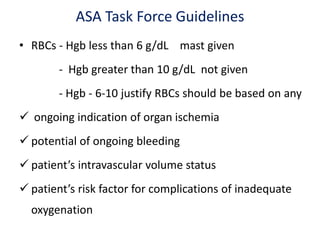
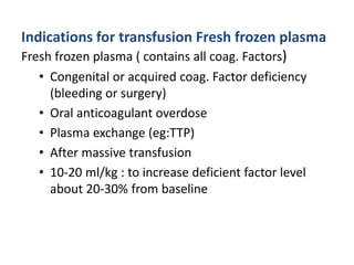
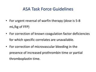
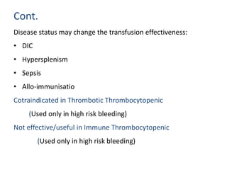
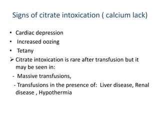
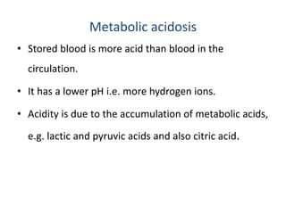
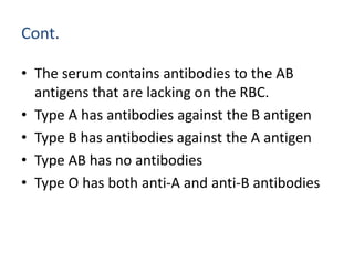
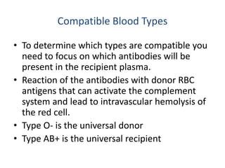
2. Blood transfusion
• It is introducing the blood of a donor or pre-donated
blood by a recipient in to the recipients' blood stream.
Purpose of Blood transfusion
• To increases the blood’s oxygen (Hgb)carrying capacity
• Reverses tissue hypoxia
• To restoring circulating volume.
• To provide clotting factors or platelets in patient with
coagulation failure.
3. Blood Products
• Whole blood- un separated blood collected in to an
approved container containing an anticoagulant
preservative solution
• Blood product-any therapeutic substance prepared
from whole blood.
- Packed Red Blood Cell
- Platelets
- Fresh Frozen Plasma
- Cryoprecipitate
4. Red Blood Cells
• Whole blood is collected in bags containing citrate-
phosphate-dextrose-adenine (CPDA) solution.
• Citrate chelates ( calcium )- prevents coagulation.
• PRBC- Prepared by centrifugation of the whole blood.
• CPDA blood has - Hct of 70-75%
- 50-70 mL of residual plasma
(TV 250-275 mL)
- A shelf live of 35 days.
5. PRBC
• Global increase in oxygen delivery with potentially
decreased microcirculatory flow
– Increased blood viscousity
– Cytokines my cause vasoconstriction
– Low levels of 2,3 DPG shifts curve left, impeding
oxygen availability
– Decreased RBC membrane deformability
– Free Hb may bind NO causing vasoconstriction
• One unit of RBCs will increase the Hb and Hct of a
1g/dL and 3% respectively.
6. Cont.
Washed RBC Any need to prevent the recipient allo-
immunisation to WBC’s , plasma antigens or any
contraindication to infuse complement
• PNH
• IgA deficiency
• Prevention of anaphylaxis
Frozen RBC’s:
• An indication for RBC transfusion +
– Autologous transfusion: rare blood groups,
– Catastrophy etc
7. Immunologic effects of RBC transfusion
• Some evidence that it may cause
– Immune suppression by altering lymphocyte
reactivity
– Pro inflammatory: cytokines in unfiltered rbc’s
might trigger SIRS or multi organ failure
8. Cont.
• An indication for RBC transfusion +
– To prevent reactions caused by WBC antibodies
• Febrile non-hemolytic transfusion reactions
– To prevent alloimmunization
– To prevent CMV transmission
• NB- White cells reduced RBC’s: < 5x10⁶ WBC’s per unit
• White cell filters (before storage or before transfusion)
9. Red Cell Transfusion Triggers
Guidelines for the clinical use of red cell transfusions
(BCSH 2001)
• Hb > 10g/dl – Transfusion not indicated
• Hb > 7-10g/dl – Transfuse only if clinically indicated
• Hb < 7g/dl – Transfusion generally
indicated
10. ASA Task Force Guidelines
• RBCs - Hgb less than 6 g/dL mast given
- Hgb greater than 10 g/dL not given
- Hgb - 6-10 justify RBCs should be based on any
ongoing indication of organ ischemia
potential of ongoing bleeding
patient’s intravascular volume status
patient’s risk factor for complications of inadequate
oxygenation
11. Fresh Frozen Plasma
• Plasma is separated from the RBC component of whole
blood by centrifugation.
• One unit has a volume of 200-250 mL and contains all the
plasma proteins (factors V and VIII).
• Preservative added at the time of collection.
• FFP is frozen promptly to preserve two labile clotting
factors (V and VIII) and thawed only immediately prior to
administration.
12. Fresh Frozen Plasma(FFP)
Given
• For coagulation factor deficiency in patients transfused
with more than one blood volume
• PT and PTT cannot be obtained in a timely fashion.
• Doses is calculated to achieve a minimum of 30% of
plasma factor concentration.
(usually achieved with 10-15mL/kg of FFP)
13. Indications for transfusion Fresh frozen plasma
Fresh frozen plasma ( contains all coag. Factors)
• Congenital or acquired coag. Factor deficiency
(bleeding or surgery)
• Oral anticoagulant overdose
• Plasma exchange (eg:TTP)
• After massive transfusion
• 10-20 ml/kg : to increase deficient factor level
about 20-30% from baseline
14. Cont.
• Immediate reversal of warfarin effect in the presence of
life threatening bleeding
• Acute DIC in the presence of bleeding and abnormal
coagulation results fibrinogen level <1g/l
• Bleeding associated with thrombolytic therapy causing
hypofibrinogenaemia
• Hypofibrinogenaemia 2o to massive transfusion
15. ASA Task Force Guidelines
• For urgent reversal of warfin therapy (dose is 5-8
mL/kg of FFP)
• For correction of known coagulation factor deficiencies
for which specific correlates are unavailable.
• For correction of microvascular bleeding in the
presence of increased prothrombin time or partial
thromboplastin time.
16. Platelets
• The platelets are separated from the plasma by
centrifugation.
• One unit of platelets will increase the platelet count
of a 70 kg adult by 5000-10,000/mm³.
• Platelet viability is optimal at 22° C but storage is
limited to 48 hours.
17. Indications for platelet transfusions
• Decreased platelet production because of bone marrow failure
or infiltration :bleeding or risk of bleeding
– Leukemia - Malignant tm infiltration
– Myelosupression - MDS
– Aplastic anemia - Myelofibrosis
• Functional platelet disease and bleeding or risk of bleeding
• Dilutional thrombocytopenia (after massive transfusion)
• Cardiac by-pass surgery
• Increased platelet destruction or consumption
– DIC, Drug induced, sepsis and ITP
18. Cont.
• Platelets:
Thrombocytopenia due to decreased platelet
production
Platelet count/mm3 Bleeding /surgery Indication for plt transfusion
> 50.000, No No
< 50.000 Yes Yes
10.000-20.000 No No
(if there is bleeding/fever/DIC/plt dysfunction) Yes
< 10.000 Yes or No Yes
19. Cont.
Disease status may change the transfusion effectiveness:
• DIC
• Hypersplenism
• Sepsis
• Allo-immunisatio
Cotraindicated in Thrombotic Thrombocytopenic
(Used only in high risk bleeding)
Not effective/useful in Immune Thrombocytopenic
(Used only in high risk bleeding)
20. Cryoprecipitate
• Cryoprecipitate is the precipitate that remains when
the FFP is thawed slowly at 4° C.
• It is a concentrated source of factor VIII, factor XIII and
fibrinogen.
• One unit of cryoprecipitate (which is the yield from
one unit of FFP) contains sufficient fibrinogen to
increase fibrinogen level 5 to 7 mg/dL.
21. Cont.
• Includes FVIII, vWF, FXIII, fibrinogen and fibronectin
• 80-120 units of FVIII, ≥150 mg fibrinogen and 20-
30 % of FXIII that is in one unit of plasma
• Can be used for the purpose of replacing the
deficient state of these factors in case of bleeding
or surgery
22. Cryoprecipitate
• It is stored at -20°C and thawed immediately prior to
use.
• Cryoprecipitate is used in the treatment of factor VIII
deficiency, hemophilia A and fibrinogen deficiencies.
23. Storage of blood
• Metabolism of glucose by erythrocytes produce lactic
acid , pyruvic acid&CO2.
• Accumulation of this substances on the storage
produce progress PH decrement.
• Acidosis impairs enzyme function &reduce cellular
viability.
24. Storage of blood
• Blood is stored at a temperature of 4 OC
- to slow the erythrocyte metabolism.
- to minimize the accumulation of acidic substance
- increase post transfusion survival of RBCs
25. Change with blood storage
• Reduced oxygen carrying capacity of blood
due to deteriorated RBC function.
• Platelet function decline rapidly with storage so that
after 48hrs of storage the platelet will not assist in
coagulation.
• Hyperkalemia due to increased potasium from
damaged RBC
• Deterioration of coagulation factors v,viii
• Acidosis
• Decreased ATP
26. Preservatives added to blood(CPDA)
• Citrate phosphate dextrose adenine(CPDA)-is an
anticoagulant preservative in which blood is stored.
• Citrate is an anticoagulant,phosphate serve as a
buffer&dextrose is a red cell energy source.
• The addition of adenine to CPD solution allows RBC to
re-synthesize ATP which extends the storage time from
21 to 35 days.
27. Sources of blood transfusion
1. Homologous/allogenic-blood transfused from donor
2. Autologous transfusion-blood is collected from the
patient &then subsequently re-infused.
• Methods of autotransfusion
Pre-deposit of blood-collection of patients own blood
prior to elective surgery.
28. • The deficit is normally corrected with in 3-5
days.
• Check the hgb conc. immediately prior to the
operation to ensure that the patient has
compensated for the deficit.
29. Advantage
i. Where there is a refusal of patient who may require
transfusion to accept blood which has been
donated by others.
ii. Patients requiring transfusion with a rare blood
group &donor of compatible blood cannot find.
iii. When there are no or only limited blood bank
facilities available.
30. Indications for blood transfusion
• Sever anemia and chronic blood loss
• Blood clotting disturbance-to provide platelet plasma
clotting factors fresh whole blood is given.
• Acute blood loss-if > 20% of the patients total
volume is lost
31. Cont.
• Estimated blood volume in different age group is
calculated as for
- Neonate 85-90ml/kg body weight
- Children 80ml/kg
- Adult (male) 70ml/kg
- Adult( female) 65ml/kg
• Value is adjusted according to preop hgb level ,cardio
respiratory status & probability of continuing blood loss.
32. Intraoperative blood loss can be estimated as
- Blood in suction machine
- Large abdominal towel=100ml
- Small gauze absorb =10ml
- Estimate blood loss on floor &drape
*Note the volume of irrigation fluid, subtract this
volume from measured blood loss
33. The need for transfusion can be minimized
- By the prevention or early dx &Rx of anemia the
condition that causes anemia.
- The use intravenous fluid replacement in cases of
acute blood loss.
- Best anesthetic and surgical management to minimize
blood loss during surgery.
- Stopping anticoagulant &anti platelet drugs before
planned surgery.
- Salvaging &re infusing surgical losses.
34. Techniques used to minimize peri-operative
blood loss.
• Anesthetic techniques
• Surgical techniques
35. Anesthetic techniques
• Avoid hypertension &tachycardia due to sympathetic
over activity by ensuring adequate level anesthesia
&analgesia.
• Avoid hypercarbia causing HTN which will increase
operative blood loss.
• Use regional anesthesia such as epidural & spinal
anesthesia when appropriate.
36. Surgical techniques
• Training, experience & care of the surgeon is the
most crucial factor.
• Meticulous attention to bleeding site use of
diathermy
• Position the level of operative site should be a little
above the heart.
e.g trendlenberg position for lower limb, pelvic &
abdominal procedures; head up posture for head
&neck surgery.
37. Cont.
• Tourniquet:- limitation pressure of tourniquet should be
approximately 100-150mmhg above systolic blood
pressure of the patient.
• Vasoconstrictors:-I nfiltration of the incision site with
adrenaline; avoid using vasoconstrictor at the end arteries
finger, toes & penis.
• Post operative adequate analgesia-post operative pain can
cause HTN &restlessness which can aggravate bleeding.
38. Before you give blood –check
- Check patient identity against patient note &
transfusion form.
- Correct blood –check label on blood &
transfusion form.
- Check the expiration date
40. Transfusion reactions
• RBCs are haemolysed or break down after introduced into
the patients circulation.
• Cause of haemolysis is incompatibility of transfused blood
with the patients blood.
• Sign of in-compatiblity may be masked by general
anesthesia.
• BP&PR of the patient must be checked every 5 minutes for
the first 15 minutes.
41. Cont.
• Intravascular haemolytic transfusion reaction-almost
always occur due to incompatibility of the ABO system.
• Very rare occurring in 1 out of 15,000-20,000 transfusions.
• GA & sedation may mask the sign &symptoms
• Occur during or shortly after (with in 24 hrs) of transfusion
42. Cont.
• In conscious patient pain at the transfusion site is the first
sign.
• In anesthesized patient the reaction is manifested by
- Urticarial rash
- Rise in body temperature
- Unexplained tachycardia
- Hypotension
- Haemoglobinuria, oliguria & jaundice
- Diffuse oozing in surgical field
• Acute circulatory collapse &renal failure are the most
serious complications & may lead to death
43. Management of sever reaction under
anesthesia
• Stop the transfusion
• Treat hypotension appropriately
- Fluids( crystalloid/colloid)
- Inotropes e.g adrenaline 1-5ml of1:10,000
- Other compatible blood if necessary
• Maintain airway give high flow oxygen
• Consider IV corticosteroid & bronchodilator
• Establish the diagnosis with the blood bank
assistance.
44. Cont.
• Treat renal vascular ischemia with
- IV Saline to keep UOP above 1ml/kg/hr insert
urinary catheter.
- Frusemide 20mg IV
- Mannitol 0.5g/kg IV
45. Massive transfusion
• Defined as the replacement of blood loss equivalent or
greater than patients total blood volume in less than
24hrs.
• Coagulation problems commonly result platelets & FFP
or plasma are required.
• Precautions when transfusing large volumes of blood
- Use 2 units of FFP for every 6 units of blood
46. Cont.
• Do platelet counts after every 6 units of blood Give
platelets if the count is <75,000/ mm3.
• Give calcium gluconate 10% (or chloride) at the rate of
10ml/6 units of blood.
• Monitor: pulse, BP, ECG, urine output, the acid base
status and serum electrolytes (if possible).
47. Transmission of infections
• Some important viral diseases may be transmitted in
transfused blood especially where screening tests are not
available.
• Viral hepatitis (jaundice may occur between 25 and 180 days).
- HIV
- Malaria
-Syphilis
48. Hyperkalaemia
• RBCs contain a very high concentration of K+
(100mmol/L).
• Plasma conta in 3.5 - 5 mmol/L K+
• Old blood (after 3 - 4 wks) - 25mmol/L of K+ in plasma.
• K+ from RBCs diffuse outs in to the plasma.
• If pt have a high serum K+ or if there is renal failure, FFP
or blood stored for less than 5 days should be used.
49. Citrate intoxication
• Whole blood is prevented from clotting (after
collection), by the use of citrate.
• Citrate forms a complex with the calcium ions in the
blood.
• Calcium is necessary for the blood to clot and the
calcium is not available (being tied up in a complex
with citrate), the blood remains fluid.
50. Cont.
• When the citrated blood is introduced into the
patient's circulation, the citrate is quickly broken down
into water and carbon dioxide and the calcium is set
free.
• If for some reason the citrate persists in the circulation
,signs of citrate intoxication(lack of calcium) are seen.
51. Signs of citrate intoxication ( calcium lack)
• Cardiac depression
• Increased oozing
• Tetany
Citrate intoxication is rare after transfusion but it
may be seen in:
- Massive transfusions,
- Transfusions in the presence of: Liver disease, Renal
disease , Hypothermia
52. Treatment
• Give 10ml of 10% calcium gluconate or chloride for
every 6 units of blood transfused.
53. Circulatory Overload
• Circulatory overload :- During rapid transfusions there is
always a risk of pulmonary edema and cardiac failure.
• It is common in
- Elderly pts
- Pts with severe anemia
- Pts with heart disease, either ischemic, valvular or HPN
- Pts receiving massive or large volume transfusions
Precautions:- Slow the transfusion if the patient's BP is normal
54. Hypothermia
• If the patient receives a significant transfusion of cold
blood, then the body temperature will fall.
• Prevented should blood warmer in the case of large
transfusions.
55. Hypothermia is associated with
Cardiovascular effects
• Reduced cardiac output
• Arrhythmias
• VF at 28°C
• Vasoconstriction
• Increased blood viscosity
• Reduced peripheral perfusion
56. Cont.
Respiratory effects
• A shift of the O2 dissociation curve to the left,
resulting in a reduced tissue O2 delivery, especially in
combination with reduced COP and vasoconstriction.
• Reduced oxygen demand and CO2 production.
58. Cont.
Other effects
• Diuresis due to inability to re-absorb sodium and
water
• Respiratory metabolic acidosis
• Increased blood glucose and potassium
• An initial increase in metabolic rate followed by a
decrease
• Decreased drug metabolism with prolonged drug
effects
• Increased platelet aggregation and
thrombocytopenia
59. Metabolic acidosis
• Stored blood is more acid than blood in the
circulation.
• It has a lower pH i.e. more hydrogen ions.
• Acidity is due to the accumulation of metabolic acids,
e.g. lactic and pyruvic acids and also citric acid.
60. Metabolic acidosis
• If large quantities of the acidic blood are introduced
into the patient's circulation, a state of metabolic
acidosis may result.
• This is usually only temporary as the citrate in stored
blood is metabolised in the circulation to
bicarbonate.
61. Compatibility Testing
Compatibility testing involves three separate
procedures involving both donor and recipient blood.
1. ABO & Rh blood type identification
2. Antibody screening of donor plasma
3. Donor/recipient cross match
62. ABO and Rhesus Typing
• Determine the ABO blood type and Rh status of both the
donor and recipient.
• Most of the fatal hemolytic transfusion reactions result
from the transfusion of ABO incompatible blood.
• Blood types are defined by the antigens present on the
surface of the RBCs.
63. Cont.
• Type A has A antigens on the surface of their
red cells.
• Type B has B antigens
• Type AB has both A and B antigens
• Type O has neither antigen
64. Cont.
• The serum contains antibodies to the AB
antigens that are lacking on the RBC.
• Type A has antibodies against the B antigen
• Type B has antibodies against the A antigen
• Type AB has no antibodies
• Type O has both anti-A and anti-B antibodies
65. Rhesus (D) Antigen
• Pts with the Rhesus (D) antigen are said to be Rh+
and those without are Rh-
• Anti-D antibodies are not constitutively present in
the serum of an Rh-negative patient.
• 60-70% of Rh- patients exposed to Rh+ RBCs will
develop anti-D antibodies
• There is a latency period before the antibodies are
synthesized.
66. Compatible Blood Types
• To determine which types are compatible you
need to focus on which antibodies will be
present in the recipient plasma.
• Reaction of the antibodies with donor RBC
antigens that can activate the complement
system and lead to intravascular hemolysis of
the red cell.
• Type O- is the universal donor
• Type AB+ is the universal recipient
67. Antibody Screen
• Antibody screen is performed to identify recipient
antibodies against RBC antigens.
• Commercially supplied RBCs which have been
selected for certain antigens they possess, are mixed
with both donor and recipient serum to screen for
the presence of unexpected antibodies.
• If the recipient plasma screen is positive, the
antibody will be identified and appropriate antigen
negative donor units will be selected.
68. Crossmatch
• Donor RBCs are mixed with recipient serum.
• The test is performed in three phases.
Phase 1 The Immediate Phase
Phase 2 The Incubation Phase
Phase 3 The Antiglobulin Phase
69. Immediate Phase
• Immediate phase serves primary to ensure that there
are no errors in the ABO typing.
• The test is performed by mixing donor RBCs and
patient serum at room temperature for macroscopic
agglutination.
• The test takes 1-5 minutes and detects ABO
incompatibility.
70. Incubation Phase
• Involves incubation of the first phase reaction at 37° C in
albumin or low-ionic strength salt solution.
• This aids the detection of incomplete antibodies that are
able to attach to a specific antigen but are unable to cause
agglutination in a saline solution.
• This phase takes 30-45 minutes to complete and primarily
detects antibodies in the Rh system.
71. Antiglobulin Phase
• Crossmatch involves the addition of antiglobulin sera
to the incubated test tubes.
• With this addition antibodies present in the sera
become attached to the antibody globulin on the
RBCs causing agglutination.
• Identifies the most incomplete antibodies from all
blood groups systems including Rh, Kell, Kidd, and
Duffy systems.
• Only performed on blood yielding a positive antibody
screen and requires 60-90 minutes.