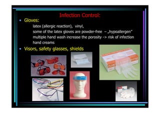
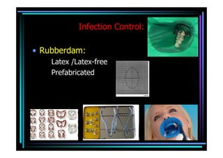
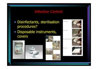
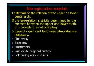
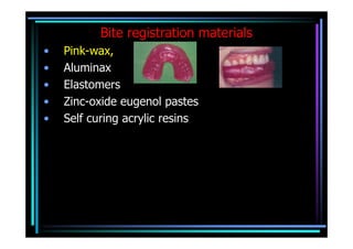
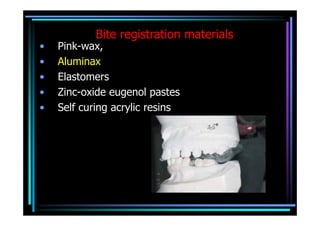
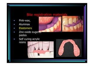
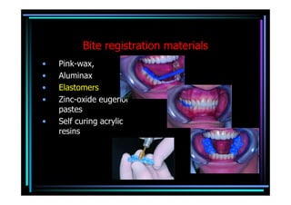
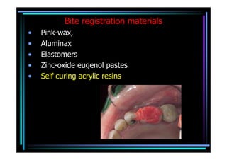
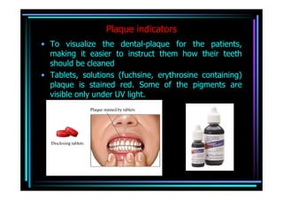
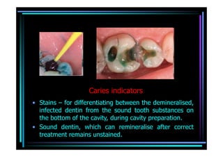
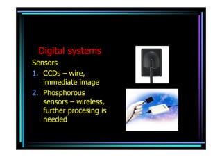
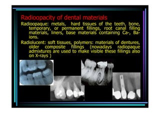
Examining, treating and
preventive methods used in
dentistry from technological
            aspect
Infection Control:
• Gloves:
       latex (allergic reaction), vinyl,
       some of the latex gloves are powder-free – „hypoallergen”
       multiple hand wash increase the porosity -> risk of infection
       hand creams
• Visors, safety glasses, shields
Infection Control:

• Rubberdam:
    Latex /Latex-free
    Prefabricated
Infection Control:

• Disinfectants, sterilisation
  procedures?
• Disposable instruments,
  covers
Instruments used for dental examinations:
•   Mouth mirror –
    •   allows indirect visualization of obscure areas of the mouth
    •   reflects light into the area being examined or treated
    •   retracts soft tissues
    •   Silvering can be on the outer surface of the glass ->less
        resistant, or on the inner surface -> double image.
Instruments used for dental examinations:
•   Dental probe: favourable with rounded end
•   Perio-probe: with depth gauge
•   Material usually stainless steel -> can be sterilized, but some
    disinfectant solutions can be harmful
Sensibility tests
The tooth, it’s dental pulp is considered vital if the blood
  supply of the pulp is intact. It could be tested directly,
  with a laser Doppler equipment.
Based on their coincidence in general, function of the
  nerves supply the dental pulp is usually tested.
• Thermal stimuli (cold, hot)
• Mild electric stimulus applied on the tooth surface (EPT)
Fixed prosthetic appliences
• Crowns, bridges, post retained cores, …
• Controlling the precision fit of cast
  metal parts:
      1. Fluids, containing stain particles (Arti-spot 2),
       2. Pigment containing aerosols (Arti-spray),
       3. Impression materials with very low viscosity
  (Fit Checker)
Removable dentures
• Complete and partial
• Controlling the precision fit of denture
  parts supported by mucosa:
To locate coarse imprinting of the prothesis, causing soft
  tissue injuries
     1. Guttapercha dissolved in chloroform, disclosing
  waxes
     2. Zinc-oxide powder mixed with Vaseline,
     3. Low viscosity impression materials
Occlusion
Occlusal indicators
To locate normal and pathologic occlusal contacts
• Occlusal foils, papers:
Optimal thickness 8-16 µm,
Irrespectively of the material of the occlusal surface
   mark the occlusal contacts, but only the real contacts
Should not change the pattern of the occlusion,
The examined surface should be dry
Different colours for the contacts in different jaw
   positions,
Occlusal indicators
To locate normal and pathologic occlusal contacts
• Occlusal foils, papers:
Occlusal indicators
To locate normal and pathologic occlusal
  contacts
• waxes
• Fluid dye
• Colour powders,
• Celluloid foils (power needed to pull out
  form the occluding surfaces)
Bite registration
Bite registration materials
To determine the relation of the upper an lower
   dental arch.
If the jaw-relation is strictly determined by the
   contacts between the upper and lower teeth,
   this procedure is not obligatory
In case of signifficant tooth-loss bite-plates are
   necessary.
• Pink-wax,
• Aluminax
• Elastomers
• Zinc-oxide eugenol pastes
• Self curing acrylic resins
Bite registration materials
•   Pink-wax,
•   Aluminax
•   Elastomers
•   Zinc-oxide eugenol pastes
•   Self curing acrylic resins
Bite registration materials
•   Pink-wax,
•   Aluminax
•   Elastomers
•   Zinc-oxide eugenol pastes
•   Self curing acrylic resins
Bite registration materials
•   Pink-wax,
•   Aluminax
•   Elastomers
•   Zinc-oxide eugenol
    pastes
•   Self curing acrylic
    resins
Bite registration materials
•   Pink-wax,
•   Aluminax
•   Elastomers
•   Zinc-oxide eugenol
    pastes
•   Self curing acrylic
    resins
Bite registration materials
•   Pink-wax,
•   Aluminax
•   Elastomers
•   Zinc-oxide eugenol pastes
•   Self curing acrylic resins
Plaque indicators
• To visualize the dental-plaque for the patients,
  making it easier to instruct them how their teeth
  should be cleaned
• Tablets, solutions (fuchsine, erythrosine containing)
  plaque is stained red. Some of the pigments are
  visible only under UV light.
Caries indicators
• Stains – for differentiating between the demineralised,
  infected dentin from the sound tooth substances on
  the bottom of the cavity, during cavity preparation.
• Sound dentin, which can remineralise after correct
  treatment remains unstained.
Plaque, debris removing
• Scaling
• Polishing: abrasive pastes that remove superficial stains but do not
  impair the enamel.
  The accidentally exposed dentine, cement is less resistant to abrasive
  particles
• „Air-polishing”: air-stream with Na+-bicarbonate particles
Materials used in dental radiology:
• Intraoral plain films: usually 3- 4 teeth
  can be seen on the image
Materials used in dental radiology:
• Occlusal films: all the teeth in the upper or lower dental
  arch and some of the surrounding soft tissues are
  depicted
Materials used in dental radiology:
• Extraoral films: In different sizes, according to the
  user’s demands, an amplifier layer can be used
  (phosphorus containing layer)
Digital systems
Sensors
1. CCDs – wire,
   immediate image
2. Phosphorous
   sensors – wireless,
   further procesing is
   needed
Radioopacity of dental materials
Radioopaque: metals, hard tissues of the teeth, bone,
   temporary, or permanent fillings, root canal filling
   materials, liners, base materials containing Ca-, Ba-
   ions.
Radiolucent: soft tissues, polymers: materials of dentures,
   older composite fillings (nowadays radiopaque
   admixtures are used to make visible these fillings also
   on X-rays )

Technological aspects perenyi

  • 1.
    Examining, treating and preventivemethods used in dentistry from technological aspect
  • 2.
    Infection Control: • Gloves: latex (allergic reaction), vinyl, some of the latex gloves are powder-free – „hypoallergen” multiple hand wash increase the porosity -> risk of infection hand creams • Visors, safety glasses, shields
  • 3.
    Infection Control: • Rubberdam: Latex /Latex-free Prefabricated
  • 4.
    Infection Control: • Disinfectants,sterilisation procedures? • Disposable instruments, covers
  • 5.
    Instruments used fordental examinations: • Mouth mirror – • allows indirect visualization of obscure areas of the mouth • reflects light into the area being examined or treated • retracts soft tissues • Silvering can be on the outer surface of the glass ->less resistant, or on the inner surface -> double image.
  • 6.
    Instruments used fordental examinations: • Dental probe: favourable with rounded end • Perio-probe: with depth gauge • Material usually stainless steel -> can be sterilized, but some disinfectant solutions can be harmful
  • 7.
    Sensibility tests The tooth,it’s dental pulp is considered vital if the blood supply of the pulp is intact. It could be tested directly, with a laser Doppler equipment. Based on their coincidence in general, function of the nerves supply the dental pulp is usually tested. • Thermal stimuli (cold, hot) • Mild electric stimulus applied on the tooth surface (EPT)
  • 8.
    Fixed prosthetic appliences •Crowns, bridges, post retained cores, …
  • 9.
    • Controlling theprecision fit of cast metal parts: 1. Fluids, containing stain particles (Arti-spot 2), 2. Pigment containing aerosols (Arti-spray), 3. Impression materials with very low viscosity (Fit Checker)
  • 10.
  • 11.
    • Controlling theprecision fit of denture parts supported by mucosa: To locate coarse imprinting of the prothesis, causing soft tissue injuries 1. Guttapercha dissolved in chloroform, disclosing waxes 2. Zinc-oxide powder mixed with Vaseline, 3. Low viscosity impression materials
  • 12.
  • 13.
    Occlusal indicators To locatenormal and pathologic occlusal contacts • Occlusal foils, papers: Optimal thickness 8-16 µm, Irrespectively of the material of the occlusal surface mark the occlusal contacts, but only the real contacts Should not change the pattern of the occlusion, The examined surface should be dry Different colours for the contacts in different jaw positions,
  • 14.
    Occlusal indicators To locatenormal and pathologic occlusal contacts • Occlusal foils, papers:
  • 15.
    Occlusal indicators To locatenormal and pathologic occlusal contacts • waxes • Fluid dye • Colour powders, • Celluloid foils (power needed to pull out form the occluding surfaces)
  • 16.
  • 17.
    Bite registration materials Todetermine the relation of the upper an lower dental arch. If the jaw-relation is strictly determined by the contacts between the upper and lower teeth, this procedure is not obligatory In case of signifficant tooth-loss bite-plates are necessary. • Pink-wax, • Aluminax • Elastomers • Zinc-oxide eugenol pastes • Self curing acrylic resins
  • 18.
    Bite registration materials • Pink-wax, • Aluminax • Elastomers • Zinc-oxide eugenol pastes • Self curing acrylic resins
  • 19.
    Bite registration materials • Pink-wax, • Aluminax • Elastomers • Zinc-oxide eugenol pastes • Self curing acrylic resins
  • 20.
    Bite registration materials • Pink-wax, • Aluminax • Elastomers • Zinc-oxide eugenol pastes • Self curing acrylic resins
  • 21.
    Bite registration materials • Pink-wax, • Aluminax • Elastomers • Zinc-oxide eugenol pastes • Self curing acrylic resins
  • 22.
    Bite registration materials • Pink-wax, • Aluminax • Elastomers • Zinc-oxide eugenol pastes • Self curing acrylic resins
  • 23.
    Plaque indicators • Tovisualize the dental-plaque for the patients, making it easier to instruct them how their teeth should be cleaned • Tablets, solutions (fuchsine, erythrosine containing) plaque is stained red. Some of the pigments are visible only under UV light.
  • 24.
    Caries indicators • Stains– for differentiating between the demineralised, infected dentin from the sound tooth substances on the bottom of the cavity, during cavity preparation. • Sound dentin, which can remineralise after correct treatment remains unstained.
  • 25.
    Plaque, debris removing •Scaling • Polishing: abrasive pastes that remove superficial stains but do not impair the enamel. The accidentally exposed dentine, cement is less resistant to abrasive particles • „Air-polishing”: air-stream with Na+-bicarbonate particles
  • 26.
    Materials used indental radiology: • Intraoral plain films: usually 3- 4 teeth can be seen on the image
  • 27.
    Materials used indental radiology: • Occlusal films: all the teeth in the upper or lower dental arch and some of the surrounding soft tissues are depicted
  • 28.
    Materials used indental radiology: • Extraoral films: In different sizes, according to the user’s demands, an amplifier layer can be used (phosphorus containing layer)
  • 29.
    Digital systems Sensors 1. CCDs– wire, immediate image 2. Phosphorous sensors – wireless, further procesing is needed
  • 30.
    Radioopacity of dentalmaterials Radioopaque: metals, hard tissues of the teeth, bone, temporary, or permanent fillings, root canal filling materials, liners, base materials containing Ca-, Ba- ions. Radiolucent: soft tissues, polymers: materials of dentures, older composite fillings (nowadays radiopaque admixtures are used to make visible these fillings also on X-rays )