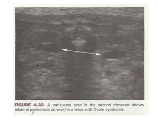
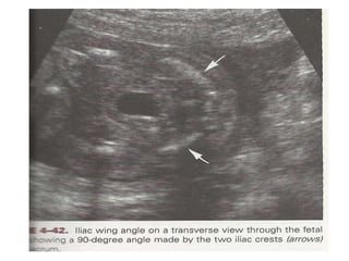
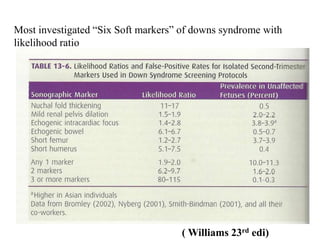
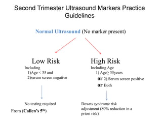
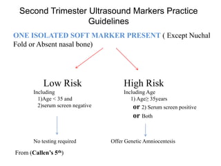
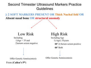
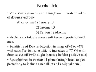
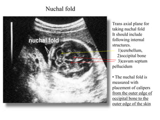
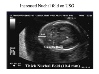
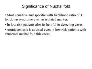
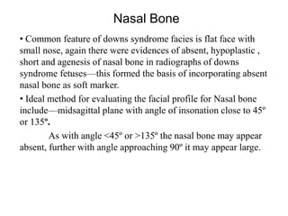
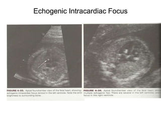
This document discusses various soft tissue markers that can be detected on a genetic sonogram to screen for fetal aneuploidies like Down syndrome. It describes markers like nuchal fold thickness, absent nasal bone, echogenic intracardiac focus, choroid plexus cysts, short long bones, pyelectasis, and single umbilical artery. It provides details on the sensitivity and significance of each marker, as well as guidelines on when genetic amniocentesis is recommended based on the number and type of markers present. The document emphasizes that while these markers can help detect aneuploidies, they are often transient and nonspecific findings that also occur commonly in euploid fetuses.
Nuchal translucency
It is a sonographic pre natal screening scan to detect cardiovascular abnormality in a fetus.
NT can also detect altered extra cellular matrix composition and limited lymphatic drainage
In this presentation we will discuss role of Doppler US in Infertility, fertilization and assisted fertilization.
we will discuss the favorable and unfavorable RI and PI.
We will discuss role of doppler us in various gynecological malignancies.
In this presentation we will focus on aetiological factors that cause infirtility. Our focus is on US depiction of these aetiological factors to help physician in the management of infirtility.
We have nothing to do with direct radiological intervention in the management of infirtility in this presentation.
Turner syndrome (gonadal dysgenesis) is one of the most common chromosomal abnormalities occuring 1 in 2500 to 1 in 3000 live-born girls. It is an important cause of short stature in girls and primary amenorrhea in young women that is usually caused by loss of part or all of an X chromosome. This review briefly summarises the current knowledge about the syndrome and the management strategies.
Lecture on prenatal genetic diagnostic techniques and their value in detection of prenatal genetic anomalies. This lecture details techniques employed in the common diagnostic interventions used in prenatal period and their usefulness.
THIS PRESENATATION IS FOR THE MEDICAL STUDENTS WHO ALSO HAVE GENETICS AND IF THEY NEED TO GIVE A SEMINAR BASED ON THIS TOPIC THIS PRESENATATION SHALL PROVE USEFUL
Adnexal masses are commonly identified in pregnancy but they are rarely malignant. Most adnexal masses either resolve spontaneously or can be managed conservatively during pregnancy. Pregnancy may alter the serum levels of tumour markers, making the interpretation of results difficult.
Manegement of adenexal masses in pregnancyWafaa Benjamin
Over the last 20 years, the use of ultrasound in pregnancy has dramatically increased the numbers of ovarian cysts diagnosed.
The majority of these ovarian cysts in pregnancy either resolve spontaneously or are due to benign conditions.
Ovarian cancer is extremely rare in women of childbearing age and thus most of these cysts can be managed conservatively.
In terms of malignancy potential, those that are malignant are likely to be borderline.
Unless there is a suspicion of malignancy or there is a significant cyst complication, such as torsion, surgery is not indicated.
MRI is a safe and useful tool to help evaluate cysts in more detail in situations where ultrasound provides an inconclusive answer.
If surgery is planned, this should take place during the second trimester to minimise the risk of miscarriage.
Whether surgery is done laparoscopically or using a traditional open approach, it is largely dependent on operator experience and patient preference.
Aspiration of ovarian cysts is only indicated where they appear simple on ultrasound and where they are causing pain or are thought to be obstructing the birth canal.
If surgery does not take place, then ultrasound follow-up during and after pregnancy may be advised accordingly.
Similar to Genetic sonogram and soft tissue markers (20)
These lecture slides, by Dr Sidra Arshad, offer a quick overview of physiological basis of a normal electrocardiogram.
Learning objectives:
1. Define an electrocardiogram (ECG) and electrocardiography
2. Describe how dipoles generated by the heart produce the waveforms of the ECG
3. Describe the components of a normal electrocardiogram of a typical bipolar leads (limb II)
4. Differentiate between intervals and segments
5. Enlist some common indications for obtaining an ECG
Study Resources:
1. Chapter 11, Guyton and Hall Textbook of Medical Physiology, 14th edition
2. Chapter 9, Human Physiology - From Cells to Systems, Lauralee Sherwood, 9th edition
3. Chapter 29, Ganong’s Review of Medical Physiology, 26th edition
4. Electrocardiogram, StatPearls - https://www.ncbi.nlm.nih.gov/books/NBK549803/
5. ECG in Medical Practice by ABM Abdullah, 4th edition
6. ECG Basics, http://www.nataliescasebook.com/tag/e-c-g-basics
These simplified slides by Dr. Sidra Arshad present an overview of the non-respiratory functions of the respiratory tract.
Learning objectives:
1. Enlist the non-respiratory functions of the respiratory tract
2. Briefly explain how these functions are carried out
3. Discuss the significance of dead space
4. Differentiate between minute ventilation and alveolar ventilation
5. Describe the cough and sneeze reflexes
Study Resources:
1. Chapter 39, Guyton and Hall Textbook of Medical Physiology, 14th edition
2. Chapter 34, Ganong’s Review of Medical Physiology, 26th edition
3. Chapter 17, Human Physiology by Lauralee Sherwood, 9th edition
4. Non-respiratory functions of the lungs https://academic.oup.com/bjaed/article/13/3/98/278874
HOT NEW PRODUCT! BIG SALES FAST SHIPPING NOW FROM CHINA!! EU KU DB BK substit...GL Anaacs
Contact us if you are interested:
Email / Skype : kefaya1771@gmail.com
Threema: PXHY5PDH
New BATCH Ku !!! MUCH IN DEMAND FAST SALE EVERY BATCH HAPPY GOOD EFFECT BIG BATCH !
Contact me on Threema or skype to start big business!!
Hot-sale products:
NEW HOT EUTYLONE WHITE CRYSTAL!!
5cl-adba precursor (semi finished )
5cl-adba raw materials
ADBB precursor (semi finished )
ADBB raw materials
APVP powder
5fadb/4f-adb
Jwh018 / Jwh210
Eutylone crystal
Protonitazene (hydrochloride) CAS: 119276-01-6
Flubrotizolam CAS: 57801-95-3
Metonitazene CAS: 14680-51-4
Payment terms: Western Union,MoneyGram,Bitcoin or USDT.
Deliver Time: Usually 7-15days
Shipping method: FedEx, TNT, DHL,UPS etc.Our deliveries are 100% safe, fast, reliable and discreet.
Samples will be sent for your evaluation!If you are interested in, please contact me, let's talk details.
We specializes in exporting high quality Research chemical, medical intermediate, Pharmaceutical chemicals and so on. Products are exported to USA, Canada, France, Korea, Japan,Russia, Southeast Asia and other countries.
Lung Cancer: Artificial Intelligence, Synergetics, Complex System Analysis, S...Oleg Kshivets
RESULTS: Overall life span (LS) was 2252.1±1742.5 days and cumulative 5-year survival (5YS) reached 73.2%, 10 years – 64.8%, 20 years – 42.5%. 513 LCP lived more than 5 years (LS=3124.6±1525.6 days), 148 LCP – more than 10 years (LS=5054.4±1504.1 days).199 LCP died because of LC (LS=562.7±374.5 days). 5YS of LCP after bi/lobectomies was significantly superior in comparison with LCP after pneumonectomies (78.1% vs.63.7%, P=0.00001 by log-rank test). AT significantly improved 5YS (66.3% vs. 34.8%) (P=0.00000 by log-rank test) only for LCP with N1-2. Cox modeling displayed that 5YS of LCP significantly depended on: phase transition (PT) early-invasive LC in terms of synergetics, PT N0—N12, cell ratio factors (ratio between cancer cells- CC and blood cells subpopulations), G1-3, histology, glucose, AT, blood cell circuit, prothrombin index, heparin tolerance, recalcification time (P=0.000-0.038). Neural networks, genetic algorithm selection and bootstrap simulation revealed relationships between 5YS and PT early-invasive LC (rank=1), PT N0—N12 (rank=2), thrombocytes/CC (3), erythrocytes/CC (4), eosinophils/CC (5), healthy cells/CC (6), lymphocytes/CC (7), segmented neutrophils/CC (8), stick neutrophils/CC (9), monocytes/CC (10); leucocytes/CC (11). Correct prediction of 5YS was 100% by neural networks computing (area under ROC curve=1.0; error=0.0).
CONCLUSIONS: 5YS of LCP after radical procedures significantly depended on: 1) PT early-invasive cancer; 2) PT N0--N12; 3) cell ratio factors; 4) blood cell circuit; 5) biochemical factors; 6) hemostasis system; 7) AT; 8) LC characteristics; 9) LC cell dynamics; 10) surgery type: lobectomy/pneumonectomy; 11) anthropometric data. Optimal diagnosis and treatment strategies for LC are: 1) screening and early detection of LC; 2) availability of experienced thoracic surgeons because of complexity of radical procedures; 3) aggressive en block surgery and adequate lymph node dissection for completeness; 4) precise prediction; 5) adjuvant chemoimmunoradiotherapy for LCP with unfavorable prognosis.
Tom Selleck Health: A Comprehensive Look at the Iconic Actor’s Wellness Journeygreendigital
Tom Selleck, an enduring figure in Hollywood. has captivated audiences for decades with his rugged charm, iconic moustache. and memorable roles in television and film. From his breakout role as Thomas Magnum in Magnum P.I. to his current portrayal of Frank Reagan in Blue Bloods. Selleck's career has spanned over 50 years. But beyond his professional achievements. fans have often been curious about Tom Selleck Health. especially as he has aged in the public eye.
Follow us on: Pinterest
Introduction
Many have been interested in Tom Selleck health. not only because of his enduring presence on screen but also because of the challenges. and lifestyle choices he has faced and made over the years. This article delves into the various aspects of Tom Selleck health. exploring his fitness regimen, diet, mental health. and the challenges he has encountered as he ages. We'll look at how he maintains his well-being. the health issues he has faced, and his approach to ageing .
Early Life and Career
Childhood and Athletic Beginnings
Tom Selleck was born on January 29, 1945, in Detroit, Michigan, and grew up in Sherman Oaks, California. From an early age, he was involved in sports, particularly basketball. which played a significant role in his physical development. His athletic pursuits continued into college. where he attended the University of Southern California (USC) on a basketball scholarship. This early involvement in sports laid a strong foundation for his physical health and disciplined lifestyle.
Transition to Acting
Selleck's transition from an athlete to an actor came with its physical demands. His first significant role in "Magnum P.I." required him to perform various stunts and maintain a fit appearance. This role, which he played from 1980 to 1988. necessitated a rigorous fitness routine to meet the show's demands. setting the stage for his long-term commitment to health and wellness.
Fitness Regimen
Workout Routine
Tom Selleck health and fitness regimen has evolved. adapting to his changing roles and age. During his "Magnum, P.I." days. Selleck's workouts were intense and focused on building and maintaining muscle mass. His routine included weightlifting, cardiovascular exercises. and specific training for the stunts he performed on the show.
Selleck adjusted his fitness routine as he aged to suit his body's needs. Today, his workouts focus on maintaining flexibility, strength, and cardiovascular health. He incorporates low-impact exercises such as swimming, walking, and light weightlifting. This balanced approach helps him stay fit without putting undue strain on his joints and muscles.
Importance of Flexibility and Mobility
In recent years, Selleck has emphasized the importance of flexibility and mobility in his fitness regimen. Understanding the natural decline in muscle mass and joint flexibility with age. he includes stretching and yoga in his routine. These practices help prevent injuries, improve posture, and maintain mobilit
micro teaching on communication m.sc nursing.pdfAnurag Sharma
Microteaching is a unique model of practice teaching. It is a viable instrument for the. desired change in the teaching behavior or the behavior potential which, in specified types of real. classroom situations, tends to facilitate the achievement of specified types of objectives.
Flu Vaccine Alert in Bangalore Karnatakaaddon Scans
As flu season approaches, health officials in Bangalore, Karnataka, are urging residents to get their flu vaccinations. The seasonal flu, while common, can lead to severe health complications, particularly for vulnerable populations such as young children, the elderly, and those with underlying health conditions.
Dr. Vidisha Kumari, a leading epidemiologist in Bangalore, emphasizes the importance of getting vaccinated. "The flu vaccine is our best defense against the influenza virus. It not only protects individuals but also helps prevent the spread of the virus in our communities," he says.
This year, the flu season is expected to coincide with a potential increase in other respiratory illnesses. The Karnataka Health Department has launched an awareness campaign highlighting the significance of flu vaccinations. They have set up multiple vaccination centers across Bangalore, making it convenient for residents to receive their shots.
To encourage widespread vaccination, the government is also collaborating with local schools, workplaces, and community centers to facilitate vaccination drives. Special attention is being given to ensuring that the vaccine is accessible to all, including marginalized communities who may have limited access to healthcare.
Residents are reminded that the flu vaccine is safe and effective. Common side effects are mild and may include soreness at the injection site, mild fever, or muscle aches. These side effects are generally short-lived and far less severe than the flu itself.
Healthcare providers are also stressing the importance of continuing COVID-19 precautions. Wearing masks, practicing good hand hygiene, and maintaining social distancing are still crucial, especially in crowded places.
Protect yourself and your loved ones by getting vaccinated. Together, we can help keep Bangalore healthy and safe this flu season. For more information on vaccination centers and schedules, residents can visit the Karnataka Health Department’s official website or follow their social media pages.
Stay informed, stay safe, and get your flu shot today!
Title: Sense of Smell
Presenter: Dr. Faiza, Assistant Professor of Physiology
Qualifications:
MBBS (Best Graduate, AIMC Lahore)
FCPS Physiology
ICMT, CHPE, DHPE (STMU)
MPH (GC University, Faisalabad)
MBA (Virtual University of Pakistan)
Learning Objectives:
Describe the primary categories of smells and the concept of odor blindness.
Explain the structure and location of the olfactory membrane and mucosa, including the types and roles of cells involved in olfaction.
Describe the pathway and mechanisms of olfactory signal transmission from the olfactory receptors to the brain.
Illustrate the biochemical cascade triggered by odorant binding to olfactory receptors, including the role of G-proteins and second messengers in generating an action potential.
Identify different types of olfactory disorders such as anosmia, hyposmia, hyperosmia, and dysosmia, including their potential causes.
Key Topics:
Olfactory Genes:
3% of the human genome accounts for olfactory genes.
400 genes for odorant receptors.
Olfactory Membrane:
Located in the superior part of the nasal cavity.
Medially: Folds downward along the superior septum.
Laterally: Folds over the superior turbinate and upper surface of the middle turbinate.
Total surface area: 5-10 square centimeters.
Olfactory Mucosa:
Olfactory Cells: Bipolar nerve cells derived from the CNS (100 million), with 4-25 olfactory cilia per cell.
Sustentacular Cells: Produce mucus and maintain ionic and molecular environment.
Basal Cells: Replace worn-out olfactory cells with an average lifespan of 1-2 months.
Bowman’s Gland: Secretes mucus.
Stimulation of Olfactory Cells:
Odorant dissolves in mucus and attaches to receptors on olfactory cilia.
Involves a cascade effect through G-proteins and second messengers, leading to depolarization and action potential generation in the olfactory nerve.
Quality of a Good Odorant:
Small (3-20 Carbon atoms), volatile, water-soluble, and lipid-soluble.
Facilitated by odorant-binding proteins in mucus.
Membrane Potential and Action Potential:
Resting membrane potential: -55mV.
Action potential frequency in the olfactory nerve increases with odorant strength.
Adaptation Towards the Sense of Smell:
Rapid adaptation within the first second, with further slow adaptation.
Psychological adaptation greater than receptor adaptation, involving feedback inhibition from the central nervous system.
Primary Sensations of Smell:
Camphoraceous, Musky, Floral, Pepperminty, Ethereal, Pungent, Putrid.
Odor Detection Threshold:
Examples: Hydrogen sulfide (0.0005 ppm), Methyl-mercaptan (0.002 ppm).
Some toxic substances are odorless at lethal concentrations.
Characteristics of Smell:
Odor blindness for single substances due to lack of appropriate receptor protein.
Behavioral and emotional influences of smell.
Transmission of Olfactory Signals:
From olfactory cells to glomeruli in the olfactory bulb, involving lateral inhibition.
Primitive, less old, and new olfactory systems with different path
2. Introduction to Genetic sonogram
•It’s a specific targeted examination for fetal aneuploidy,
most specific for Downs syndrome, that searches for the
presence of
1)Fetal structural anomalies
2)Aneuploidy markers and
3)Abnormal biometry. (Callen’s 5th)
• These aneuploidy markers are called as “soft” markers
and these are variations in normal anatomy that, except for
their relationship to aneuploidy, are unlikely to be
clinically significant. (Callen’s 5th)
• soft markers are often transient and nonspecific findings
which can also occur frequently in euploid fetuses.
3. Genetic Sonogram
•Sensitivity of genetic sonogram ranges from 84%
among women with advanced maternal age to 90%
among women younger than 35 years with abnormal
triple test.
• Performed between 18 to 20 weeks of gestation.
• Major advantage is, it reduces the invasive testing rate
and can optimize the selection of candidate for invasive
testing in order to minimize the procedure related losses
of normal fetuses without significantly decreasing
detection rate.
(Callen’s 5th)
4. Candidate for Genetic sonogram
Low risk vs. high risk population
• Inappropriate for LOW RISK population
1)High false positive rate of 12 to 15% in high risk
population (which is probably more in low risk).
2)Each marker by itself has only low to moderate
sensitivity for Downs, when found in isolation it not necessarily
increase the risk but increases cost and anxiety in LOW RISK.
3) In LOW RISK patient the prior risk may be so low that
Presence of one marker will not qualify a patient for
Amniocentesis.
4) Further the accuracy of soft markers has only been
studied in HIGH RISK population and generalizing the result in
low risk is not appropriate.
8. Most investigated “Six Soft markers” of downs syndrome with
likelihood ratio
( Williams 23rd edi)
9. Second Trimester Ultrasound Markers Practice
Guidelines
Normal Ultrasound (No marker present)
Low Risk
Including
1)Age < 35 and
2)serum screen negative
High Risk
Including Age
1) Age≥ 35years
or 2) Serum screen positive
or Both
No testing required Downs syndrome risk
adjustment (80% reduction in a
priori risk)
From (Callen’s 5th)
10. Second Trimester Ultrasound Markers Practice
Guidelines
ONE ISOLATED SOFT MARKER PRESENT ( Except Nuchal
Fold or Absent nasal bone)
Low Risk
Including
1)Age < 35 and
2)serum screen negative
High Risk
Including Age
1) Age≥ 35years
or 2) Serum screen positive
or Both
No testing required Offer Genetic Amniocentesis
From (Callen’s 5th)
11. Second Trimester Ultrasound Markers Practice
Guidelines
≥ 2 SOFT MARKERS PRESENT OR Thick Nuchal fold OR
Absent nasal bone OR structural anomaly
Low Risk
Including
1)Age < 35 and
2)serum screen negative
High Risk
Including Age
1) Age≥ 35years
or 2) Serum screen positive
or Both
Offer Genetic AmniocentesisOffer Genetic Amniocentesis
From (Callen’s 5th)
13. Nuchal fold
• Most sensitive and specific single midtrimester marker
of downs syndrome.
Also seen in 1) trisomy 18
2) trisomy 13
3) Turners syndrome.
• Nuchal skin folds is excess soft tissue in posterior neck
area.
• Sensitivity of Downs detection in range of 42 to 43%
with cut off as 6mm, sensitivity increases to 77.8% with
5mm as cut off (with slight increase in false positive rate)
• Best obtained in trans axial plane through head, angled
posteriorly to include cerebellum and occipital bone.
14. Trans axial plane for
taking nuchal fold
It should include
following internal
structures.
1)cerebellum,
2)occipital bone
3)cavum septum
pellucidum
• The nuchal fold is
measured with
placement of calipers
from the outer edge of
occipital bone to the
outer edge of the skin
Nuchal fold
16. Significance of Nuchal fold
• Most sensitive and specific with likelihood ratio of 11
for down syndrome even as isolated marker.
• In low risk patients also its helpful in detecting cases.
• Amniocentesis is advised even in low risk patients with
abnormal nuchal fold thickness.
17. Nasal Bone
• Common feature of downs syndrome facies is flat face with
small nose, again there were evidences of absent, hypoplastic ,
short and agenesis of nasal bone in radiographs of downs
syndrome fetuses—this formed the basis of incorporating absent
nasal bone as soft marker.
• Ideal method for evaluating the facial profile for Nasal bone
include—midsagittal plane with angle of insonation close to 45º
or 135º.
As with angle <45º or >135º the nasal bone may appear
absent, further with angle approaching 90º it may appear large.
19. Nasal Bone
• Sensitivity for absent nasal bone have ranged from 28
to 66%, with likelihood ratio of 83 for Downs.
(this approximates that of nuchal fold thickening).
• After addition of absent nasal bone as soft marker, the
sensitivity of genetic sonogram has increased from 87%
to 92.8% without increasing false +ve rate.
• Short or hypoplastic nasal bone as soft marker requires
sonographic normograms which are specific to local
population and based on it cutoff are decided for better
results.
•Further BPD/NBL ratio >10 can also be used, with
81% sensitivity for downs syndrome.
20. Echogenic Intracardiac Focus (EIF)
• Echogenic cardiac foci (EIF) is commonly seen on 2nd
trimester USG –in 3 to 4% of normal fetuses.
• EIF-its discrete brightly echogenic spot with brightness
similar to bone, due to reflection from coarse intra
myocardial calcification of papillary muscles and chordae
tendinae surrounded by myocardial fibrosis.
• Best visualized in apical 4 chamber view with apex
towards or away from transducer.
• Resolves by 3rd trimester
•Most frequently seen in the left ventricle, but also can be
seen in right ventricle or bilaterally.
•Likelihood ratio of EIF for Downs syndrome ranges
between 1.8 to 4.2.
22. Significance of EIF
• EIF is associated with aneuploidy trisomy 21 and 13.
• In high risk populations EIF increases the risk of
downs syndrome, but in low risk EIF is considered a
normal variant with absence of other major or minor
USG anomalies.
• M/C in left ventricle but right side or bilateral EIF-
higher risk of aneuploidy.
• Multiple(67%) and large in size-higher risk of
aneuploidy.
• Right vs. Left EIF-higher risk of aneuploidy in right
side EIF.
24. Choroid plexus Cysts (CPCs)
• Lateral ventricles contain sonolucent CSF and with in this
lies brightly echogenic choroid plexus that normally fills the
atrium and may contains cyst.
• Prevalence among normal is variable from 0.3% to 3.6%.
• CPCs seen between 16 to 21 weeks of GA and are transient,
by 23rd weeks undergo resolutions and uncommon after 25 to
26 weeks.
• CPCs can be unilateral/bilateral/ unilocular or multilocular.
•Commonly they are multilocular and range from 0.5 to 2cm,
occasionally large as to fill almost entire lateral ventricle.
25. Significance of Choroid plexus Cysts
•CPCs always resolve, Larger cysts may take longer time.
•CPCs are associated with fetal aneuploidy- Trisomy 18.
•Prevalence of CPCs in trisomy 18 is 53% when other soft tissue
marker is also present, isolated CPCs are not associated with
trisomy 18.
• Larger cyst (>1cm) compared to smaller increases the risk of
trisomy 18.
• Isolated presence of CPCs in low risk patient is not considered
clinically significant, does not change risk category.
• CPCs in absence of other minor or major soft markers & in
absence of other risk factors considered to be a “normal variant”
and no further evaluation is needed.
• Recommendations are if CPCs is present detailed USG for other
soft markers to be done, and if isolated and they always resolve
repeat scan add no value to decision making.
29. Hyper echogenic bowel
• Non specific and can be seen in normal fetuses (0.5%).
• Normally in 2nd trimester bowel is homogenous with
same echogenicity as rest of abdomen, if it has
echogenicity similar to iliac bones-
HYPERECHOGENIC.
• Hyper echogenic bowel is seen with increased
frequency in trisomy 21, it increases risk by six to
sevenfold ( with other risk factors like age, other soft
markers).
•Risk of trisomy 21 with isolated Hyper echogenic bowel
is 1.4%.
30. Significance of Hyper echogenic bowel
• Other conditions were Hyper echogenic bowel is seen
1) congenital infections- Cytomegalovirus, herpes
simplex, parvo virus.
2) meconium ileus (due to cystic fibrosis).
3) Bowel obstructions/ Atresia / malformations.
4) Prenatal vaginal bleeding and fetal swallowing
of blood from inatraamniotic hemorrhage.
• There is also increases risk of fetal demise, IUGR and
placenta related complications.
• Risk of both cystic fibrosis and aneuploidy increases
with degree of echogenicity.
31. Hyper echogenic bowel
Recommendations-
1)consider maternal
studies for congenital
infection, cystic
fibrosis carrier status.
2)invasive testing for
fetal karyotype.
3)serial scan for
IUGR and to rule out
bowel malformations
( evident mostly in
3rd trimester).
32. Short Long Bones
•Somatic and visceral growth profiles of midtrimester
trisomy 21 fetuses have shown short limbs which are
discordant with head circumference (HC).
• Short Femur length – BPD/Femur length ratio or
observed to expected FL ratio—associated with downs
syndrome.
•Assessed between 15 to 23 weeks, but most helpful in
detecting trisomy 21 between 17 to 19 weeks.
•Ideal method—FL measured with diaphysis located
horizontally in image ( as vertical measurement a/w
high false + rate).
33. Short Long Bones
• Different studies showing different sensitivity rates for trisomy
21 detection.
• Most accepted studies out of all is by Lockwood et al showing
sensitivity of 51% with BPD/FL ratio and by Benacerraf et al
showing sensitivity of 68%.
34. Short Long Bones
• Humeral length--short humeral length also associated
with trisomy 21.
• with measured to expected HL >0.90 as cutoff
sensitivity is 50%.
• Short HL is less useful then FL as screening marker.
35. Significance of Short Long Bones
• Isolated Short FL is not much sensitive rather
combining it with other USG markers.
•Combining both HL and FL assessments—it carry a 11
fold higher risk of having trisomy 21.
• Gender influences the long bones length, affected
male fetuses had more short FL and HL than affected
female fetuses.
36. Pyelectasis
• Renal pyelectasis ( pelvic dilation) is measured in AP
dimension as fluid filled renal pelvis.
• Common finding on USG seen in 2 to 2.8% of normal
fetuses.
• Measured in view with kidney and spine are positioned
directly toward or away from transducer.
• Cut off is >4mm.
• sensitivity is 25% with cut off > 4mm.
• Isolated pyelectasis carries slightly increased risk but not
enough to justify amniocentesis in low risks and to be used
with other USG markers.
• When present in high risk as isolated it increased priori
risk for fetal trisomy 21.
•
37.
38. Lliac Wing angle
• Trisomy 21 fetuses have pelvic dysplasia and wider
lateral span of iliac wing as compared to normal thus
causing a widened iliac angle.
• Its measured in transverse view of fetal pelvis, the
iliac angle made by two iliac crests at pivot point of
sacral spine is measured.
•Mean iliac angle in downs is 80± 19.7 ºas compare to
63.1±0.3ºin normal, thus by taking 90º as cut off—
sensitivity is 36.8%.
39.
40. Significance of Lliac Wing angle
• It is non reproducible due to variation in measurement
of angle.
Depending on 1) actual transverse level
2) intra and interobserver variation.
3) Orientation of spine, axial level
and GA.
• Not a useful marker of trisomy 21.
41. Ear Length
• Abnormally small ears is one of the consistent clinical
finding in newborns and infants with aneuploidy
including downs syndrome.
•Its always found sonographically in cases with trisomy
18 and 13, and in half of the cases of trisomy 21.
•As a result Short ear length on midtrimester USG, is
taken as one of the soft markers of aneuploidy.
• Sensitivity of short ear length (<10th percentile of GA)
in midtrimester is 71%. ( as per Lettieri et al).
• Measured to expected ear length ratio < 0.8 had
sensitivity of 75% for downs and 83.3% for trisomy 18.
(Callens 5th edi)
42.
43. Ear Length
• Further studies has shown that sensitivity of ear length
for downs syndrome is 41%, for trisomy 18 is 96% and
for trisomy 13 its 100%.
•
44. Single umbilical artery
• Single Umbilical artery (SUA) is associated with
cytogenetic abnormality – with incidence of 17% with
SUA.
• Trisomy 18 is most common aneuploidy associated
with SUA, then trisomy 13, Turners syndrome and
triploidy, but Downs syndrome is not commonly
associated with SUA.
• In midtrimester USG transverse views of a free loop of
cord with adequate magnification is examined, In SUA
only two vessels one large (vein) and smaller (artery)
will be seen.
45. Single umbilical artery
• Single Umbilical artery (SUA) is associated with
cytogenetic abnormality – with incidence of 17% with
SUA.
• Trisomy 18 is most common aneuploidy associated
with SUA, then trisomy 13, Turners syndrome and
triploidy, but Downs syndrome is not commonly
associated with SUA.
• In midtrimester USG transverse views of a free loop of
cord with adequate magnification is examined, In SUA
only two vessels one large (vein) and smaller (artery)
will be seen.
46.
47. Single umbilical artery
• Overall SUA has incidence of 11.3% among
cytogenetically abnormal pregnancies, it can be found in
10 to 50% of trisomy 18 and 13 fetuses.
• It has also been reported to be associated with
1) Congenital anomalies involving various organ
system.(most common system involved-CVS, GIT, CNS).
2) Genetic syndrome ( VATER, Meckel Gruber syndrome)
3) Fetal growth restriction.
4) Prematurity.
5) Increased perinatal mortality rate.
48. Significance of Single umbilical artery
• In fetuses with isolated SUA there is no increased
incidence for chromosomal abnormality.
• With isolated SUA a detailed sonogram by experience
personnel for other anomalies to be done, once
confirmed isolated and in absence of high risk factors
no need of invasive analysis.
• If other multiple malformations are detected with SUA
Patient should be offered prenatal karyotyping
49. Sandal Gap
• Sandal gap is separation of great toe from second toe
causing a wide gap between two phalanges.
•