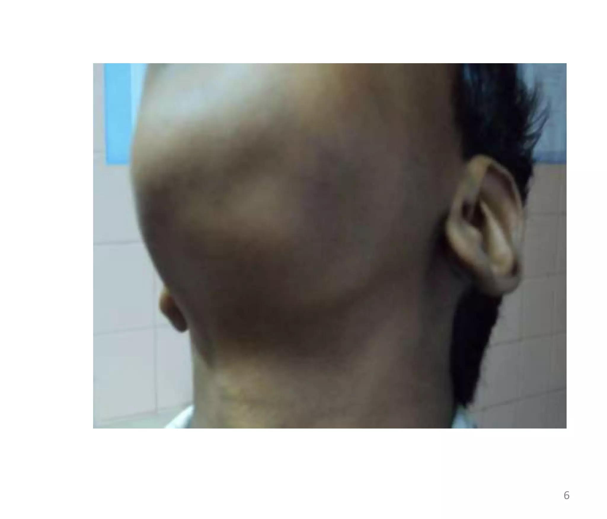
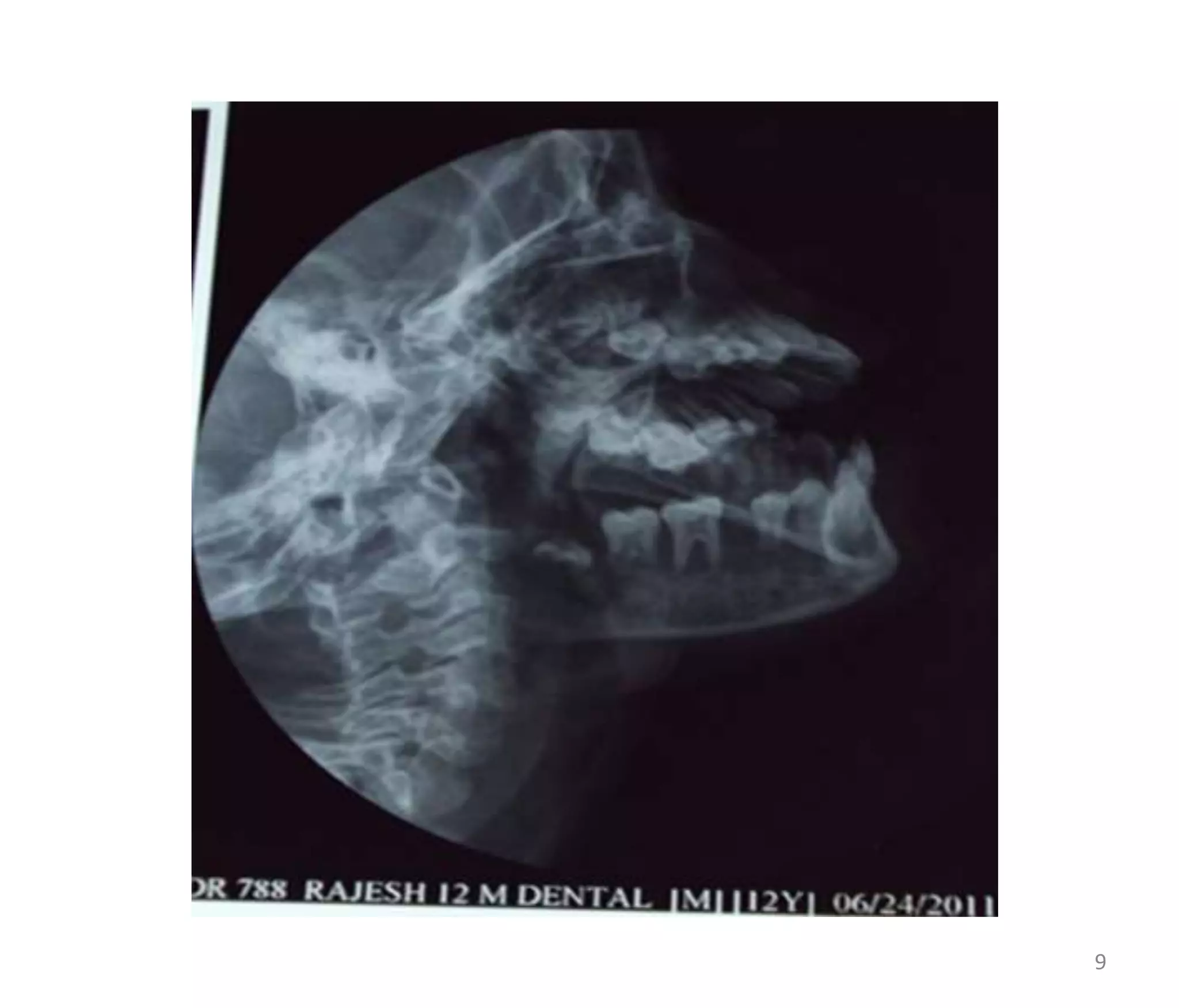
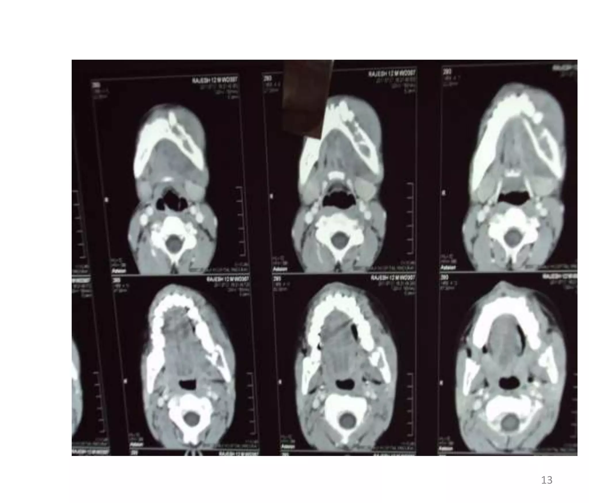
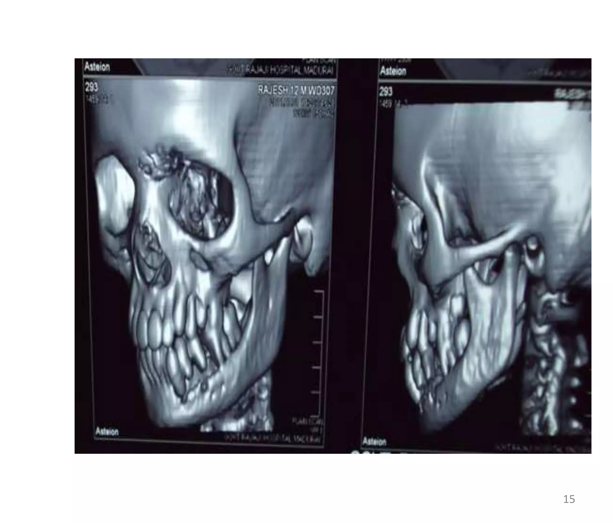
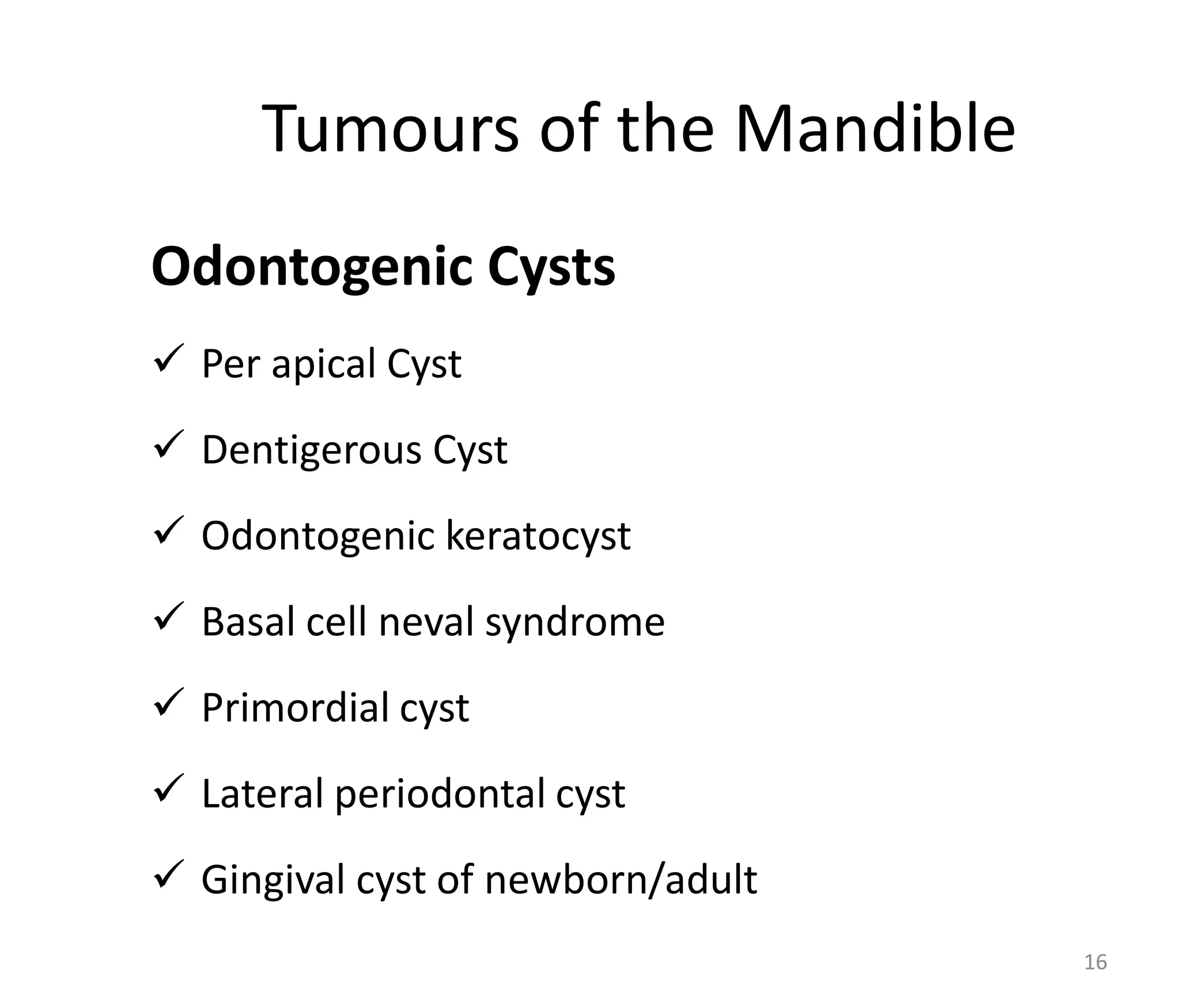
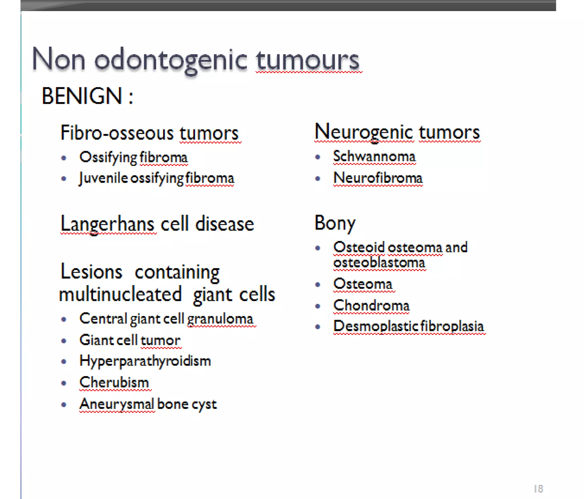
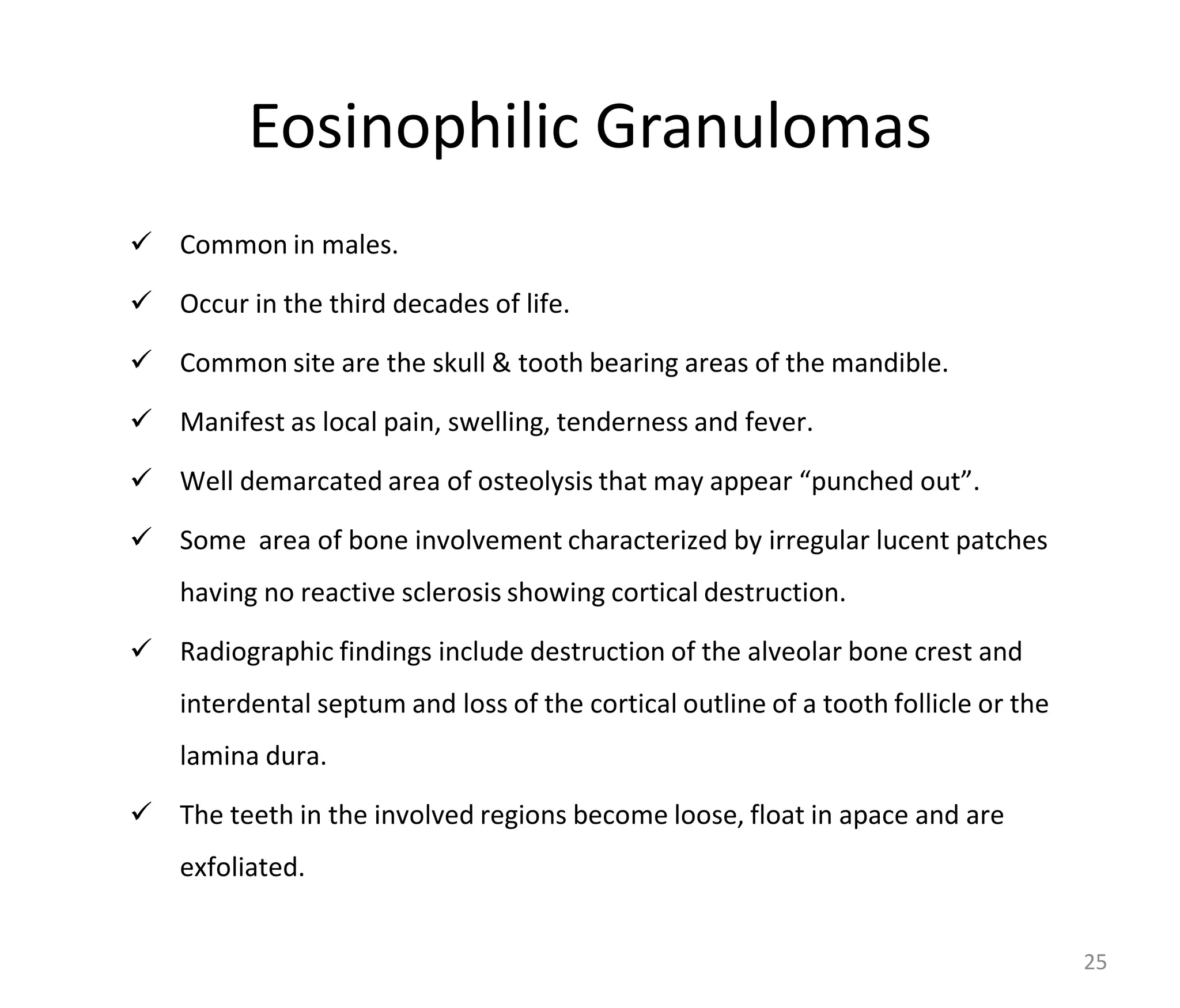
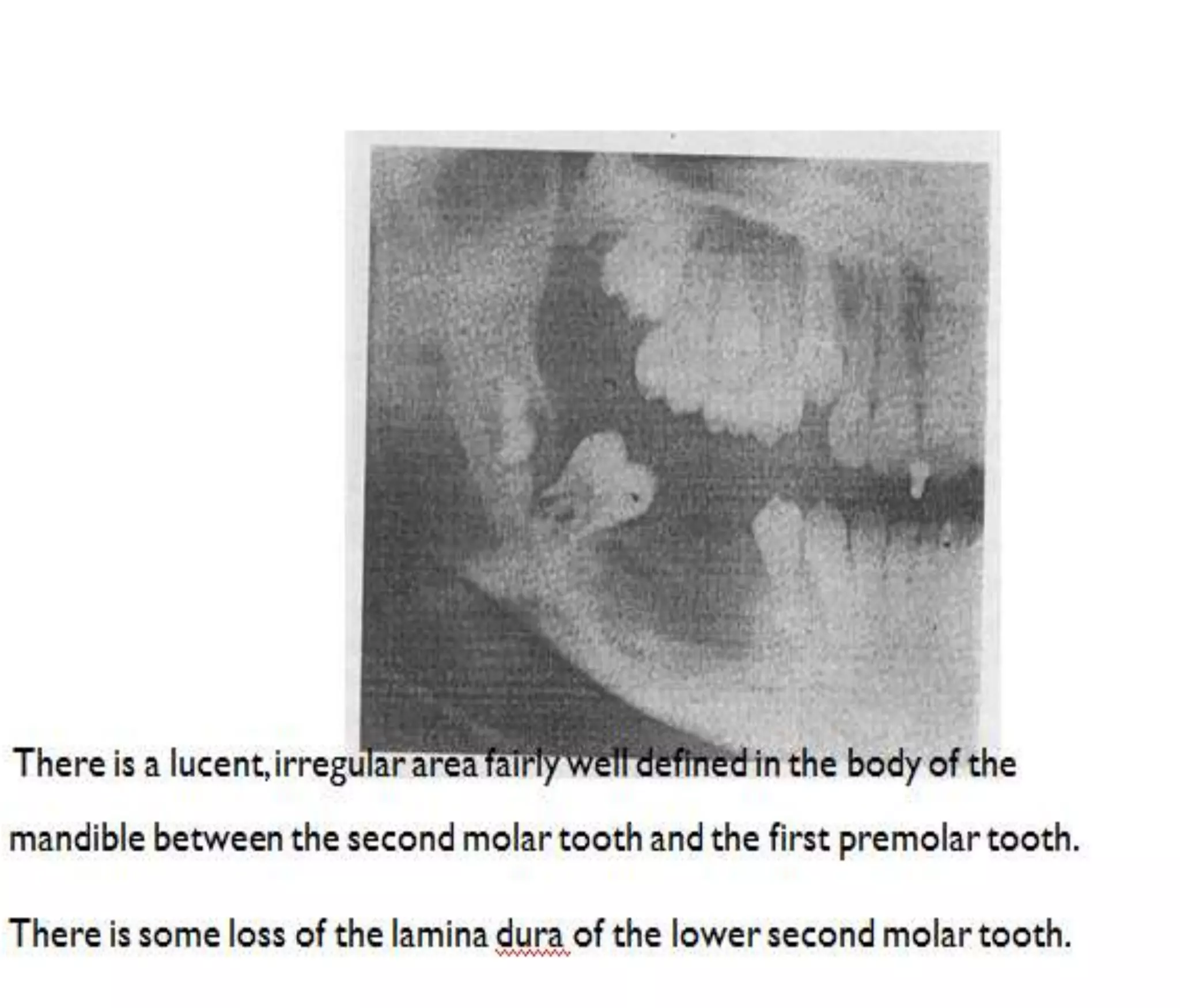
I sincerely thank my
colleague for providing
the slides to me
Dr.M.Shanthi., I Yr - DMRD PG
2
A CASE PRESENTATION
 Rajesh, 12/M
 C/O Swelling - Left Jaw - 1 Month
 Starts as a Small Swelling
3
4
5
6
EXAMINATION
 A 5/4 swelling – Lt body of mandible involving both
sides of alveolar margin.
 Resorption of tooth present.
 Multiple cervical LN present.
7
8
9
X-RAY MANDIBLE LT-OBLIQUE VIEW
 E/O ill-defined lytic lesion showing permeative
pattern of destruction involving horizontal ramus &
angle of mandible.
 Lamina dura &alveolar margin shows evidence of
destruction resulting in exfoliation of teeth.
 Impacted 3rd molar seen.
 Evidence of ill-defined soft tissue adjacent to mass
seen.
10
11
CT - PLAIN
• E/O lytic destruction involving outer horizontal
ramus,angle of the mandible wt large soft tissue
component which extend on both alveolar&buccal
aspect.
• No E/O expansion of bone/periosteal
reaction/calcification.
• E/O destruction of lamina dura &alveolar margin wt
exfoliation of teeth.
12
13
CT - CONTRAST
 Moderate heterogenous enchancement.
14
15
Tumours of the Mandible
Odontogenic Cysts
 Per apical Cyst
 Dentigerous Cyst
 Odontogenic keratocyst
 Basal cell neval syndrome
 Primordial cyst
 Lateral periodontal cyst
 Gingival cyst of newborn/adult
16
ODONTOGENIC TUMOURS
EPITHELIAL TUMORS
Benign:
• Ameloblastoma
• Calcifying epithelial
odontogenic tumor
• Adenomatoid odontogenic tumor
• Squamous odontogenic tumor
Malignant:
• Malignant ameloblastoma
• Clear cell odontogenic carcinoma
• Odontogenic carcinoma
MESENCHYMAL TUMORS
Benign:
• Odontogenic fibroma
• Cementoblastoma
• Odontogenic myxoma
• Cementifying fibroma
Malignant:
• Ameloblastic
fibrosarcoma
17
18
NON ODONTOGENIC TUMOURS
MALIGNANT
• Ewing’s sarcoma
• Burkitt’s lymphoma
• Osteosarcoma
• Chondrosarcoma
• Malignant fibrous histiocytoma
• Multiple myeloma
• Solitary plasmacytoma of bone
• Malignant peripheral nerve sheath tumor
• Postradiation sarcoma of bone
19
Differential Diagnosis
1. Lymphoma
2. Giant Cell Granuloma
3. Eosilophilic Granuloma
4. Ewings sarcoma
20
AMELOBLASTOMA
• Equal frequency in men and women.
• Peak incidence in the 3rd and 4th decades of life.
• Slowing growing painless mass.
• Swelling is the presenting symptom.
• Radiographically, it is radiolucent, either multilocular or unilocular.
• multilocular form shows honeycomb or bubble like appearance.
• It has a tendency to break thro the cortex with tumour ext into soft
tissue.
• There can be bony expansion with scalloped margin and there is no
periosteal reaction.
• Loss of the lamina dura, erosion of the tooth apex, displacement of
teeth seen.
21
22
FIBROSSEOUS TUMOUR
• This tumour produce ground glass opacity and
areas of calcification, hence possibility of this
tumour is less likely.
23
24
Eosinophilic Granulomas
 Common in males.
 Occur in the third decades of life.
 Common site are the skull & tooth bearing areas of the mandible.
 Manifest as local pain, swelling, tenderness and fever.
 Well demarcated area of osteolysis that may appear “punched out”.
 Some area of bone involvement characterized by irregular lucent patches
having no reactive sclerosis showing cortical destruction.
 Radiographic findings include destruction of the alveolar bone crest and
interdental septum and loss of the cortical outline of a tooth follicle or the
lamina dura.
 The teeth in the involved regions become loose, float in apace and are
exfoliated.
25
26
Central Giant Cell Granuloma (Giant Cell Reparative Cyst).
 Occurs in girls and young women during the second and third
decades of life.
 The majority of these lesions are found in the anterior mandible.
 Initially, a giant cell reparative cyst manifests as a small, unilocular
radiolucent lesion that can mimic an odontogenic cyst.
 With development, however, the lesion becomes multilocular,
exhibiting a honeycomb appearance.
 Tiny bone septa are present traversing the lesion.
 There may be evidence of expansion, root resorption, and erosion
or remodeling of the cortex. In addition, the lesion may cross the
midline of the mandible.
27
 Central giant cell granuloma in a 34-year-old man.
 CT scan (bone windowing) demonstrates a cystic lesion (arrows) within
the mandible. Note the erosion of the mandibular cortex.
28
HYPERPARATHROIDISM
• In both forms of hyperparathyroidism, excess
PTH levels stimulate osteoclast-mediated bone
resorption, which may produce a focal bone
lesion known as a brown tumor of
hyperparathyroidism.
• This lesion appears radiographically as a well-
defined unilocular or multilocular radiolucency
and it commonly occurs in the jaws.
• These lesions may be solitary or multiple.
• They are histologically identical to central giant29
30
CHERUBISM
• It commonly begin to manifest as painless, bilateral, symmetric
expansion of the jaws between 2 and 5 years of age .
• The lesions are confined to the mandible and maxilla. The regions
most often affected are the mandibular angle, ascending ramus,
retromolar region, and maxillary tuberosity. The mandibular
condyles are always spared.
• With involvement of the maxillary contribution to the orbital floor,
the globes may be displaced upward, resulting in scleralhow.
• With eyes that appear to be turned upward and a round face,
children with a severe form of this condition appear like cherubs
• Radiographically, the involved bones show multilocular
radiolucencies with thin and expanded cortices.
• There may be premature exfoliation of primary teeth, as well as
unerupted and displaced permanent teeth. 31
32
NEUROGENIC TUMOUR
• Well defined soft tissue structure with well
corticated margin.
• There is often displacement of adjacent structures
with root resorption.
• Widening of inferior alveolar canal is characteristic.
33
BURKITTS LYMPHOMA
• The endemic (African) and sporadic (American) forms of Burkitt’s
lymphoma are characterized by the activation of the c-myc oncogene
• THE ENDEMIC FORM has a peak incidence between 3rd and 8 years
of age.
• The maxilla is involved more frequently than the mandible,
although all four quadrants may be involved.
• In contrast, THE SPORADIC FORM OCCURS in a slightly older age
group with a peak incidence between 10 and 12 years of age.
• The lesions are more localized, most commonly involving one
quadrant; and the mandible is affected more frequently than the
maxilla.
• Jaw lesions can progress rapidly, appearing as a facial swelling or
exophytic mass. It is associated with mobility of teeth pain and
paresthesia.
• Radiographically, an osteolytic process with ragged, ll-defined
34
Histologically, Burkitt’s lymphoma represents an
undifferentiated small, noncleaved B-cell
lymphoma.
BURKITT’S LYMPHOMA-cont
35
Lymphoma Mandible
 Primary Lymphoma of bone may occur in the
mandible and maxilla.
 Such bone Lymphomas are predominantly
histiocytic (large cell) Lymphomas; they occur more
frequently in the mandible than in the maxilla.
 There is a predominance in males.
 Radiographically there are ill-defined, lytic
destructive areas of variable size.
36
37
Ewing’s Sarcoma
• Diffuse, irregular, lytic bone lesion.
• Cortical expansion variable.
• Radiographs often show “moth-eaten” appearance
and laminar periosteal bone reaction.
• Cortex may be eroded or expanded.
38
39
Osteosarcoma
Radiographic Findings
• EARLY INTRAORAL FINDINGS
• Displacement of teeth
• Root resorption
• Absent or attenuated lamina dura
• Uniformly widened periodontal membrane space
• LATER JAW BONE FINDINGS
• Lytic, “moth-eaten” destruction
• Cortical destruction
• Soft tissue extension
• Erosion of mandibular canal
• 25% of cases have “sunburst effect” (radiating radiopaque spicules)
40
41
THANK YOU
42

Mandibular tumour

  • 1.
    I sincerely thankmy colleague for providing the slides to me
  • 2.
  • 3.
    A CASE PRESENTATION Rajesh, 12/M  C/O Swelling - Left Jaw - 1 Month  Starts as a Small Swelling 3
  • 4.
  • 5.
  • 6.
  • 7.
    EXAMINATION  A 5/4swelling – Lt body of mandible involving both sides of alveolar margin.  Resorption of tooth present.  Multiple cervical LN present. 7
  • 8.
  • 9.
  • 10.
    X-RAY MANDIBLE LT-OBLIQUEVIEW  E/O ill-defined lytic lesion showing permeative pattern of destruction involving horizontal ramus & angle of mandible.  Lamina dura &alveolar margin shows evidence of destruction resulting in exfoliation of teeth.  Impacted 3rd molar seen.  Evidence of ill-defined soft tissue adjacent to mass seen. 10
  • 11.
  • 12.
    CT - PLAIN •E/O lytic destruction involving outer horizontal ramus,angle of the mandible wt large soft tissue component which extend on both alveolar&buccal aspect. • No E/O expansion of bone/periosteal reaction/calcification. • E/O destruction of lamina dura &alveolar margin wt exfoliation of teeth. 12
  • 13.
  • 14.
    CT - CONTRAST Moderate heterogenous enchancement. 14
  • 15.
  • 16.
    Tumours of theMandible Odontogenic Cysts  Per apical Cyst  Dentigerous Cyst  Odontogenic keratocyst  Basal cell neval syndrome  Primordial cyst  Lateral periodontal cyst  Gingival cyst of newborn/adult 16
  • 17.
    ODONTOGENIC TUMOURS EPITHELIAL TUMORS Benign: •Ameloblastoma • Calcifying epithelial odontogenic tumor • Adenomatoid odontogenic tumor • Squamous odontogenic tumor Malignant: • Malignant ameloblastoma • Clear cell odontogenic carcinoma • Odontogenic carcinoma MESENCHYMAL TUMORS Benign: • Odontogenic fibroma • Cementoblastoma • Odontogenic myxoma • Cementifying fibroma Malignant: • Ameloblastic fibrosarcoma 17
  • 18.
  • 19.
    NON ODONTOGENIC TUMOURS MALIGNANT •Ewing’s sarcoma • Burkitt’s lymphoma • Osteosarcoma • Chondrosarcoma • Malignant fibrous histiocytoma • Multiple myeloma • Solitary plasmacytoma of bone • Malignant peripheral nerve sheath tumor • Postradiation sarcoma of bone 19
  • 20.
    Differential Diagnosis 1. Lymphoma 2.Giant Cell Granuloma 3. Eosilophilic Granuloma 4. Ewings sarcoma 20
  • 21.
    AMELOBLASTOMA • Equal frequencyin men and women. • Peak incidence in the 3rd and 4th decades of life. • Slowing growing painless mass. • Swelling is the presenting symptom. • Radiographically, it is radiolucent, either multilocular or unilocular. • multilocular form shows honeycomb or bubble like appearance. • It has a tendency to break thro the cortex with tumour ext into soft tissue. • There can be bony expansion with scalloped margin and there is no periosteal reaction. • Loss of the lamina dura, erosion of the tooth apex, displacement of teeth seen. 21
  • 22.
  • 23.
    FIBROSSEOUS TUMOUR • Thistumour produce ground glass opacity and areas of calcification, hence possibility of this tumour is less likely. 23
  • 24.
  • 25.
    Eosinophilic Granulomas  Commonin males.  Occur in the third decades of life.  Common site are the skull & tooth bearing areas of the mandible.  Manifest as local pain, swelling, tenderness and fever.  Well demarcated area of osteolysis that may appear “punched out”.  Some area of bone involvement characterized by irregular lucent patches having no reactive sclerosis showing cortical destruction.  Radiographic findings include destruction of the alveolar bone crest and interdental septum and loss of the cortical outline of a tooth follicle or the lamina dura.  The teeth in the involved regions become loose, float in apace and are exfoliated. 25
  • 26.
  • 27.
    Central Giant CellGranuloma (Giant Cell Reparative Cyst).  Occurs in girls and young women during the second and third decades of life.  The majority of these lesions are found in the anterior mandible.  Initially, a giant cell reparative cyst manifests as a small, unilocular radiolucent lesion that can mimic an odontogenic cyst.  With development, however, the lesion becomes multilocular, exhibiting a honeycomb appearance.  Tiny bone septa are present traversing the lesion.  There may be evidence of expansion, root resorption, and erosion or remodeling of the cortex. In addition, the lesion may cross the midline of the mandible. 27
  • 28.
     Central giantcell granuloma in a 34-year-old man.  CT scan (bone windowing) demonstrates a cystic lesion (arrows) within the mandible. Note the erosion of the mandibular cortex. 28
  • 29.
    HYPERPARATHROIDISM • In bothforms of hyperparathyroidism, excess PTH levels stimulate osteoclast-mediated bone resorption, which may produce a focal bone lesion known as a brown tumor of hyperparathyroidism. • This lesion appears radiographically as a well- defined unilocular or multilocular radiolucency and it commonly occurs in the jaws. • These lesions may be solitary or multiple. • They are histologically identical to central giant29
  • 30.
  • 31.
    CHERUBISM • It commonlybegin to manifest as painless, bilateral, symmetric expansion of the jaws between 2 and 5 years of age . • The lesions are confined to the mandible and maxilla. The regions most often affected are the mandibular angle, ascending ramus, retromolar region, and maxillary tuberosity. The mandibular condyles are always spared. • With involvement of the maxillary contribution to the orbital floor, the globes may be displaced upward, resulting in scleralhow. • With eyes that appear to be turned upward and a round face, children with a severe form of this condition appear like cherubs • Radiographically, the involved bones show multilocular radiolucencies with thin and expanded cortices. • There may be premature exfoliation of primary teeth, as well as unerupted and displaced permanent teeth. 31
  • 32.
  • 33.
    NEUROGENIC TUMOUR • Welldefined soft tissue structure with well corticated margin. • There is often displacement of adjacent structures with root resorption. • Widening of inferior alveolar canal is characteristic. 33
  • 34.
    BURKITTS LYMPHOMA • Theendemic (African) and sporadic (American) forms of Burkitt’s lymphoma are characterized by the activation of the c-myc oncogene • THE ENDEMIC FORM has a peak incidence between 3rd and 8 years of age. • The maxilla is involved more frequently than the mandible, although all four quadrants may be involved. • In contrast, THE SPORADIC FORM OCCURS in a slightly older age group with a peak incidence between 10 and 12 years of age. • The lesions are more localized, most commonly involving one quadrant; and the mandible is affected more frequently than the maxilla. • Jaw lesions can progress rapidly, appearing as a facial swelling or exophytic mass. It is associated with mobility of teeth pain and paresthesia. • Radiographically, an osteolytic process with ragged, ll-defined 34
  • 35.
    Histologically, Burkitt’s lymphomarepresents an undifferentiated small, noncleaved B-cell lymphoma. BURKITT’S LYMPHOMA-cont 35
  • 36.
    Lymphoma Mandible  PrimaryLymphoma of bone may occur in the mandible and maxilla.  Such bone Lymphomas are predominantly histiocytic (large cell) Lymphomas; they occur more frequently in the mandible than in the maxilla.  There is a predominance in males.  Radiographically there are ill-defined, lytic destructive areas of variable size. 36
  • 37.
  • 38.
    Ewing’s Sarcoma • Diffuse,irregular, lytic bone lesion. • Cortical expansion variable. • Radiographs often show “moth-eaten” appearance and laminar periosteal bone reaction. • Cortex may be eroded or expanded. 38
  • 39.
  • 40.
    Osteosarcoma Radiographic Findings • EARLYINTRAORAL FINDINGS • Displacement of teeth • Root resorption • Absent or attenuated lamina dura • Uniformly widened periodontal membrane space • LATER JAW BONE FINDINGS • Lytic, “moth-eaten” destruction • Cortical destruction • Soft tissue extension • Erosion of mandibular canal • 25% of cases have “sunburst effect” (radiating radiopaque spicules) 40
  • 41.
  • 42.