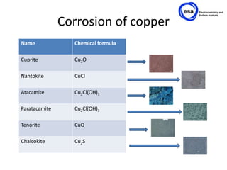
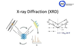
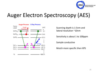
It’s what’s on the outside that counts, true story!


                                                                                                                    1
Promotor: Pieter-Jan Sabbe & Dr. A. Adriaens   Bachelorstudents: Nils De Rybel, Nicky Huys, Maria Gomez Rodriguez
Outline

•   Why study surfaces?
•   Goal of project
•   Corrosion layers of copper
•   Spectroscopic techniques used
•   Conclusion


                                    2
Goal
•   Make copper corrosion layers
•   Study surface specific techniques
•   Apply surface specific techniques
•   Compare the layers


                                        3
Why study surfaces?

                    Polymer biodegradation             Surface tension control
                        Catalysis                             Adsorption, desorption
                                                                            Sensors


Surface hardening                                                           Friction control
                                    Behaviour determined
                                          Applications
                                         by surface


                                                                       Corrosion
            Crystal growth
Controlled release polymers
                                    Optical recording media

                                                                   Anti-reflection coatings
       Magnetic recording media
                                                                                               4
Corrosion of copper
Name             Chemical formula

Cuprite          Cu2O

Nantokite        CuCl

Atacamite        Cu2Cl(OH)3

Paratacamite     Cu2Cl(OH)3

Tenorite         CuO

Chalcokite       Cu2S
Optical Microscopy
Optical Microscopy
• Cupper coupon:


• Chalcokite
Optical Microscopy
• Cuprite.


• Nantokite
Optical microscopy
• Atacamite.


• Paratacamite
Optical microscope
• Tenorite


• Chalcokite
X-ray Diffraction (XRD)


Paratacamite
Copper
Nantokite
X-ray Diffraction (XRD)


Paratacamite
Copper
Nantokite
X-ray Diffraction (XRD)


Chalcokite
Copper
X-ray Photoelectron Spectroscopy
              (XPS)


                                   16
X-ray Photoelectron
Spectroscopy (XPS)

               S-Probe Monochromatized XPS
               spectrometer from Surface
               Science Instruments (VG)

               AlKα x-ray (1486.6 eV)
               1x10⁻⁹ mbar
               Analysis surface : 250 x 1000 μm


                                            17
Standard spectra


                   18
Spectra
                  300000
                                                            Copper peaks
                  250000


                  200000
Intensity (CPS)


                                                                                                 Tenorite (CuO)
                  150000                                                                         Nantokite (CuCl)
                                                                                                 Cuprite (Cu2O)

                  100000                                                                         Chalcocite (Cu2S)
                                                                                                 Paratacamite (Cu2Cl(OH)3)

                   50000


                       0
                           900   910   920   930      940      950       960   970   980   990
                                                   Binding energy (eV)


                                                                                                                             19
Auger Electron Spectroscopy (AES)
                   Scanning depth is 1-5nm and
                   lateral resolution ~10nm

                   Sensitivity is about 1 to 100ppm

                   Sample conductive

                   Mutch more specific then XPS


                                                      20
X-ray Excited Optical Luminescence
                 (XEOL)
• How it works


                                       21
X-ray Excited Optical Luminescence
                 (XEOL)
• Source


                                       22
X-ray Excited Optical Luminescence
               (XEOL)                                             Nantokite
                                                                  Paratacamite(?)
                        20.00


                        18.00                                                        Ref. Nantokite (CuCl)


                        16.00
                                                                                                    Nantokite
                                                                                                    Paratacamite(?)
                        14.00
 Normalized Intensity


                        12.00


                        10.01                                                                       Ref. Paratacamite (Cu2Cl(OH)3)


                        08.01


                        06.01


                        04.01


                        02.01


                        00.01
                            889.20   891.00   892.79   894.59   896.38     898.18      899.97   901.77       903.56   905.36    907.15
                                                                                    -2
                                                                     Energy / keV 10


                                                                                                                                         23
Scanning Electron Microscopy (SEM)


                                     24
Scanning Electron Microscopy (SEM)


                                     25
Scanning Electron Microscopy (SEM)


                                     26
Scanning Electron Microscopy (SEM)

          1 - 30 keV


        0.5 - 10 nm
                                     27
Scanning Electron Microscopy (SEM)
• Secondary electrons


                                      28
Scanning Electron Microscopy (SEM)
• Backscattered electrons


                                      29
Scanning Electron Microscopy (SEM)


                                     30

Surface analysisversion3

Editor's Notes