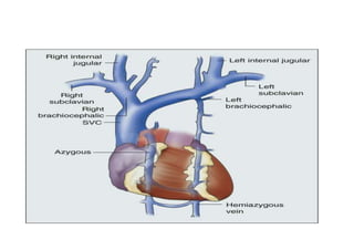
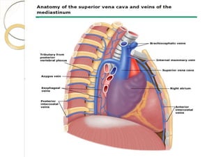
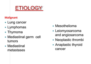
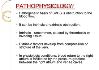
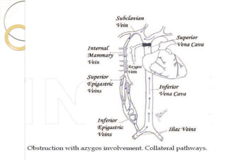
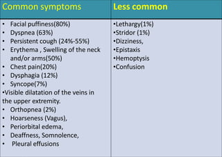
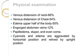
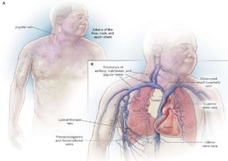
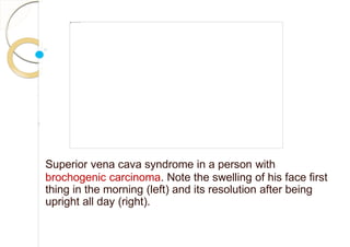
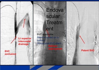
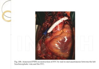
SlideShare a Scribd company logo
SUPERIOR VENA CAVA
SYNDROME
Dr. SHAILESH GUPTA
26-3-2018
MEDIASTINUM
Mediastinum
• Bulky septum
between the
pleural cavities
& lungs
Extends-
• Root of the
neck to
diaphragm
• From the
sternum to
vertebral
column
Divisions of mediastinum
• Divided by an imaginary horizontal plane from
the sternal angle to the intervertebral disc
between 4th & 5th vertebra in to
• superior mediastinum
• Inferior mediastinum –
Anterior
Middle
Posterior
Superior mediastinum
• Posterior to the sternum & anterior to the
bodies of first four thoracic vertebra
Contents of superior mediastinum
• Thymus
• Rt & Lt brachiocephalic
veins
• Left superior intercostal vein
• Superior vena cava
• Arch of aorta & it’s three
branches
• Trachea
• Esophagus
• Phrenic nerves
• Vagus nerves
• Lt recurrent laryngeal nerve
• Thoracic duct
• Other small structures
Thymus
• Asymmetric bilobed gland
• Upper extent may reach as high as thyroid gland
• Involved in early development of immune system
• Large in children
• Atrophies after puberty
Brachiocephalic veins
•Located immediately
posterior to thymus
•Formed at the jn. of
IJ & subclavian veins.
• Begins post to clavicle
•Tributaries: vertebral,
first post. Intercostal,
int. thoracic,
inf. Thyroid,
thymic veins
Left superior intercostal vein
• Drains upper two or
three intercostal
veins, left bronchial
veins & left
pericardiophrenic
veins
• Drains in to left
brachiocephalic
veins
Superior vena cava
• Vertically oriented
• Begins posterior
to the lower edge
of first costal
cartilage
• terminates at the
lower border of
right third costal
cartilage
Arch of aorta & it’s branches
• Begins when ascending
aorta emerges from
pericardial sac
• Terminates on left side
of vertebral level
between Th 4th - 5th
Branches-
Brachiocephalic trunk
Left CCA
Left subclavian
Thyroid ima
Trachea
• Midline structure
• Present anterior to
esophagus
• Swallowing, breathing,
disease & specialized
instrumentation causes
shift of trachea
• divides just inferior to
sternal angle (T3- T4)
into Right & Left principal
bronchus
Thoracic duct
• Major lymphatic vessel
of the body
• Enters sup.
mediastinum from
below at the level of
T4/5
• continues through the
superior med. posterior
to arch of aorta
between the esophagus
& Lt pleura
Esophagus
• Starts at the C6th &
terminates at cardiac
opening of stomach
(T12)
• Descends anterior to
vertebral bodies in
midline
• Crossed laterally by the
azygos vein on the right
side & arch of aorta on
the left side
SVC SYNDROME
Superior vena cava syndrome (SVCS) is obstruction of
blood flow through the superior vena cava (SVC).
HISTORY
 First recorded description of SVC obstruction
(SVCO) - 1757 when William Hunter
described the entity in a patient with a
syphilitic aortic aneurysm.
 For nearly two centuries- nonmalignant
processes such as aortic aneurysms,
syphilitic aortitis, or chronic mediastinitis due
to tuberculosis were the predominant
etiologic factors.
 Now Rare
 In the preantibiotic era-
◦ syphilitic thoracic aortic aneurysms,
 fibrosing mediastinitis,
 untreated infection were frequent causes of the SVC
syndrome.
 Subsequently, malignancy became the most
common cause, accounting for 90 percent of
cases by the 1980s.
 More recently, the incidence of SVC
syndrome due to thrombosis has risen,
largely because of increased use of
intravascular devices such as catheters and
pacemakers.
ANATOMY
 SVC originates in the chest, behind the first right
sternocostal articulation, from the confluence of two
main collector vessels: Right and Left
brachiocephalic veins which receive the ipsilateral
internal jugular and subclavian veins.
 Internal jugular vein drains-----head and deep
sections of the neck
 Subclavian vein--- upper limbs, superior chest and
superficial head and neck.
 After the brachiocephalic convergence, the SVC
follows the right lateral margin of the sternum in an
inferoposterior direction.
 Finally, it enters the pericardium superiorly and
opens into the right atrium
 The SVC’s length ranges from 6 to 8 cm.
 Its diameter is usually 20-22 mm.
 The blood pressure ranges from -5 to 5
mmHg and the flow is discontinuous
depending on the heart pulse cycle.
 The SVC receives a single affluent vein:
the azygos vein.
 The azygos vein joins the SVC from the
right side, at its mid length, above the
right bronchus.
 No valve divides the superior vena
cava from the right atrium. As a result,
the right atrial and right ventricular
contractions are conducted up into
the internal jugular vein and, through
the sternocleidomastoid muscle,
can be seen as the jugular venous
pressure.
AZYGOS VEIN
 Azygos vein transports deoxygenated blood from the posterior
walls of the thorax and abdomen into the superior
venacava.
It is formed by the union of
 Ascending lumbar veins with
 Right subcostal veins
At the level of the 12th thoracic vertebra
 Ascending in the posterior mediastinum, and arching over the
right main bronchus posteriorly at the root of the right lung to
join the superior vena cava.
 A major tributary is the hemiazygos vein, a similar
structure on the opposite side of the vertebral
column.
Other tributaries include
 Bronchial veins,
 Pericardial veins, and
 Posterior right intercostal veins.
 It communicates with the vertebral venous plexuses.
HEMIAZYGOS VEIN
 It runs superiorly in the lower thoracic region, just
to the left side of the vertebral column.
 Hemiazygos vein and the accessory hemiazygos
vein, when taken together, essentially serve as the
left-sided equivalent of the azygos vein.
 It usually begins in the left ascending lumbar vein
or renal vein, and passes upward through the left
crus of the diaphragm to enter the thorax.
 It continues ascending on the left side of the
vertebral column, and at the level of the 9th
thoracic vertebra, it passes rightward across the
vertebral column, behind the aorta, esophagus, and
thoracic duct, to end in the azygos vein.
 The hemiazygos may or may not be continuous
superiorly with the accessory hemiazygos vein.
 It receives the 9th, 10th, and 11th posterior
intercostal veins and the subcostal vein of the left
side, and some esophageal and mediastinal veins.
ACCESSORY HEMIAZYGOS
VEIN
 Receives the posterior intercostal veins from the
4th, 5th, 6th, and 7th ICS.
 It either crosses the body of 8th thoracic vertebra
to join the azygous vein or ends in the
hemiazygos.
 When this vein is small, or altogether absent, the
left superior intercostal vein may extend as low as
the 5th or 6th ICS.
Posterior intercostal veins
There are eleven posterior intercostal veins on each side.
 The 1st posterior intercostal vein, drains into the
brachiocephalic vein or the vertebral vein.
 The 2nd and 3rd (and often 4th) posterior intercostal
veins drain into the superior intercostal vein.
 The remaining posterior intercostal veins drain into the
azygos vein on the right, or the hemiazygos vein and
accessory hemiazygous on the left.
SVC OBSTRUCTION
 In SVC obstruction, the azygos vein is
responsible for the most important
collateral circulation.
According to the expected collateral pathways,
the SVC can be divided into two segments:
 Supra-azygos or preazygos and
 Infra-azygos or postazygos SVC.
 There are four possible collateral systems---
They are represented by
1. Azygos venous system,
2. Internal thoracic venous system,
3. Vertebral venous system and
4. External thoracic venous system.
1. Azygos venous system is the only direct path
into the SVC.
2. Internal thoracic vein is the collector between
SVC and inferior vena cava (IVC) via epigastric
and iliac veins.
3. Vertebral veins with intercostals, lumbar and
sacral veins, represent the posterior network
between SVC and IVC.
4. External thoracic vein system is the most
superficial and it is represented by axillary,
lateral thoracic and superficial epigastric
veins.
ETIOLOGY
Malignant
 Lung cancer
 Lymphomas
 Thymoma
 Mediastinal germ cell
tumors
 Mediastinal
metastases
 Mesothelioma
 Leiomyosarcoma
and angiosarcoma
 Neoplastic thrombi
 Anaplastic thyroid
cancer
Benign
 Fibrosing mediastinitis
(idiopathic or radiation-
induced)
 Infectious diseases –
1. Tuberculosis,
2. Histoplasmosis,
3. Echinococcosis,
4. Syphilis,
5. Aspergillosis,
6. Blastomycosis,
7. Filariasis,
8. Nocardiosis.
 Thrombosis (non-
neoplastic)
 Lymphadenopathies
1. sarcoidosis,
2. Behçet’s syndrome,
3. Castleman’s disease
 Aortic aneurysm
 Substernal goiter
 Pericardial, thymic,
bronchogenic cysts
 Iatrogenic
1. Pacemaker and
defibrillator placement
2. Central venous
catheters
PATHOPHYSIOLOGY
 Pathogenetic basis of SVCS is obstruction to the
blood flow.
 It can be intrinsic or extrinsic obstruction.
 Intrinsic—uncommon, caused by thrombosis or
invading tissue.
 Extrinsic factors develop from compression or
stricture of the vein.
 In physiologic conditions, blood return to the right
atrium is facilitated by the pressure gradient
between the right atrium and venae cavae.
 When obstruction of the SVC occurs, the vascular
resistances rise and the venous return decreases.
 When SVC shows a significant stenosis (3/5 of the
lumen or more), blood flow is redirected through the
collateral circulation in order to bypass the obstruction
and restore the venous return.
 In acute impairments, the blood flow is not
rapidly distributed through the collateral network
so symptoms arise markedly.
 In the case of slow-growing diseases, the
collateral venous network has enough time to
expand in order to receive the circulating volume.
 For this reason, long-lasting, severe SVC
obstruction can sometimes be found without
significant symptoms
The clinical seriousness is related to several factors:
1. Level of obstruction and rapidity of
development, determining the effectiveness of
collateral circulation
2. Impairment of lymphatic drainage (pulmonary
interstitial edema or pleural effusion)
3. Involvement of other mediastinal structures
(compression or invasion of heart, pulmonary
artery and central airways, phrenic nerve
paralysis)
 Superficial dilated vascular routes are the main sign of
collateral circulation and appear swollen and non-
pulsating.
 In case of marked obesity, superficial veins can be
missing at inspection.
 Variety of collateral circulation and the differences in the
venous rearrangement are expression of the SVC
obstruction site.
Anatomic classification includes three levels of
obstruction:
1. Obstruction of the upper SVC, proximal to (above the
level of) azygos entry point.
2. Obstruction with azygos involvement.
3. Obstruction of the lower SVC, distal to (below the level
of) azygos entry point.
 In this situation, there is no impediment to normal
blood flow through the azygos vein which opens
into the patent tract of the SVC.
 Venous drainage coming from the head neck,
shoulders and arms cannot directly reach the
right atrium.
 From the superior tract of the SVC, blood flow
is reversed and directed to the azygos, mainly
through the right superior intercostal vein.
 In this case, the azygos vein cannot be used as
collateral pathway and the only viable blood return is
carried by minor vessels to IVC (cava-cava or
anazygotic circulation).
 From the internal thoracic veins, blood is forced to the
intercostal veins, then to azygos and hemiazygos veins.
 The flow is thus reversed into the ascending lumbar
veins to the iliac veins.
 Direct anastomosis between the azygos’ origin and
the IVC and between hemiazygos and left renal vein
are also active.
 In addition, the internal thoracic veins can flow into
the superior epigastric veins.
 From the superior epigastric veins, blood is carried to
the inferior epigastric veins across the superficial
system of the cutaneous abdominal veins and finally
to the iliac veins.
 Another course is between the thoraco-epigastric
vein (collateral of the axillary vein) and the external
iliac vein.
 In these conditions, the collateral circulation is partly
deep and partly superficial.
 The reversed circulation through the described
pathways, remains less efficient than the azygos
system and venous hypertension is usually more
severe.
 For this reason, this kind of SVC obstruction is often
related to important symptoms, dyspnea and pleural
effusion.
 In this condition, the obstruction is just below
the azygos arch.
 The blood flow is distributed from the
superior body into the azygos and
hemiazygos veins, in which the flow is
inverted, to the IVC tributaries.
 In this type of obstruction, the superficial collateral
system is not always evident but the azygos and
hemiazygos congestion and dilatation are usually
important.
 The hemodynamic changes lead to edema and
cyanosis of the upper chest and pleural effusion.
Pleural effusion is often slowly-growing and
rightsided, probably due to anatomical reasons:
 There is a wider anastomosis between hemiazygos
and IVC than between azygos and IVC.
Classification of SVCS
 There are three main classification proposals which
follow different methods of categorization.
Doty and Standford’s classification (anatomical)
1. Type I: stenosis of up to 90% of the supra-
azygos SVC
2. Type II: stenosis of more than 90% of the supra-
azygos SVC
3. Type III: complete occlusion of SVC with azygos
reverse blood flow
4. Type IV: complete occlusion of SVC with the
involvement of the major tributaries and azygos
vein
Yu’s classification (clinical)
1. Grade 0: asymptomatic (imaging evidence of SVC
obstruction)
2. Grade 1: mild (plethora, cyanosis, head and neck
edema)
3. Grade 2: moderate (grade 1 evidence + functional
impairment)
4. Grade 3: severe (mild/moderate cerebral or
laryngeal edema, limited cardiac reserve)
5. Grade 4: life-threatening (significant cerebral or
laryngeal edema, cardiac failure)
6. Grade 5: fatal
Bigsby’s classification (operative risk)
 Low risk
 High risk
The low risk patients present with
 No dyspnea at rest,
 No facial cyanosis in the upright position,
 No change of dyspnea, No worsening of facial
edema and Cyanosis during the supine
position.
 The high risk patients present with facial
cyanosis or dyspnea at rest in the sitting
position.
• Diagnosis is made by history, physical
examination, and lab studies
• findings will depend on
1. The degree of occlusion
2. The rapidity of development
3. Presence or absence of collateral
circulation
PRESENTING SYMPTOM S OF SVCO
positional changes such as bendingThese symptoms may be worsened by
forward, stooping, or lying down.
Common symptoms Less common
• Facial puffiness(80%)
• Dyspnea (63%)
• Persistent cough (24%-55%)
• Erythema , Swelling of the neck
and/or arms(50%)
• Chest pain(20%)
• Dysphagia (12%)
• Syncope(7%)
•Visible dilatation of the veins in
the upper extremity.
• Orthopnea (2%)
• Hoarseness (Vagus),
• Periorbital edema,
• Deaffness, Somnolence,
• Pleural effusions
•Lethargy(1%)
•Stridor (1%)
•Dizziness,
•Epistaxis
•Hemoptysis
•Confusion
 Venous distension of neck-66%
 Venous distension of Chest-54%
 Edema upper half of the body-50%
 Engorged abdomen veins-12%
 Papilledema, stupor, and even coma.
 Cyanosis and edema are aggravated by
horizontal position and relived by upright
position
Superior vena cava syndrome in a person with
brochogenic carcinoma. Note the swelling of his face first
thing in the morning (left) and its resolution after being
upright all day (right).
DIFFERENTIAL DIAGNOSIS
 Heart failure and pericardial tamponade
 Nephrotic syndrome
Diagnosis
 Diagnosis of SVCS can be made simply on physical
examination.
 When the extent of disease is minimal, the physical
findings may not be prominent then it is difficult to
diagnose.
 Establishing the underlying etiology is more
important because certain disorders that cause
SVCS may be more amenable to specific treatment
regimens.
RADIOLOGICAL STUDIES
a) CHEST RADIOGRAPHY
 The initial diagnostic test for suspected SVCS.
 Is not specific for SVCS.
 helpful in identifying the cause of the disorder.
 Right sided findings are common.
Chest radiography…..
X-Ray findings suggestive of underlying malignancy,
Mediastinal widening
Pleural effusion(s)
Right hilar mass
Cardiomegaly
Calcified paratracheal lymph nodes-
Granulomatous disease
 Anterior mediastinal mass
 Normal(16%)
 In the absence of previous catheterization or surgery, a
normal result on chest radiography in a patient with
SVCS is almost pathognomonic of chronic fibrous
mediastinitis.
ULTRASONOGRAPHY
 SVC cannot be imaged because of
poor acoustic window
 Patency can be indirectly determined with
normal wave forms in brachiocephalic and
subclavian veins.
 Exclusion of thrombus in upper extremity,
subclavian, brachiocephalic and axillary
veins.
Computed tomography scanning
 CT-Provides an effective, noninvasive evaluation of
the superior vena cava and its collateral circulation.
 CT scanning provides
1) Anatomic details of the mediastinal and thoracic
organs
2) Allows identification of the cause and extent of
the obstruction,
3) Documents collateral circulation,
4) Provides guidance for Percutaneous biopsies
5) Guides the formulation for radiotherapy.
Magnetic resonance imaging (MRI)
 MRI is often important in determining the cause of
SVCS.
 MRI, by virtue of its multidimensional capabilities,
shows the relationships of vessels, lymph nodes,
and other mediastinal structures.
 It is an acceptable alternative for patients with renal
failure or those with contrast allergies.
Contrast venography
 An x-ray test that provides an image of the veins after a
contrast dye is injected into a vein.
Advantages:-
 The extent and site of obstruction.
 The nature and degree of obstruction.
 Patency of the superior vena cava.
 Differentiation between intrinsic and extrinsic obstruction.
 Assessment of collateral vessels
 the degree of venous distension of the neck and arms
Contrast venography…..
 Measurement of actual venous pressure
 The presence of the internal jugular vein reflux.
 Is essential prior to planning any surgical bypass
operation.
 Surgical bypass operations are easier to accomplish
when the brachiocephalic veins are not involved.
 However, if all the intrathoracic veins are obstructed,
extrathoracic bypass operations can be undertaken,
 Very helpful in documenting obstructions caused by
thrombus formation. When thrombosis is present,
treatment with fibrinolytic agents (eg, urokinase,
streptokinase) is pursued and
 Repeat venography can be used to evaluate
treatment efficacy.
Venographic classification
1. Type I: stenosis of up to 90% of the
supra-azygos SVC
2. Type II: stenosis of more than 90%
of the supra-azygos SVC
3. Type III: complete occlusion of SVC
with azygos reverse blood flow
4. Type IV: complete occlusion of SVC
with the involvement of the major
tributaries and azygos vein
Radionuclide venography.
 This test is less invasive than contrast
venography
 is less specific in defining Patency and
flow.
 Radionuclide venography may be of
value in long-term follow-up studies.
Diagnostic surgery.
 When all other diagnostic procedures fail to provide
information about the cause of SVCS, Exploratory
thoracotomy be the last alternative.
 Advantages—surgery allows direct visualization of
the underlying disease process, assessment of the
extent of disease involvement, and accessibility for
tissue biopsy
 However, this procedure is the most invasive and is
associated with increased risks.
TREATMENT OF SVCO
Treatment Of SVCO
 Depending on the underlying condition,
multiple treatment options are available
for superior vena cava obstruction. The
primary treatment options include
 Medical Care
 Radiation
 Chemotherapy
 Thrombolytic therapy
 Anticoagulation
 Stents and balloon angioplasty and
 Surgery.
Medical Care
 The goals of SVCS management are to relieve
symptoms and to attempt cure of the primary
malignant
Conservative treatment -symptomatic improvement
 including elevation of the head end of the bed
and supplemental oxygen.
 Emergency treatment (Corticosteroids and
diuretics )
 For Brain edema,
 decreased cardiac output,
or upper airway edema.
Dexamethasone (Decadron,
Dexasone)
For symptomatic management in tumor-
associated edema.
 8-40 mg IV once initially, followed by
4-6 mg IV/PO q6-8h
Other Medications
 Loop Diuretic agents
 Salt Restriction.
 Oxygen
Radiation therapy
Indications.
 The majority of cases of SVCS are caused by
malignancy; thus, most patients receive radiation
treatment at some point in their illness.
Emergency radiation treatment
 To life-threatening cerebral or laryngeal edema
prior to a tissue diagnosis of malignancy.
 To relieve obstructive symptoms
 Inappropriate for the treatment of an underlying
thrombosis or granulomatosis causing the
obstruction
Radiation Dosage
 Initiated at high dose daily for the first few days.
followed by conventional low daily doses. total dose
is dependent on tumor histology.
 Lymphomas (3000 to 4000 cGy,)
 Carcinomas require (4000 to 5000 cGy or more)
Lower doses of radiation treatment
 When systemic disease is present and short- term
palliation is the goal.
 Radiation to Heart and Spinal cord.
 who are receiving chemotherapeutic agents such as
doxorubicin, which can enhance radiation toxicity.
Response to RT
3 to 4 days- Resolution of facial edema and
venous distension of the upper extremities .
1 to 3 weeks- Radiographic improvement .
Not effective -Thrombosis is cause for SVCO
 When RT successfully completed in pts of
SVCS with malignancies, 10% to 20% survive
more than 2 years.
Side effects of RT.
 Persistent fever,
 Bleeding,
 SVC perforation at the site of tumor
invasion,
 Nausea, Vomiting,
 Anorexia,
 Leukopenia,
Hemoptysis,
Late Complications
 Skin irritation;
 Esophagitis;
 Pulmonary or mediastinal fibrosis;
Chemotherapy
 Chemotherapy may be used as a
primary therapy or as an adjunct to
radiotherapy
 treatment of choice for SVCS caused
by Mediastinal lymphoma is a
combination of chemotherapy and
radiotherapy.
Thrombolytic Therapy
 Pericatheter thrombosis is seen in
approximately 50% of Non-
anticoagulated patients with long term
Central vein Catheters
 In Acute Cases- excellent results with
thrombolytic therapy.
Benefits of Thrombolytic therapy
 Fast dissolution of emboli,
 Quickened recovery,
 Prevention of recurrent thrombus
formation,
 Rapid restoration of hemodynamic
disturbances.
Urokinase
Action:-
 Converts plasminogen to plasmin,
which degrades fibrin clots, fibrinogen,
and other plasma proteins.
Adult Dose:
Loading dose: 4400 U/kg IV over 10
min and increase to 6000 U/kg/h
Maintenance dose: 4400-6000 U/kg/h
IV
Anticoagulation
 Patients with SVCS are at increased risk for
deep vein thrombosis and pulmonary embolism.
 In patients for whom thrombosis is the cause of
SVCS, anticoagulation therapy should be
administered after successful thrombolytic
treatment.
 Once the symptoms subside after
thrombolytic therapy, anticoagulation should
be maintained as long as the central venous
catheter is present.
Anticoagulants - Heparin
Action:-
 Inhibits thrombosis by inactivating activated
factor X and inhibiting conversion of prothrombin
to thrombin.
Adult:-
 5000 U IV bolus, then infusion to maintain aPTT
2-3 times the reference range
Pediatric:-
 Initial dose: 50 U/kg IV
Maintenance infusion: 15-25 U/kg/h IV
Increase dose by 2-4 U/kg/h IV q6-8h using aPTT
Anticoagulants -Warfarin
(Coumarin)
Action:-
 Inhibits synthesis of vitamin K–dependent
coagulation factors (factors II, VII, IX, X).
Adult:-
 Initial: 5-10 mg PO
Maintenance: 2-10 mg PO qd to maintain
INR of 2-3.
Stents
 Recent advances in interventional radiology have
contributed expandable wire stents and balloon
angioplasty.
 can be placed across the stenotic portion.
 stents have little thrombogenic potential
 After thrombolytic therapy, stent placement has
been noted to be a more successful approach.
 After stent, patients experience instantaneous relief of
symptoms.
 The placement of stents is performed under local
anesthesia.
Balloon Angioplasty
 For localized lesions, balloon angioplasty
with or without stenting has also been
shown to significantly reduce the
symptoms of SVCS
Lt superior
intercostal
drainage
SVC
occlusion
Endova
scular
Treatm
ent
SVC syndrome
Stent mounted on a
balloon
Status post SVC stent
Balloon
deployment
Patent SVC
Surgical Treatment
 Surgical bypass is an additional alternative to relieve SVCS.
 is usually recommended to benign disease and to only a few
patients with malignancy.
 Patients selected for surgery should have the Category-IV
venographic signs, i.e, total vena caval obstruction.
 Surgery in cases of fibrosing mediastinitis can be extremely
complicated, because of the extensive collateral circulation
under high venous pressure.
 Advantage is definitive removal of the obstruction and direct
tissue diagnosis.
 Long-term results after surgical bypass are lacking, because
their life expectancy is short.
PROGNOSIS
Benign disease-life expectancy unchanged
Malignant obstruction of SVC
 Untreated - 30 days average life
expectancy
 Treated - < 7 month average life
expectancy
- 20% 1-year survival for lung cancer
-NSCLC-poor prognosis, palliative care+RT
- 50% 2-year survival for lymphoma

More Related Content

What's hot

MANAGEMENT OF EARLY STAGE NON SMALL CELL LUNG CARCINOMA
MANAGEMENT OF EARLY STAGE NON SMALL CELL LUNG CARCINOMAMANAGEMENT OF EARLY STAGE NON SMALL CELL LUNG CARCINOMA
MANAGEMENT OF EARLY STAGE NON SMALL CELL LUNG CARCINOMA
Isha Jaiswal
 
Management of Primary CNS Lymphoma (PCNSL)
Management of Primary CNS Lymphoma (PCNSL)Management of Primary CNS Lymphoma (PCNSL)
Management of Primary CNS Lymphoma (PCNSL)
Narayan Adhikari
 
Carcinoma cervix brachytherapy- dr upasna
Carcinoma cervix   brachytherapy- dr upasnaCarcinoma cervix   brachytherapy- dr upasna
Carcinoma cervix brachytherapy- dr upasna
Upasna Saxena
 
Ca lung
Ca lung Ca lung
Ca lung
kilichophy
 
Lytic leisons of the skull
Lytic leisons of the skullLytic leisons of the skull
Lytic leisons of the skull
Milan Silwal
 
Small Cell Lung Cancer Management by Dr.Tinku Joseph
Small Cell Lung Cancer Management by Dr.Tinku JosephSmall Cell Lung Cancer Management by Dr.Tinku Joseph
Small Cell Lung Cancer Management by Dr.Tinku Joseph
Dr.Tinku Joseph
 
BASICS OF NEUROANATOMY & BRAIN TUMOR RADIOLOGY
BASICS OF NEUROANATOMY & BRAIN TUMOR RADIOLOGYBASICS OF NEUROANATOMY & BRAIN TUMOR RADIOLOGY
BASICS OF NEUROANATOMY & BRAIN TUMOR RADIOLOGY
Kanhu Charan
 
Carotid artery stenosis
Carotid artery stenosisCarotid artery stenosis
Carotid artery stenosis
Ramachandra Barik
 
Radiation Pneumonitis
Radiation PneumonitisRadiation Pneumonitis
Radiation Pneumonitis
HEBAGOMAA1984
 
LANDMARK TRIALS IN BREAST CANCER
LANDMARK TRIALS IN BREAST CANCERLANDMARK TRIALS IN BREAST CANCER
LANDMARK TRIALS IN BREAST CANCER
Aaditya Prakash
 
Targeted therapy and immunotherapy in lung cancer
Targeted therapy and immunotherapy in lung cancerTargeted therapy and immunotherapy in lung cancer
Targeted therapy and immunotherapy in lung cancer
Alok Gupta
 
Small cell lung cancer
Small cell lung cancerSmall cell lung cancer
Small cell lung cancer
DrAyush Garg
 
Non Small Cell Lung Cancer
Non Small Cell Lung CancerNon Small Cell Lung Cancer
Non Small Cell Lung Cancerfondas vakalis
 
Maliganant spinal cord compression main
Maliganant spinal cord compression mainMaliganant spinal cord compression main
Maliganant spinal cord compression main
Sasikumar Sambasivam
 
SVC syndrome - a surgical perspective
SVC syndrome - a surgical perspectiveSVC syndrome - a surgical perspective
SVC syndrome - a surgical perspective
SrikanthK120
 
high grade glioma
high grade gliomahigh grade glioma
high grade glioma
HardikSharma590779
 
Lung Cancer: Diagnosis, Staging, and Treatment
Lung Cancer: Diagnosis, Staging, and TreatmentLung Cancer: Diagnosis, Staging, and Treatment
Lung Cancer: Diagnosis, Staging, and Treatment
Dene W. Daugherty
 
Cardiac tumours
Cardiac tumoursCardiac tumours
Cardiac tumours
Ankur Batra
 
Thymoma
ThymomaThymoma
Thymoma
Rawa Muhsin
 

What's hot (20)

MANAGEMENT OF EARLY STAGE NON SMALL CELL LUNG CARCINOMA
MANAGEMENT OF EARLY STAGE NON SMALL CELL LUNG CARCINOMAMANAGEMENT OF EARLY STAGE NON SMALL CELL LUNG CARCINOMA
MANAGEMENT OF EARLY STAGE NON SMALL CELL LUNG CARCINOMA
 
Management of Primary CNS Lymphoma (PCNSL)
Management of Primary CNS Lymphoma (PCNSL)Management of Primary CNS Lymphoma (PCNSL)
Management of Primary CNS Lymphoma (PCNSL)
 
Carcinoma cervix brachytherapy- dr upasna
Carcinoma cervix   brachytherapy- dr upasnaCarcinoma cervix   brachytherapy- dr upasna
Carcinoma cervix brachytherapy- dr upasna
 
Radiation for Lung Cancer
Radiation for Lung CancerRadiation for Lung Cancer
Radiation for Lung Cancer
 
Ca lung
Ca lung Ca lung
Ca lung
 
Lytic leisons of the skull
Lytic leisons of the skullLytic leisons of the skull
Lytic leisons of the skull
 
Small Cell Lung Cancer Management by Dr.Tinku Joseph
Small Cell Lung Cancer Management by Dr.Tinku JosephSmall Cell Lung Cancer Management by Dr.Tinku Joseph
Small Cell Lung Cancer Management by Dr.Tinku Joseph
 
BASICS OF NEUROANATOMY & BRAIN TUMOR RADIOLOGY
BASICS OF NEUROANATOMY & BRAIN TUMOR RADIOLOGYBASICS OF NEUROANATOMY & BRAIN TUMOR RADIOLOGY
BASICS OF NEUROANATOMY & BRAIN TUMOR RADIOLOGY
 
Carotid artery stenosis
Carotid artery stenosisCarotid artery stenosis
Carotid artery stenosis
 
Radiation Pneumonitis
Radiation PneumonitisRadiation Pneumonitis
Radiation Pneumonitis
 
LANDMARK TRIALS IN BREAST CANCER
LANDMARK TRIALS IN BREAST CANCERLANDMARK TRIALS IN BREAST CANCER
LANDMARK TRIALS IN BREAST CANCER
 
Targeted therapy and immunotherapy in lung cancer
Targeted therapy and immunotherapy in lung cancerTargeted therapy and immunotherapy in lung cancer
Targeted therapy and immunotherapy in lung cancer
 
Small cell lung cancer
Small cell lung cancerSmall cell lung cancer
Small cell lung cancer
 
Non Small Cell Lung Cancer
Non Small Cell Lung CancerNon Small Cell Lung Cancer
Non Small Cell Lung Cancer
 
Maliganant spinal cord compression main
Maliganant spinal cord compression mainMaliganant spinal cord compression main
Maliganant spinal cord compression main
 
SVC syndrome - a surgical perspective
SVC syndrome - a surgical perspectiveSVC syndrome - a surgical perspective
SVC syndrome - a surgical perspective
 
high grade glioma
high grade gliomahigh grade glioma
high grade glioma
 
Lung Cancer: Diagnosis, Staging, and Treatment
Lung Cancer: Diagnosis, Staging, and TreatmentLung Cancer: Diagnosis, Staging, and Treatment
Lung Cancer: Diagnosis, Staging, and Treatment
 
Cardiac tumours
Cardiac tumoursCardiac tumours
Cardiac tumours
 
Thymoma
ThymomaThymoma
Thymoma
 

Similar to Svc syndrome,

Congenital heart disease
Congenital heart diseaseCongenital heart disease
Congenital heart disease
Zahra Khan
 
Mediastinum 2020.pdf
Mediastinum 2020.pdfMediastinum 2020.pdf
Mediastinum 2020.pdf
Fatimakabara1
 
Aorta, SVC, PULMONARY TRUNK.pptx
Aorta, SVC, PULMONARY TRUNK.pptxAorta, SVC, PULMONARY TRUNK.pptx
Aorta, SVC, PULMONARY TRUNK.pptx
MovieDownload83
 
USMLE CVS 005 Blood vessels – Arteries and veins.pdf
USMLE   CVS  005 Blood vessels – Arteries and veins.pdfUSMLE   CVS  005 Blood vessels – Arteries and veins.pdf
USMLE CVS 005 Blood vessels – Arteries and veins.pdf
AHMED ASHOUR
 
Subclavian artery and it's branches
Subclavian artery and it's branchesSubclavian artery and it's branches
Subclavian artery and it's branches
Harshal Shinde
 
Cynotic heart disease plane radiography
Cynotic heart disease plane radiography Cynotic heart disease plane radiography
Cynotic heart disease plane radiography
Dr-Girish Gunari
 
Mediastinum.pptx
Mediastinum.pptxMediastinum.pptx
Mediastinum.pptx
Abhishek840813
 
IE, RHD.pptx
IE, RHD.pptxIE, RHD.pptx
IE, RHD.pptx
SumanjitDas1
 
Blood supply of head and neck.pdf
Blood supply of head and neck.pdfBlood supply of head and neck.pdf
Blood supply of head and neck.pdf
ParulGarg894243
 
Vascular anomalies chest
Vascular anomalies chestVascular anomalies chest
Vascular anomalies chest
fenderhm
 
Azygos system of veins
Azygos system of veinsAzygos system of veins
Azygos system of veins
Idris Siddiqui
 
USMLE CVS 001 Mediastinum anatomy medical chest .pdf
USMLE   CVS  001 Mediastinum anatomy medical chest .pdfUSMLE   CVS  001 Mediastinum anatomy medical chest .pdf
USMLE CVS 001 Mediastinum anatomy medical chest .pdf
AHMED ASHOUR
 
Cardiac embryology
Cardiac embryologyCardiac embryology
Cardiac embryology
Rezaul hayat
 
USMLE CVS 006 007 Development of the heart anatomy .pdf
USMLE   CVS  006 007 Development of the heart anatomy .pdfUSMLE   CVS  006 007 Development of the heart anatomy .pdf
USMLE CVS 006 007 Development of the heart anatomy .pdf
AHMED ASHOUR
 
Anatomy of heart by dr rajesh-1.pptx
Anatomy of heart by dr rajesh-1.pptxAnatomy of heart by dr rajesh-1.pptx
Anatomy of heart by dr rajesh-1.pptx
Rajesh Munigial
 
MEDISTINUM and The HEART.pptx1111111111111111111
MEDISTINUM and The HEART.pptx1111111111111111111MEDISTINUM and The HEART.pptx1111111111111111111
MEDISTINUM and The HEART.pptx1111111111111111111
marrahmohamed33
 
CVS Anaesthesiology.pptx
CVS Anaesthesiology.pptxCVS Anaesthesiology.pptx
CVS Anaesthesiology.pptx
Sandeep Singh Jadon
 

Similar to Svc syndrome, (20)

SVC SYNDROME
SVC SYNDROMESVC SYNDROME
SVC SYNDROME
 
Congenital heart disease
Congenital heart diseaseCongenital heart disease
Congenital heart disease
 
Mediastinum 2020.pdf
Mediastinum 2020.pdfMediastinum 2020.pdf
Mediastinum 2020.pdf
 
Aorta, SVC, PULMONARY TRUNK.pptx
Aorta, SVC, PULMONARY TRUNK.pptxAorta, SVC, PULMONARY TRUNK.pptx
Aorta, SVC, PULMONARY TRUNK.pptx
 
USMLE CVS 005 Blood vessels – Arteries and veins.pdf
USMLE   CVS  005 Blood vessels – Arteries and veins.pdfUSMLE   CVS  005 Blood vessels – Arteries and veins.pdf
USMLE CVS 005 Blood vessels – Arteries and veins.pdf
 
Subclavian artery and it's branches
Subclavian artery and it's branchesSubclavian artery and it's branches
Subclavian artery and it's branches
 
Cynotic heart disease plane radiography
Cynotic heart disease plane radiography Cynotic heart disease plane radiography
Cynotic heart disease plane radiography
 
Mediastinum.pptx
Mediastinum.pptxMediastinum.pptx
Mediastinum.pptx
 
IE, RHD.pptx
IE, RHD.pptxIE, RHD.pptx
IE, RHD.pptx
 
Blood supply of head and neck.pdf
Blood supply of head and neck.pdfBlood supply of head and neck.pdf
Blood supply of head and neck.pdf
 
Vascular anomalies chest
Vascular anomalies chestVascular anomalies chest
Vascular anomalies chest
 
Azygos system of veins
Azygos system of veinsAzygos system of veins
Azygos system of veins
 
USMLE CVS 001 Mediastinum anatomy medical chest .pdf
USMLE   CVS  001 Mediastinum anatomy medical chest .pdfUSMLE   CVS  001 Mediastinum anatomy medical chest .pdf
USMLE CVS 001 Mediastinum anatomy medical chest .pdf
 
Cardiac embryology
Cardiac embryologyCardiac embryology
Cardiac embryology
 
USMLE CVS 006 007 Development of the heart anatomy .pdf
USMLE   CVS  006 007 Development of the heart anatomy .pdfUSMLE   CVS  006 007 Development of the heart anatomy .pdf
USMLE CVS 006 007 Development of the heart anatomy .pdf
 
Anatomy of heart by dr rajesh-1.pptx
Anatomy of heart by dr rajesh-1.pptxAnatomy of heart by dr rajesh-1.pptx
Anatomy of heart by dr rajesh-1.pptx
 
17 Heart
17 Heart17 Heart
17 Heart
 
MEDISTINUM and The HEART.pptx1111111111111111111
MEDISTINUM and The HEART.pptx1111111111111111111MEDISTINUM and The HEART.pptx1111111111111111111
MEDISTINUM and The HEART.pptx1111111111111111111
 
CVS Anaesthesiology.pptx
CVS Anaesthesiology.pptxCVS Anaesthesiology.pptx
CVS Anaesthesiology.pptx
 
Truncus
TruncusTruncus
Truncus
 

Recently uploaded

Report Back from SGO 2024: What’s the Latest in Cervical Cancer?
Report Back from SGO 2024: What’s the Latest in Cervical Cancer?Report Back from SGO 2024: What’s the Latest in Cervical Cancer?
Report Back from SGO 2024: What’s the Latest in Cervical Cancer?
bkling
 
How to Give Better Lectures: Some Tips for Doctors
How to Give Better Lectures: Some Tips for DoctorsHow to Give Better Lectures: Some Tips for Doctors
How to Give Better Lectures: Some Tips for Doctors
LanceCatedral
 
263778731218 Abortion Clinic /Pills In Harare ,
263778731218 Abortion Clinic /Pills In Harare ,263778731218 Abortion Clinic /Pills In Harare ,
263778731218 Abortion Clinic /Pills In Harare ,
sisternakatoto
 
Light House Retreats: Plant Medicine Retreat Europe
Light House Retreats: Plant Medicine Retreat EuropeLight House Retreats: Plant Medicine Retreat Europe
Light House Retreats: Plant Medicine Retreat Europe
Lighthouse Retreat
 
ARTHROLOGY PPT NCISM SYLLABUS AYURVEDA STUDENTS
ARTHROLOGY PPT NCISM SYLLABUS AYURVEDA STUDENTSARTHROLOGY PPT NCISM SYLLABUS AYURVEDA STUDENTS
ARTHROLOGY PPT NCISM SYLLABUS AYURVEDA STUDENTS
Dr. Vinay Pareek
 
planning for change nursing Management ppt
planning for change nursing Management pptplanning for change nursing Management ppt
planning for change nursing Management ppt
Thangamjayarani
 
BRACHYTHERAPY OVERVIEW AND APPLICATORS
BRACHYTHERAPY OVERVIEW  AND  APPLICATORSBRACHYTHERAPY OVERVIEW  AND  APPLICATORS
BRACHYTHERAPY OVERVIEW AND APPLICATORS
Krishan Murari
 
Sex determination from mandible pelvis and skull
Sex determination from mandible pelvis and skullSex determination from mandible pelvis and skull
Sex determination from mandible pelvis and skull
ShashankRoodkee
 
Physiology of Special Chemical Sensation of Taste
Physiology of Special Chemical Sensation of TastePhysiology of Special Chemical Sensation of Taste
Physiology of Special Chemical Sensation of Taste
MedicoseAcademics
 
Triangles of Neck and Clinical Correlation by Dr. RIG.pptx
Triangles of Neck and Clinical Correlation by Dr. RIG.pptxTriangles of Neck and Clinical Correlation by Dr. RIG.pptx
Triangles of Neck and Clinical Correlation by Dr. RIG.pptx
Dr. Rabia Inam Gandapore
 
Maxilla, Mandible & Hyoid Bone & Clinical Correlations by Dr. RIG.pptx
Maxilla, Mandible & Hyoid Bone & Clinical Correlations by Dr. RIG.pptxMaxilla, Mandible & Hyoid Bone & Clinical Correlations by Dr. RIG.pptx
Maxilla, Mandible & Hyoid Bone & Clinical Correlations by Dr. RIG.pptx
Dr. Rabia Inam Gandapore
 
Superficial & Deep Fascia of the NECK.pptx
Superficial & Deep Fascia of the NECK.pptxSuperficial & Deep Fascia of the NECK.pptx
Superficial & Deep Fascia of the NECK.pptx
Dr. Rabia Inam Gandapore
 
How STIs Influence the Development of Pelvic Inflammatory Disease.pptx
How STIs Influence the Development of Pelvic Inflammatory Disease.pptxHow STIs Influence the Development of Pelvic Inflammatory Disease.pptx
How STIs Influence the Development of Pelvic Inflammatory Disease.pptx
FFragrant
 
MANAGEMENT OF ATRIOVENTRICULAR CONDUCTION BLOCK.pdf
MANAGEMENT OF ATRIOVENTRICULAR CONDUCTION BLOCK.pdfMANAGEMENT OF ATRIOVENTRICULAR CONDUCTION BLOCK.pdf
MANAGEMENT OF ATRIOVENTRICULAR CONDUCTION BLOCK.pdf
Jim Jacob Roy
 
basicmodesofventilation2022-220313203758.pdf
basicmodesofventilation2022-220313203758.pdfbasicmodesofventilation2022-220313203758.pdf
basicmodesofventilation2022-220313203758.pdf
aljamhori teaching hospital
 
Pharma Pcd Franchise in Jharkhand - Yodley Lifesciences
Pharma Pcd Franchise in Jharkhand - Yodley LifesciencesPharma Pcd Franchise in Jharkhand - Yodley Lifesciences
Pharma Pcd Franchise in Jharkhand - Yodley Lifesciences
Yodley Lifesciences
 
Ozempic: Preoperative Management of Patients on GLP-1 Receptor Agonists
Ozempic: Preoperative Management of Patients on GLP-1 Receptor Agonists  Ozempic: Preoperative Management of Patients on GLP-1 Receptor Agonists
Ozempic: Preoperative Management of Patients on GLP-1 Receptor Agonists
Saeid Safari
 
Knee anatomy and clinical tests 2024.pdf
Knee anatomy and clinical tests 2024.pdfKnee anatomy and clinical tests 2024.pdf
Knee anatomy and clinical tests 2024.pdf
vimalpl1234
 
Tom Selleck Health: A Comprehensive Look at the Iconic Actor’s Wellness Journey
Tom Selleck Health: A Comprehensive Look at the Iconic Actor’s Wellness JourneyTom Selleck Health: A Comprehensive Look at the Iconic Actor’s Wellness Journey
Tom Selleck Health: A Comprehensive Look at the Iconic Actor’s Wellness Journey
greendigital
 
ANATOMY AND PHYSIOLOGY OF URINARY SYSTEM.pptx
ANATOMY AND PHYSIOLOGY OF URINARY SYSTEM.pptxANATOMY AND PHYSIOLOGY OF URINARY SYSTEM.pptx
ANATOMY AND PHYSIOLOGY OF URINARY SYSTEM.pptx
Swetaba Besh
 

Recently uploaded (20)

Report Back from SGO 2024: What’s the Latest in Cervical Cancer?
Report Back from SGO 2024: What’s the Latest in Cervical Cancer?Report Back from SGO 2024: What’s the Latest in Cervical Cancer?
Report Back from SGO 2024: What’s the Latest in Cervical Cancer?
 
How to Give Better Lectures: Some Tips for Doctors
How to Give Better Lectures: Some Tips for DoctorsHow to Give Better Lectures: Some Tips for Doctors
How to Give Better Lectures: Some Tips for Doctors
 
263778731218 Abortion Clinic /Pills In Harare ,
263778731218 Abortion Clinic /Pills In Harare ,263778731218 Abortion Clinic /Pills In Harare ,
263778731218 Abortion Clinic /Pills In Harare ,
 
Light House Retreats: Plant Medicine Retreat Europe
Light House Retreats: Plant Medicine Retreat EuropeLight House Retreats: Plant Medicine Retreat Europe
Light House Retreats: Plant Medicine Retreat Europe
 
ARTHROLOGY PPT NCISM SYLLABUS AYURVEDA STUDENTS
ARTHROLOGY PPT NCISM SYLLABUS AYURVEDA STUDENTSARTHROLOGY PPT NCISM SYLLABUS AYURVEDA STUDENTS
ARTHROLOGY PPT NCISM SYLLABUS AYURVEDA STUDENTS
 
planning for change nursing Management ppt
planning for change nursing Management pptplanning for change nursing Management ppt
planning for change nursing Management ppt
 
BRACHYTHERAPY OVERVIEW AND APPLICATORS
BRACHYTHERAPY OVERVIEW  AND  APPLICATORSBRACHYTHERAPY OVERVIEW  AND  APPLICATORS
BRACHYTHERAPY OVERVIEW AND APPLICATORS
 
Sex determination from mandible pelvis and skull
Sex determination from mandible pelvis and skullSex determination from mandible pelvis and skull
Sex determination from mandible pelvis and skull
 
Physiology of Special Chemical Sensation of Taste
Physiology of Special Chemical Sensation of TastePhysiology of Special Chemical Sensation of Taste
Physiology of Special Chemical Sensation of Taste
 
Triangles of Neck and Clinical Correlation by Dr. RIG.pptx
Triangles of Neck and Clinical Correlation by Dr. RIG.pptxTriangles of Neck and Clinical Correlation by Dr. RIG.pptx
Triangles of Neck and Clinical Correlation by Dr. RIG.pptx
 
Maxilla, Mandible & Hyoid Bone & Clinical Correlations by Dr. RIG.pptx
Maxilla, Mandible & Hyoid Bone & Clinical Correlations by Dr. RIG.pptxMaxilla, Mandible & Hyoid Bone & Clinical Correlations by Dr. RIG.pptx
Maxilla, Mandible & Hyoid Bone & Clinical Correlations by Dr. RIG.pptx
 
Superficial & Deep Fascia of the NECK.pptx
Superficial & Deep Fascia of the NECK.pptxSuperficial & Deep Fascia of the NECK.pptx
Superficial & Deep Fascia of the NECK.pptx
 
How STIs Influence the Development of Pelvic Inflammatory Disease.pptx
How STIs Influence the Development of Pelvic Inflammatory Disease.pptxHow STIs Influence the Development of Pelvic Inflammatory Disease.pptx
How STIs Influence the Development of Pelvic Inflammatory Disease.pptx
 
MANAGEMENT OF ATRIOVENTRICULAR CONDUCTION BLOCK.pdf
MANAGEMENT OF ATRIOVENTRICULAR CONDUCTION BLOCK.pdfMANAGEMENT OF ATRIOVENTRICULAR CONDUCTION BLOCK.pdf
MANAGEMENT OF ATRIOVENTRICULAR CONDUCTION BLOCK.pdf
 
basicmodesofventilation2022-220313203758.pdf
basicmodesofventilation2022-220313203758.pdfbasicmodesofventilation2022-220313203758.pdf
basicmodesofventilation2022-220313203758.pdf
 
Pharma Pcd Franchise in Jharkhand - Yodley Lifesciences
Pharma Pcd Franchise in Jharkhand - Yodley LifesciencesPharma Pcd Franchise in Jharkhand - Yodley Lifesciences
Pharma Pcd Franchise in Jharkhand - Yodley Lifesciences
 
Ozempic: Preoperative Management of Patients on GLP-1 Receptor Agonists
Ozempic: Preoperative Management of Patients on GLP-1 Receptor Agonists  Ozempic: Preoperative Management of Patients on GLP-1 Receptor Agonists
Ozempic: Preoperative Management of Patients on GLP-1 Receptor Agonists
 
Knee anatomy and clinical tests 2024.pdf
Knee anatomy and clinical tests 2024.pdfKnee anatomy and clinical tests 2024.pdf
Knee anatomy and clinical tests 2024.pdf
 
Tom Selleck Health: A Comprehensive Look at the Iconic Actor’s Wellness Journey
Tom Selleck Health: A Comprehensive Look at the Iconic Actor’s Wellness JourneyTom Selleck Health: A Comprehensive Look at the Iconic Actor’s Wellness Journey
Tom Selleck Health: A Comprehensive Look at the Iconic Actor’s Wellness Journey
 
ANATOMY AND PHYSIOLOGY OF URINARY SYSTEM.pptx
ANATOMY AND PHYSIOLOGY OF URINARY SYSTEM.pptxANATOMY AND PHYSIOLOGY OF URINARY SYSTEM.pptx
ANATOMY AND PHYSIOLOGY OF URINARY SYSTEM.pptx
 

Svc syndrome,

  • 1. SUPERIOR VENA CAVA SYNDROME Dr. SHAILESH GUPTA 26-3-2018
  • 3. Mediastinum • Bulky septum between the pleural cavities & lungs Extends- • Root of the neck to diaphragm • From the sternum to vertebral column
  • 4. Divisions of mediastinum • Divided by an imaginary horizontal plane from the sternal angle to the intervertebral disc between 4th & 5th vertebra in to • superior mediastinum • Inferior mediastinum – Anterior Middle Posterior
  • 5. Superior mediastinum • Posterior to the sternum & anterior to the bodies of first four thoracic vertebra
  • 6. Contents of superior mediastinum • Thymus • Rt & Lt brachiocephalic veins • Left superior intercostal vein • Superior vena cava • Arch of aorta & it’s three branches • Trachea • Esophagus • Phrenic nerves • Vagus nerves • Lt recurrent laryngeal nerve • Thoracic duct • Other small structures
  • 7. Thymus • Asymmetric bilobed gland • Upper extent may reach as high as thyroid gland • Involved in early development of immune system • Large in children • Atrophies after puberty
  • 8. Brachiocephalic veins •Located immediately posterior to thymus •Formed at the jn. of IJ & subclavian veins. • Begins post to clavicle •Tributaries: vertebral, first post. Intercostal, int. thoracic, inf. Thyroid, thymic veins
  • 9. Left superior intercostal vein • Drains upper two or three intercostal veins, left bronchial veins & left pericardiophrenic veins • Drains in to left brachiocephalic veins
  • 10. Superior vena cava • Vertically oriented • Begins posterior to the lower edge of first costal cartilage • terminates at the lower border of right third costal cartilage
  • 11. Arch of aorta & it’s branches • Begins when ascending aorta emerges from pericardial sac • Terminates on left side of vertebral level between Th 4th - 5th Branches- Brachiocephalic trunk Left CCA Left subclavian Thyroid ima
  • 12. Trachea • Midline structure • Present anterior to esophagus • Swallowing, breathing, disease & specialized instrumentation causes shift of trachea • divides just inferior to sternal angle (T3- T4) into Right & Left principal bronchus
  • 13. Thoracic duct • Major lymphatic vessel of the body • Enters sup. mediastinum from below at the level of T4/5 • continues through the superior med. posterior to arch of aorta between the esophagus & Lt pleura
  • 14. Esophagus • Starts at the C6th & terminates at cardiac opening of stomach (T12) • Descends anterior to vertebral bodies in midline • Crossed laterally by the azygos vein on the right side & arch of aorta on the left side
  • 15. SVC SYNDROME Superior vena cava syndrome (SVCS) is obstruction of blood flow through the superior vena cava (SVC).
  • 16. HISTORY  First recorded description of SVC obstruction (SVCO) - 1757 when William Hunter described the entity in a patient with a syphilitic aortic aneurysm.  For nearly two centuries- nonmalignant processes such as aortic aneurysms, syphilitic aortitis, or chronic mediastinitis due to tuberculosis were the predominant etiologic factors.  Now Rare
  • 17.  In the preantibiotic era- ◦ syphilitic thoracic aortic aneurysms,  fibrosing mediastinitis,  untreated infection were frequent causes of the SVC syndrome.  Subsequently, malignancy became the most common cause, accounting for 90 percent of cases by the 1980s.  More recently, the incidence of SVC syndrome due to thrombosis has risen, largely because of increased use of intravascular devices such as catheters and pacemakers.
  • 18. ANATOMY  SVC originates in the chest, behind the first right sternocostal articulation, from the confluence of two main collector vessels: Right and Left brachiocephalic veins which receive the ipsilateral internal jugular and subclavian veins.  Internal jugular vein drains-----head and deep sections of the neck  Subclavian vein--- upper limbs, superior chest and superficial head and neck.
  • 19.
  • 20.  After the brachiocephalic convergence, the SVC follows the right lateral margin of the sternum in an inferoposterior direction.  Finally, it enters the pericardium superiorly and opens into the right atrium  The SVC’s length ranges from 6 to 8 cm.  Its diameter is usually 20-22 mm.
  • 21.  The blood pressure ranges from -5 to 5 mmHg and the flow is discontinuous depending on the heart pulse cycle.  The SVC receives a single affluent vein: the azygos vein.  The azygos vein joins the SVC from the right side, at its mid length, above the right bronchus.
  • 22.  No valve divides the superior vena cava from the right atrium. As a result, the right atrial and right ventricular contractions are conducted up into the internal jugular vein and, through the sternocleidomastoid muscle, can be seen as the jugular venous pressure.
  • 23.
  • 24. AZYGOS VEIN  Azygos vein transports deoxygenated blood from the posterior walls of the thorax and abdomen into the superior venacava. It is formed by the union of  Ascending lumbar veins with  Right subcostal veins At the level of the 12th thoracic vertebra  Ascending in the posterior mediastinum, and arching over the right main bronchus posteriorly at the root of the right lung to join the superior vena cava.
  • 25.  A major tributary is the hemiazygos vein, a similar structure on the opposite side of the vertebral column. Other tributaries include  Bronchial veins,  Pericardial veins, and  Posterior right intercostal veins.  It communicates with the vertebral venous plexuses.
  • 26. HEMIAZYGOS VEIN  It runs superiorly in the lower thoracic region, just to the left side of the vertebral column.  Hemiazygos vein and the accessory hemiazygos vein, when taken together, essentially serve as the left-sided equivalent of the azygos vein.  It usually begins in the left ascending lumbar vein or renal vein, and passes upward through the left crus of the diaphragm to enter the thorax.
  • 27.  It continues ascending on the left side of the vertebral column, and at the level of the 9th thoracic vertebra, it passes rightward across the vertebral column, behind the aorta, esophagus, and thoracic duct, to end in the azygos vein.  The hemiazygos may or may not be continuous superiorly with the accessory hemiazygos vein.  It receives the 9th, 10th, and 11th posterior intercostal veins and the subcostal vein of the left side, and some esophageal and mediastinal veins.
  • 28. ACCESSORY HEMIAZYGOS VEIN  Receives the posterior intercostal veins from the 4th, 5th, 6th, and 7th ICS.  It either crosses the body of 8th thoracic vertebra to join the azygous vein or ends in the hemiazygos.  When this vein is small, or altogether absent, the left superior intercostal vein may extend as low as the 5th or 6th ICS.
  • 29. Posterior intercostal veins There are eleven posterior intercostal veins on each side.  The 1st posterior intercostal vein, drains into the brachiocephalic vein or the vertebral vein.  The 2nd and 3rd (and often 4th) posterior intercostal veins drain into the superior intercostal vein.  The remaining posterior intercostal veins drain into the azygos vein on the right, or the hemiazygos vein and accessory hemiazygous on the left.
  • 30. SVC OBSTRUCTION  In SVC obstruction, the azygos vein is responsible for the most important collateral circulation. According to the expected collateral pathways, the SVC can be divided into two segments:  Supra-azygos or preazygos and  Infra-azygos or postazygos SVC.
  • 31.  There are four possible collateral systems--- They are represented by 1. Azygos venous system, 2. Internal thoracic venous system, 3. Vertebral venous system and 4. External thoracic venous system.
  • 32. 1. Azygos venous system is the only direct path into the SVC. 2. Internal thoracic vein is the collector between SVC and inferior vena cava (IVC) via epigastric and iliac veins. 3. Vertebral veins with intercostals, lumbar and sacral veins, represent the posterior network between SVC and IVC. 4. External thoracic vein system is the most superficial and it is represented by axillary, lateral thoracic and superficial epigastric veins.
  • 33. ETIOLOGY Malignant  Lung cancer  Lymphomas  Thymoma  Mediastinal germ cell tumors  Mediastinal metastases  Mesothelioma  Leiomyosarcoma and angiosarcoma  Neoplastic thrombi  Anaplastic thyroid cancer
  • 34. Benign  Fibrosing mediastinitis (idiopathic or radiation- induced)  Infectious diseases – 1. Tuberculosis, 2. Histoplasmosis, 3. Echinococcosis, 4. Syphilis, 5. Aspergillosis, 6. Blastomycosis, 7. Filariasis, 8. Nocardiosis.  Thrombosis (non- neoplastic)  Lymphadenopathies 1. sarcoidosis, 2. Behçet’s syndrome, 3. Castleman’s disease  Aortic aneurysm  Substernal goiter  Pericardial, thymic, bronchogenic cysts  Iatrogenic 1. Pacemaker and defibrillator placement 2. Central venous catheters
  • 35. PATHOPHYSIOLOGY  Pathogenetic basis of SVCS is obstruction to the blood flow.  It can be intrinsic or extrinsic obstruction.  Intrinsic—uncommon, caused by thrombosis or invading tissue.  Extrinsic factors develop from compression or stricture of the vein.  In physiologic conditions, blood return to the right atrium is facilitated by the pressure gradient between the right atrium and venae cavae.
  • 36.  When obstruction of the SVC occurs, the vascular resistances rise and the venous return decreases.  When SVC shows a significant stenosis (3/5 of the lumen or more), blood flow is redirected through the collateral circulation in order to bypass the obstruction and restore the venous return.
  • 37.  In acute impairments, the blood flow is not rapidly distributed through the collateral network so symptoms arise markedly.  In the case of slow-growing diseases, the collateral venous network has enough time to expand in order to receive the circulating volume.  For this reason, long-lasting, severe SVC obstruction can sometimes be found without significant symptoms
  • 38. The clinical seriousness is related to several factors: 1. Level of obstruction and rapidity of development, determining the effectiveness of collateral circulation 2. Impairment of lymphatic drainage (pulmonary interstitial edema or pleural effusion) 3. Involvement of other mediastinal structures (compression or invasion of heart, pulmonary artery and central airways, phrenic nerve paralysis)
  • 39.  Superficial dilated vascular routes are the main sign of collateral circulation and appear swollen and non- pulsating.  In case of marked obesity, superficial veins can be missing at inspection.  Variety of collateral circulation and the differences in the venous rearrangement are expression of the SVC obstruction site. Anatomic classification includes three levels of obstruction: 1. Obstruction of the upper SVC, proximal to (above the level of) azygos entry point. 2. Obstruction with azygos involvement. 3. Obstruction of the lower SVC, distal to (below the level of) azygos entry point.
  • 40.  In this situation, there is no impediment to normal blood flow through the azygos vein which opens into the patent tract of the SVC.  Venous drainage coming from the head neck, shoulders and arms cannot directly reach the right atrium.  From the superior tract of the SVC, blood flow is reversed and directed to the azygos, mainly through the right superior intercostal vein.
  • 41.
  • 42.  In this case, the azygos vein cannot be used as collateral pathway and the only viable blood return is carried by minor vessels to IVC (cava-cava or anazygotic circulation).  From the internal thoracic veins, blood is forced to the intercostal veins, then to azygos and hemiazygos veins.  The flow is thus reversed into the ascending lumbar veins to the iliac veins.
  • 43.  Direct anastomosis between the azygos’ origin and the IVC and between hemiazygos and left renal vein are also active.  In addition, the internal thoracic veins can flow into the superior epigastric veins.  From the superior epigastric veins, blood is carried to the inferior epigastric veins across the superficial system of the cutaneous abdominal veins and finally to the iliac veins.  Another course is between the thoraco-epigastric vein (collateral of the axillary vein) and the external iliac vein.
  • 44.
  • 45.  In these conditions, the collateral circulation is partly deep and partly superficial.  The reversed circulation through the described pathways, remains less efficient than the azygos system and venous hypertension is usually more severe.  For this reason, this kind of SVC obstruction is often related to important symptoms, dyspnea and pleural effusion.
  • 46.  In this condition, the obstruction is just below the azygos arch.  The blood flow is distributed from the superior body into the azygos and hemiazygos veins, in which the flow is inverted, to the IVC tributaries.
  • 47.  In this type of obstruction, the superficial collateral system is not always evident but the azygos and hemiazygos congestion and dilatation are usually important.  The hemodynamic changes lead to edema and cyanosis of the upper chest and pleural effusion. Pleural effusion is often slowly-growing and rightsided, probably due to anatomical reasons:  There is a wider anastomosis between hemiazygos and IVC than between azygos and IVC.
  • 48.
  • 49. Classification of SVCS  There are three main classification proposals which follow different methods of categorization. Doty and Standford’s classification (anatomical) 1. Type I: stenosis of up to 90% of the supra- azygos SVC 2. Type II: stenosis of more than 90% of the supra- azygos SVC 3. Type III: complete occlusion of SVC with azygos reverse blood flow 4. Type IV: complete occlusion of SVC with the involvement of the major tributaries and azygos vein
  • 50. Yu’s classification (clinical) 1. Grade 0: asymptomatic (imaging evidence of SVC obstruction) 2. Grade 1: mild (plethora, cyanosis, head and neck edema) 3. Grade 2: moderate (grade 1 evidence + functional impairment) 4. Grade 3: severe (mild/moderate cerebral or laryngeal edema, limited cardiac reserve) 5. Grade 4: life-threatening (significant cerebral or laryngeal edema, cardiac failure) 6. Grade 5: fatal
  • 51. Bigsby’s classification (operative risk)  Low risk  High risk The low risk patients present with  No dyspnea at rest,  No facial cyanosis in the upright position,  No change of dyspnea, No worsening of facial edema and Cyanosis during the supine position.  The high risk patients present with facial cyanosis or dyspnea at rest in the sitting position.
  • 52. • Diagnosis is made by history, physical examination, and lab studies • findings will depend on 1. The degree of occlusion 2. The rapidity of development 3. Presence or absence of collateral circulation
  • 53. PRESENTING SYMPTOM S OF SVCO positional changes such as bendingThese symptoms may be worsened by forward, stooping, or lying down. Common symptoms Less common • Facial puffiness(80%) • Dyspnea (63%) • Persistent cough (24%-55%) • Erythema , Swelling of the neck and/or arms(50%) • Chest pain(20%) • Dysphagia (12%) • Syncope(7%) •Visible dilatation of the veins in the upper extremity. • Orthopnea (2%) • Hoarseness (Vagus), • Periorbital edema, • Deaffness, Somnolence, • Pleural effusions •Lethargy(1%) •Stridor (1%) •Dizziness, •Epistaxis •Hemoptysis •Confusion
  • 54.  Venous distension of neck-66%  Venous distension of Chest-54%  Edema upper half of the body-50%  Engorged abdomen veins-12%  Papilledema, stupor, and even coma.  Cyanosis and edema are aggravated by horizontal position and relived by upright position
  • 55.
  • 56.
  • 57. Superior vena cava syndrome in a person with brochogenic carcinoma. Note the swelling of his face first thing in the morning (left) and its resolution after being upright all day (right).
  • 58. DIFFERENTIAL DIAGNOSIS  Heart failure and pericardial tamponade  Nephrotic syndrome
  • 59. Diagnosis  Diagnosis of SVCS can be made simply on physical examination.  When the extent of disease is minimal, the physical findings may not be prominent then it is difficult to diagnose.  Establishing the underlying etiology is more important because certain disorders that cause SVCS may be more amenable to specific treatment regimens.
  • 60. RADIOLOGICAL STUDIES a) CHEST RADIOGRAPHY  The initial diagnostic test for suspected SVCS.  Is not specific for SVCS.  helpful in identifying the cause of the disorder.  Right sided findings are common.
  • 61. Chest radiography….. X-Ray findings suggestive of underlying malignancy, Mediastinal widening Pleural effusion(s) Right hilar mass Cardiomegaly Calcified paratracheal lymph nodes- Granulomatous disease  Anterior mediastinal mass  Normal(16%)  In the absence of previous catheterization or surgery, a normal result on chest radiography in a patient with SVCS is almost pathognomonic of chronic fibrous mediastinitis.
  • 62. ULTRASONOGRAPHY  SVC cannot be imaged because of poor acoustic window  Patency can be indirectly determined with normal wave forms in brachiocephalic and subclavian veins.  Exclusion of thrombus in upper extremity, subclavian, brachiocephalic and axillary veins.
  • 63. Computed tomography scanning  CT-Provides an effective, noninvasive evaluation of the superior vena cava and its collateral circulation.  CT scanning provides 1) Anatomic details of the mediastinal and thoracic organs 2) Allows identification of the cause and extent of the obstruction, 3) Documents collateral circulation, 4) Provides guidance for Percutaneous biopsies 5) Guides the formulation for radiotherapy.
  • 64. Magnetic resonance imaging (MRI)  MRI is often important in determining the cause of SVCS.  MRI, by virtue of its multidimensional capabilities, shows the relationships of vessels, lymph nodes, and other mediastinal structures.  It is an acceptable alternative for patients with renal failure or those with contrast allergies.
  • 65. Contrast venography  An x-ray test that provides an image of the veins after a contrast dye is injected into a vein. Advantages:-  The extent and site of obstruction.  The nature and degree of obstruction.  Patency of the superior vena cava.  Differentiation between intrinsic and extrinsic obstruction.  Assessment of collateral vessels  the degree of venous distension of the neck and arms
  • 66. Contrast venography…..  Measurement of actual venous pressure  The presence of the internal jugular vein reflux.  Is essential prior to planning any surgical bypass operation.  Surgical bypass operations are easier to accomplish when the brachiocephalic veins are not involved.  However, if all the intrathoracic veins are obstructed, extrathoracic bypass operations can be undertaken,  Very helpful in documenting obstructions caused by thrombus formation. When thrombosis is present, treatment with fibrinolytic agents (eg, urokinase, streptokinase) is pursued and  Repeat venography can be used to evaluate treatment efficacy.
  • 67. Venographic classification 1. Type I: stenosis of up to 90% of the supra-azygos SVC 2. Type II: stenosis of more than 90% of the supra-azygos SVC 3. Type III: complete occlusion of SVC with azygos reverse blood flow 4. Type IV: complete occlusion of SVC with the involvement of the major tributaries and azygos vein
  • 68. Radionuclide venography.  This test is less invasive than contrast venography  is less specific in defining Patency and flow.  Radionuclide venography may be of value in long-term follow-up studies.
  • 69. Diagnostic surgery.  When all other diagnostic procedures fail to provide information about the cause of SVCS, Exploratory thoracotomy be the last alternative.  Advantages—surgery allows direct visualization of the underlying disease process, assessment of the extent of disease involvement, and accessibility for tissue biopsy  However, this procedure is the most invasive and is associated with increased risks.
  • 71. Treatment Of SVCO  Depending on the underlying condition, multiple treatment options are available for superior vena cava obstruction. The primary treatment options include  Medical Care  Radiation  Chemotherapy  Thrombolytic therapy  Anticoagulation  Stents and balloon angioplasty and  Surgery.
  • 72. Medical Care  The goals of SVCS management are to relieve symptoms and to attempt cure of the primary malignant Conservative treatment -symptomatic improvement  including elevation of the head end of the bed and supplemental oxygen.  Emergency treatment (Corticosteroids and diuretics )  For Brain edema,  decreased cardiac output, or upper airway edema.
  • 73. Dexamethasone (Decadron, Dexasone) For symptomatic management in tumor- associated edema.  8-40 mg IV once initially, followed by 4-6 mg IV/PO q6-8h
  • 74. Other Medications  Loop Diuretic agents  Salt Restriction.  Oxygen
  • 75. Radiation therapy Indications.  The majority of cases of SVCS are caused by malignancy; thus, most patients receive radiation treatment at some point in their illness. Emergency radiation treatment  To life-threatening cerebral or laryngeal edema prior to a tissue diagnosis of malignancy.  To relieve obstructive symptoms  Inappropriate for the treatment of an underlying thrombosis or granulomatosis causing the obstruction
  • 76. Radiation Dosage  Initiated at high dose daily for the first few days. followed by conventional low daily doses. total dose is dependent on tumor histology.  Lymphomas (3000 to 4000 cGy,)  Carcinomas require (4000 to 5000 cGy or more) Lower doses of radiation treatment  When systemic disease is present and short- term palliation is the goal.  Radiation to Heart and Spinal cord.  who are receiving chemotherapeutic agents such as doxorubicin, which can enhance radiation toxicity.
  • 77. Response to RT 3 to 4 days- Resolution of facial edema and venous distension of the upper extremities . 1 to 3 weeks- Radiographic improvement . Not effective -Thrombosis is cause for SVCO  When RT successfully completed in pts of SVCS with malignancies, 10% to 20% survive more than 2 years.
  • 78. Side effects of RT.  Persistent fever,  Bleeding,  SVC perforation at the site of tumor invasion,  Nausea, Vomiting,  Anorexia,  Leukopenia, Hemoptysis, Late Complications  Skin irritation;  Esophagitis;  Pulmonary or mediastinal fibrosis;
  • 79. Chemotherapy  Chemotherapy may be used as a primary therapy or as an adjunct to radiotherapy  treatment of choice for SVCS caused by Mediastinal lymphoma is a combination of chemotherapy and radiotherapy.
  • 80. Thrombolytic Therapy  Pericatheter thrombosis is seen in approximately 50% of Non- anticoagulated patients with long term Central vein Catheters  In Acute Cases- excellent results with thrombolytic therapy.
  • 81. Benefits of Thrombolytic therapy  Fast dissolution of emboli,  Quickened recovery,  Prevention of recurrent thrombus formation,  Rapid restoration of hemodynamic disturbances.
  • 82. Urokinase Action:-  Converts plasminogen to plasmin, which degrades fibrin clots, fibrinogen, and other plasma proteins. Adult Dose: Loading dose: 4400 U/kg IV over 10 min and increase to 6000 U/kg/h Maintenance dose: 4400-6000 U/kg/h IV
  • 83. Anticoagulation  Patients with SVCS are at increased risk for deep vein thrombosis and pulmonary embolism.  In patients for whom thrombosis is the cause of SVCS, anticoagulation therapy should be administered after successful thrombolytic treatment.  Once the symptoms subside after thrombolytic therapy, anticoagulation should be maintained as long as the central venous catheter is present.
  • 84. Anticoagulants - Heparin Action:-  Inhibits thrombosis by inactivating activated factor X and inhibiting conversion of prothrombin to thrombin. Adult:-  5000 U IV bolus, then infusion to maintain aPTT 2-3 times the reference range Pediatric:-  Initial dose: 50 U/kg IV Maintenance infusion: 15-25 U/kg/h IV Increase dose by 2-4 U/kg/h IV q6-8h using aPTT
  • 85. Anticoagulants -Warfarin (Coumarin) Action:-  Inhibits synthesis of vitamin K–dependent coagulation factors (factors II, VII, IX, X). Adult:-  Initial: 5-10 mg PO Maintenance: 2-10 mg PO qd to maintain INR of 2-3.
  • 86. Stents  Recent advances in interventional radiology have contributed expandable wire stents and balloon angioplasty.  can be placed across the stenotic portion.  stents have little thrombogenic potential  After thrombolytic therapy, stent placement has been noted to be a more successful approach.  After stent, patients experience instantaneous relief of symptoms.  The placement of stents is performed under local anesthesia.
  • 87. Balloon Angioplasty  For localized lesions, balloon angioplasty with or without stenting has also been shown to significantly reduce the symptoms of SVCS
  • 88. Lt superior intercostal drainage SVC occlusion Endova scular Treatm ent SVC syndrome Stent mounted on a balloon Status post SVC stent Balloon deployment Patent SVC
  • 89. Surgical Treatment  Surgical bypass is an additional alternative to relieve SVCS.  is usually recommended to benign disease and to only a few patients with malignancy.  Patients selected for surgery should have the Category-IV venographic signs, i.e, total vena caval obstruction.  Surgery in cases of fibrosing mediastinitis can be extremely complicated, because of the extensive collateral circulation under high venous pressure.  Advantage is definitive removal of the obstruction and direct tissue diagnosis.  Long-term results after surgical bypass are lacking, because their life expectancy is short.
  • 90.
  • 91.
  • 92.
  • 93.
  • 94. PROGNOSIS Benign disease-life expectancy unchanged Malignant obstruction of SVC  Untreated - 30 days average life expectancy  Treated - < 7 month average life expectancy - 20% 1-year survival for lung cancer -NSCLC-poor prognosis, palliative care+RT - 50% 2-year survival for lymphoma