This document provides an overview of ocular motility disorders, including:
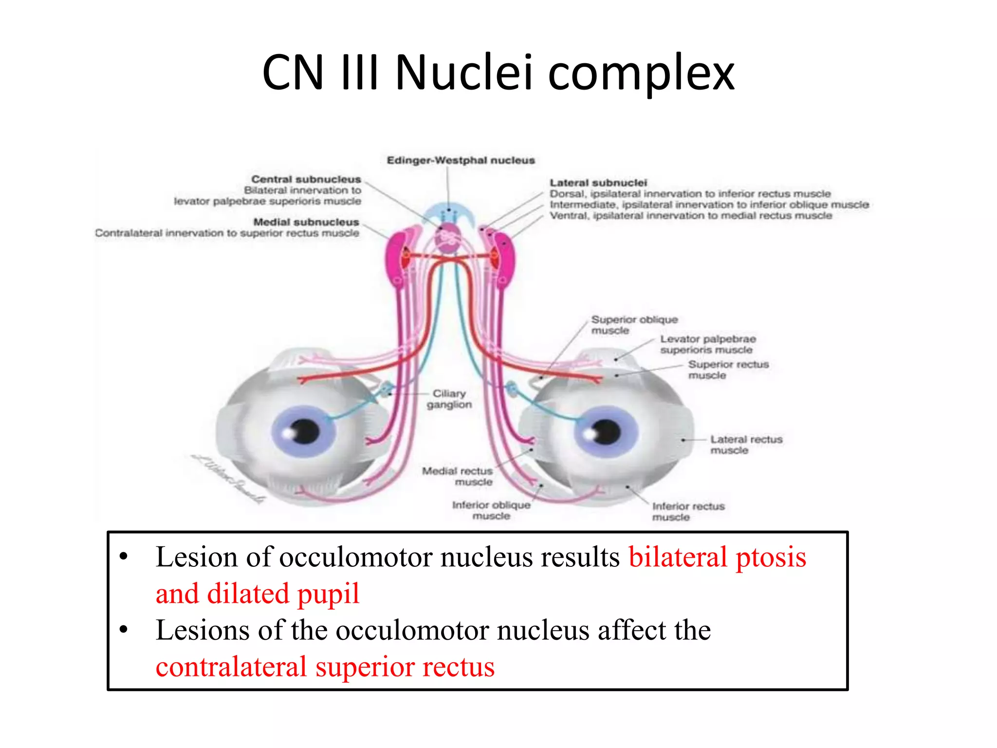
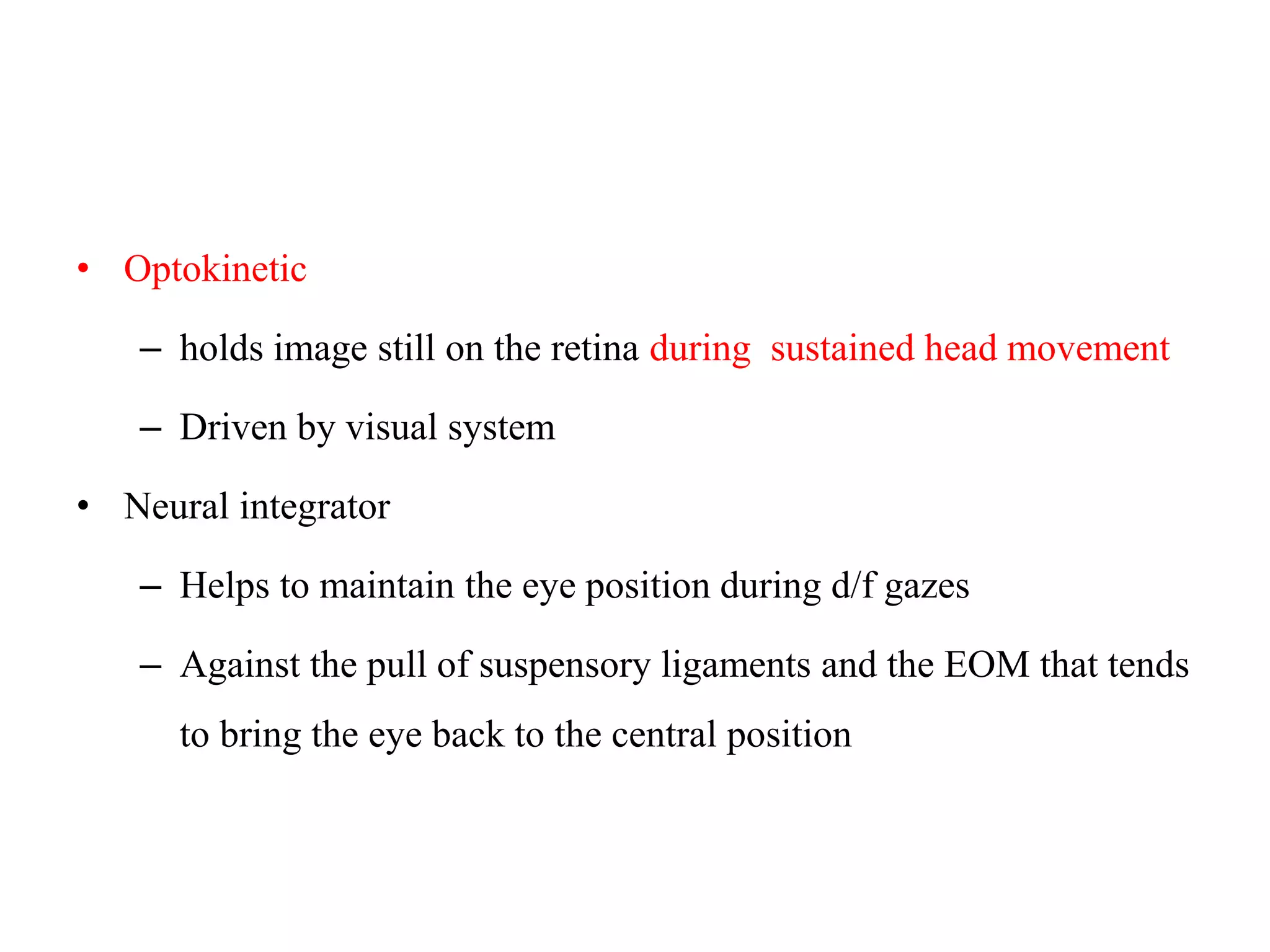
1. The anatomy and function of the extraocular muscles and neural control centers.
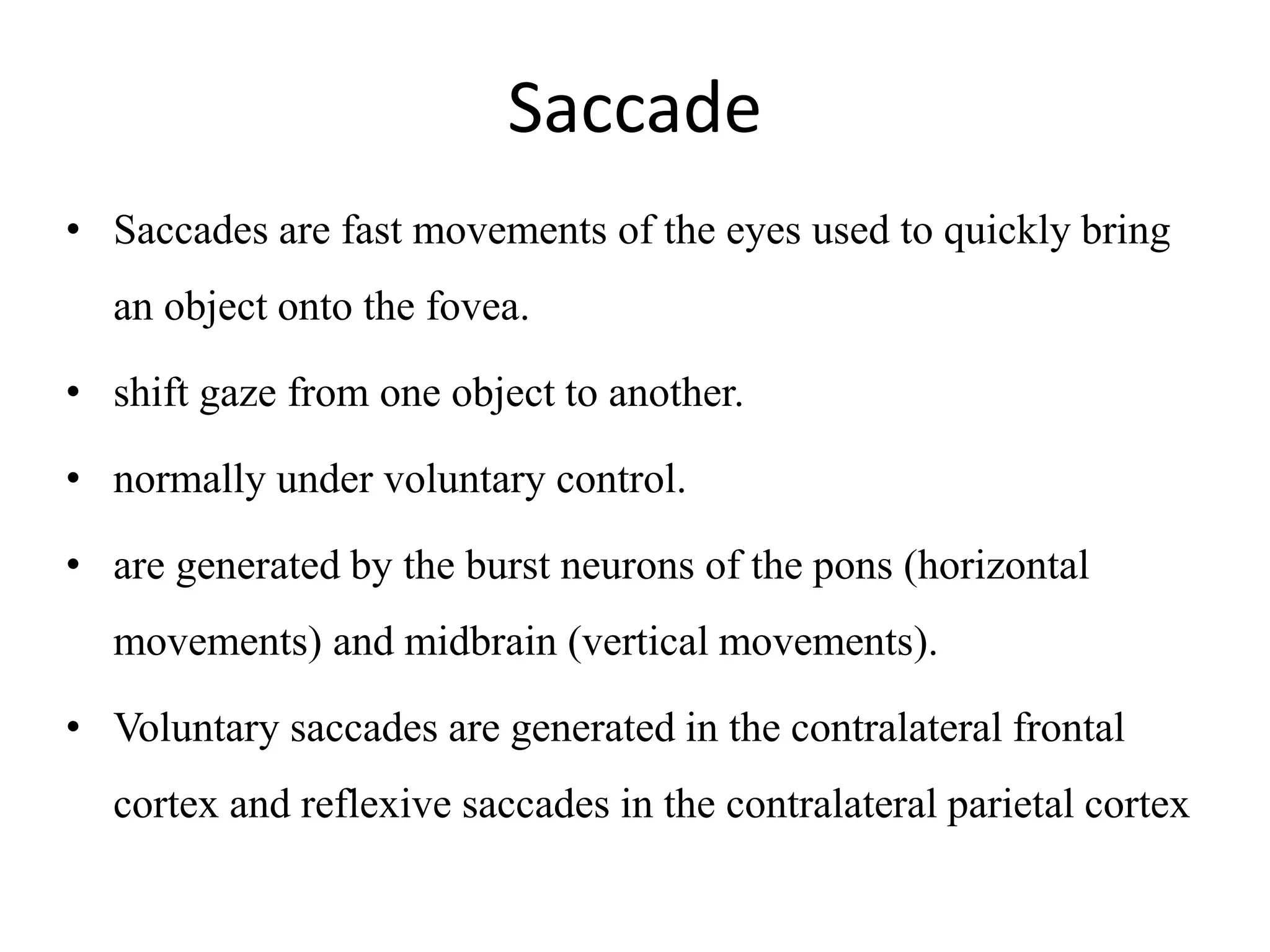
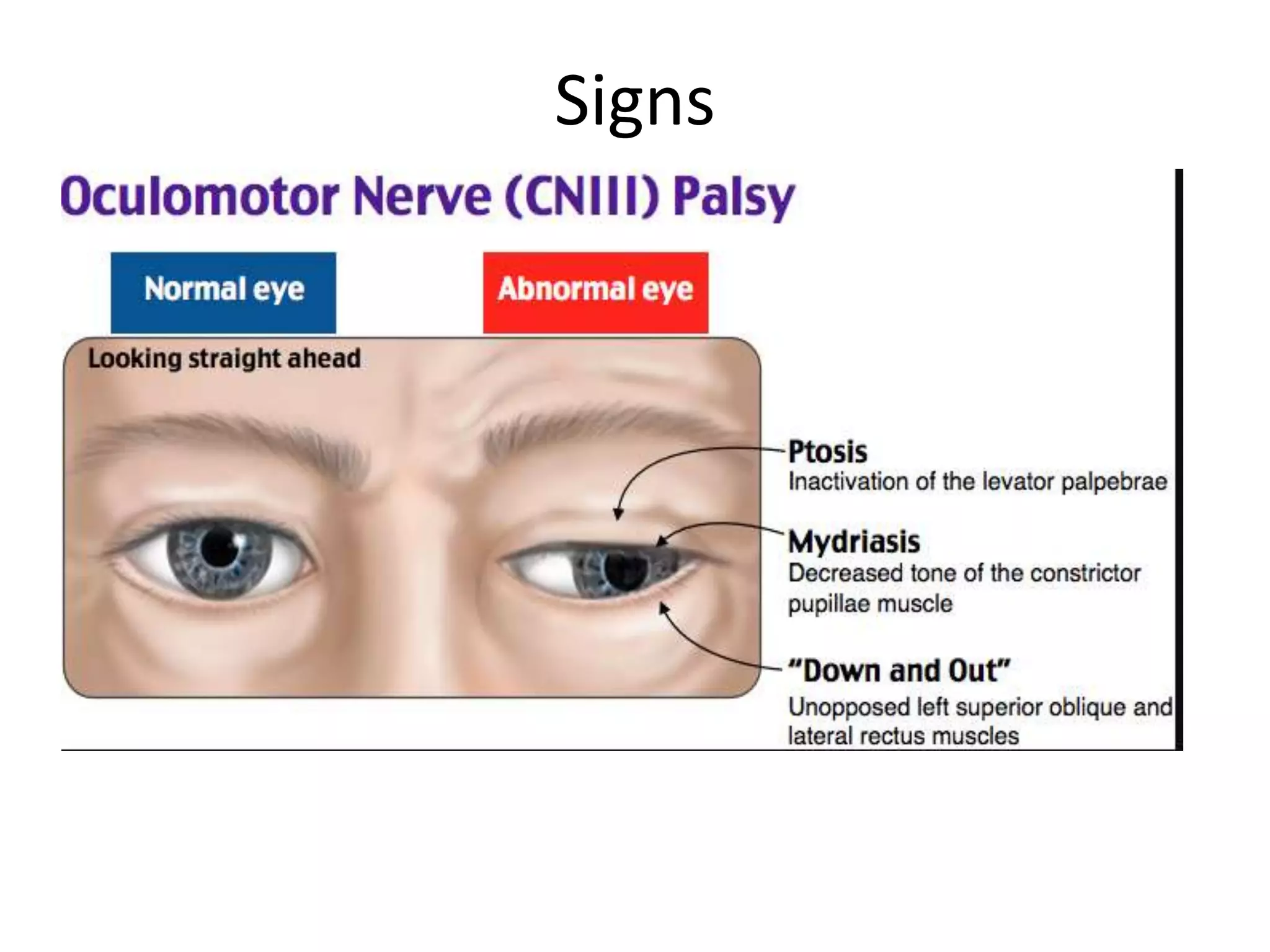
2. Classification of abnormal eye movements including types (e.g. saccades, smooth pursuit) and causes (e.g. myopathies, nerve palsies).
3. Specific disorders are described in detail such as internuclear ophthalmoplegia, Parinaud's syndrome, and progressive supranuclear palsy.
4. Nystagmus is classified and mechanisms are discussed.
3rd,4th, 6th nerves
Extraocular muscles
How to examine for ocular motility
Ophthalmoplegia
Diplopia and related disorders
Gaze pathway
How to examine for gaze
Gaze palsy
Types of eye movements
How to examine for EM
Nystagmus and non nystagmus ocular oscillation
Metabolic optic neuropathies
The three subcategories of metabolic optic neuropathies are
•Heredodegenerative (such as leber's hereditary optic neuropathy).
•Nutritional deficiencies (such as vitamins B12 or folic acid).
•Toxicities (such as ethambutol or cyanide).
Paralytic squint. Here 4th nerve and 6th nerve palsy have been explained briefly. Even few important point related to its investigation and management is explained.
a brief yet interesting topic.
i am a dm neurology senior resident , the presentation has been created after going through three main books of neurology, Dejong , Bradley and Brazis . the ppt contains both descriptive videos and pictures of the various domains of ocular motility disorders. and a brief description of 3rd cranial nerve disorder too.
Unit 8 - Information and Communication Technology (Paper I).pdfThiyagu K
This slides describes the basic concepts of ICT, basics of Email, Emerging Technology and Digital Initiatives in Education. This presentations aligns with the UGC Paper I syllabus.
Synthetic Fiber Construction in lab .pptxPavel ( NSTU)
Synthetic fiber production is a fascinating and complex field that blends chemistry, engineering, and environmental science. By understanding these aspects, students can gain a comprehensive view of synthetic fiber production, its impact on society and the environment, and the potential for future innovations. Synthetic fibers play a crucial role in modern society, impacting various aspects of daily life, industry, and the environment. ynthetic fibers are integral to modern life, offering a range of benefits from cost-effectiveness and versatility to innovative applications and performance characteristics. While they pose environmental challenges, ongoing research and development aim to create more sustainable and eco-friendly alternatives. Understanding the importance of synthetic fibers helps in appreciating their role in the economy, industry, and daily life, while also emphasizing the need for sustainable practices and innovation.
How to Split Bills in the Odoo 17 POS ModuleCeline George
Bills have a main role in point of sale procedure. It will help to track sales, handling payments and giving receipts to customers. Bill splitting also has an important role in POS. For example, If some friends come together for dinner and if they want to divide the bill then it is possible by POS bill splitting. This slide will show how to split bills in odoo 17 POS.
The Art Pastor's Guide to Sabbath | Steve ThomasonSteve Thomason
What is the purpose of the Sabbath Law in the Torah. It is interesting to compare how the context of the law shifts from Exodus to Deuteronomy. Who gets to rest, and why?
Instructions for Submissions thorugh G- Classroom.pptxJheel Barad
This presentation provides a briefing on how to upload submissions and documents in Google Classroom. It was prepared as part of an orientation for new Sainik School in-service teacher trainees. As a training officer, my goal is to ensure that you are comfortable and proficient with this essential tool for managing assignments and fostering student engagement.
The French Revolution, which began in 1789, was a period of radical social and political upheaval in France. It marked the decline of absolute monarchies, the rise of secular and democratic republics, and the eventual rise of Napoleon Bonaparte. This revolutionary period is crucial in understanding the transition from feudalism to modernity in Europe.
For more information, visit-www.vavaclasses.com
How to Create Map Views in the Odoo 17 ERPCeline George
The map views are useful for providing a geographical representation of data. They allow users to visualize and analyze the data in a more intuitive manner.
Students, digital devices and success - Andreas Schleicher - 27 May 2024..pptxEduSkills OECD
Andreas Schleicher presents at the OECD webinar ‘Digital devices in schools: detrimental distraction or secret to success?’ on 27 May 2024. The presentation was based on findings from PISA 2022 results and the webinar helped launch the PISA in Focus ‘Managing screen time: How to protect and equip students against distraction’ https://www.oecd-ilibrary.org/education/managing-screen-time_7c225af4-en and the OECD Education Policy Perspective ‘Students, digital devices and success’ can be found here - https://oe.cd/il/5yV
The Roman Empire A Historical Colossus.pdfkaushalkr1407
The Roman Empire, a vast and enduring power, stands as one of history's most remarkable civilizations, leaving an indelible imprint on the world. It emerged from the Roman Republic, transitioning into an imperial powerhouse under the leadership of Augustus Caesar in 27 BCE. This transformation marked the beginning of an era defined by unprecedented territorial expansion, architectural marvels, and profound cultural influence.
The empire's roots lie in the city of Rome, founded, according to legend, by Romulus in 753 BCE. Over centuries, Rome evolved from a small settlement to a formidable republic, characterized by a complex political system with elected officials and checks on power. However, internal strife, class conflicts, and military ambitions paved the way for the end of the Republic. Julius Caesar’s dictatorship and subsequent assassination in 44 BCE created a power vacuum, leading to a civil war. Octavian, later Augustus, emerged victorious, heralding the Roman Empire’s birth.
Under Augustus, the empire experienced the Pax Romana, a 200-year period of relative peace and stability. Augustus reformed the military, established efficient administrative systems, and initiated grand construction projects. The empire's borders expanded, encompassing territories from Britain to Egypt and from Spain to the Euphrates. Roman legions, renowned for their discipline and engineering prowess, secured and maintained these vast territories, building roads, fortifications, and cities that facilitated control and integration.
The Roman Empire’s society was hierarchical, with a rigid class system. At the top were the patricians, wealthy elites who held significant political power. Below them were the plebeians, free citizens with limited political influence, and the vast numbers of slaves who formed the backbone of the economy. The family unit was central, governed by the paterfamilias, the male head who held absolute authority.
Culturally, the Romans were eclectic, absorbing and adapting elements from the civilizations they encountered, particularly the Greeks. Roman art, literature, and philosophy reflected this synthesis, creating a rich cultural tapestry. Latin, the Roman language, became the lingua franca of the Western world, influencing numerous modern languages.
Roman architecture and engineering achievements were monumental. They perfected the arch, vault, and dome, constructing enduring structures like the Colosseum, Pantheon, and aqueducts. These engineering marvels not only showcased Roman ingenuity but also served practical purposes, from public entertainment to water supply.
2. Outlines
• EOM Anatomy and function
• Regulatory Mechanisms of eye movement
• Different types of eye movement
• Classification of abnormal eye movement
• References
3. Anatomy of EOMs
• Six extraocular muscles
– 4 recti + 2 oblique
• Origin of extraocular muscles
– All recti extraocular muscles originate from the orbital apex –
annulus of Zinn( common tendinous ring ).
– IQ originates from medial side of orbital floor
– SO originates from sphenoid bone
4.
5. CN III Nuclei complex
• Lesion of occulomotor nucleus results bilateral ptosis
and dilated pupil
• Lesions of the occulomotor nucleus affect the
contralateral superior rectus
6. • Eye movement =to bring and maintain the image on to the
fovea by finding, fixating ,maintaining on to the retina
• Systems to hold eyes steady on the fovea
– Ocular fixation
– Vestibular system
– Optokinetic
– CNS Neural integrator
7. • Ocular fixation
– Helps to keep the image of the object remain stationary on
the fovea when the head is not moving
– Also suppresses any unwanted saccades
– Occipital lobe
• Vestibular system
– holds image still on the retina during brief head movement
– VOR
8. • Optokinetic
– holds image still on the retina during sustained head movement
– Driven by visual system
• Neural integrator
– Helps to maintain the eye position during d/f gazes
– Against the pull of suspensory ligaments and the EOM that tends
to bring the eye back to the central position
9. Law
• Hering’s law
– The law of equal innervation
– States that the muscles yoked for conjugate eye movement
receives equal innervation .
• Sherrington’s law/Law of reciprocal innervation
– An agonist muscles receive a neural impulse to contract
– an equivalent inhibitory impulse is sent to the neurons
supplying the appropriate antagonist muscle resulting in
relative relaxation of the antagonists
10. Eye Movement Control Center
Supranuclear
• Cortical
– Frontal lobe –FEF,SEF
,DMFC
– Parietal lobe –PEF
– Occipital lobe –Visual
Cortex
• Basal ganglia
• Thalamus
• Cerebellum
Nuclear
• Midbrain
--Superior colliculus
--nucleus of Cajal
---riMLF
--Nuclei of CN-III & IV
• Pons
---PPRF
---Nuclei of CN-VI
----Vestibular nuclei
Infranuclear
•Oculomotor nerve
•EOM
13. • PTO cortex are primarily responsible for smooth pursuit
movements in the ipsilateral direction, via connections with
the vestibular nuclei, cerebellum, and PPRF,
14. Vertical Conjugate Eye Movements
• riMLF equivalent to PPRF and
INC to Step cells
• The upgaze centers lie more
dorsally
• The downgaze centers lie more
ventrally
15. Types of Eye movement
Eye movement can be classified according to several systems:
• Duction =movement of one eye
• Version =movement of two eyes in same direction(conjugate )
• Vergence =Binocular dysconjugate movement
18. Saccade
• Saccades are fast movements of the eyes used to quickly bring
an object onto the fovea.
• shift gaze from one object to another.
• normally under voluntary control.
• are generated by the burst neurons of the pons (horizontal
movements) and midbrain (vertical movements).
• Voluntary saccades are generated in the contralateral frontal
cortex and reflexive saccades in the contralateral parietal cortex
19. Smooth Pursuit
• refers to the voluntary movement of the eyes used to track a
target moving at a low velocity
• It functions to keep the moving object on the fovea to
maximize vision
• Patients with impaired smooth pursuit require frequent small
saccades to keep up with the target ===saccadic pursuit
20. Abnormal eye movements
• Disorders can be broadly divided into
– i) peripheral (infranuclear and nuclear) and
– ii) central (internuclear and supranuclear).
21. PERIPHERAL DISORDERS OF OCULAR
MOTILITY
• Disturbances of ocular motility may result from processes
involving the
– Orbit-- causing mechanical limitation of eye movement
– From ocular myopathies
– Neuromuscular transmission disorders
– A palsy of an individual ocular motor nerve.
22. Muscle Disease
Ocular myopathies causes
– Weakness of EOM
– Ptosis
– Weakness of eye closure
• Due to myopathic involvement of facial muscles
– The common causes
• Chronic progressive external ophthalmoplegia (CPEO)
• Oculopharyngeal muscular dystrophy.
– Rarely
• Inflammatory myositis
23. Restrictive orbitopathies
– Restriction of movement
– Most common restrictive orbitinopathy is TED ( thyroid eye
disease)
– Restrictive myopathy is easily confused with weakness of
antagonist muscle (e.g.. restrictive myopathy of MR mimic
weakness of LR)==do Forced duction test
– Abnormal forced duction
• Inferior rectus muscle is the most commonly involved muscle
in TED followed by medial rectus and superior rectus
24. CPEO
• Is a syndrome that has numerous etiologies,
– It is most often due to mitochondrial myopathy.
• Progressive ptosis and symmetric eye muscle
weakness(immobility),painless ,pupil spared
• Patients do not typically have diplopia.
• Examination shows loss of both voluntary and reflex eye
movements;
25. Oculopharyngeal dystrophy
• Rare autosomal dominant
• Causes marked progressive bilateral ptosis and dysphagia ±
proximal muscle weakness
• Usually begin in the fifth or sixth decade.
• The signature of the disease
– Ocular myopathy + pharyngeal weakness.
26. Neuromuscular Transmission
Disorders
• MG
– Frequently involve EOMs(50-70% early,,90% late)
– Typically present with fluctuating binocular diplopia, ptosis
, opthalmoparesis or both
– Can mimic gaze palsy or the pattern of any individual nerve.
– On tensilon test hypermetric saccade due to central
adaptation
• LEMS
– Rarely involve extraocular muscles except mild ptosis
• Botulism
– Ophthalmoplegia + Fixed dilated pupils
27. INDIVIDUAL NERVE PALSIES
• Most common cause of 4th nerve palsy is trauma
• Most common cause of 3rd & 6th nerve palsy
– Microangiopathic vascular disease
– Followed by trauma
28. Oculomotor Nerve Palsy
Results varying degree and combination of EOM weakness ,
ptosis and pupil involvement
• Opthalmoplagia
– Internal -- Limited to the pupillary sphincter and ciliary
muscle
– external --- Only the extraocular muscles
– complete -- Both
• Ptosis
29. Signs
• Nuclear/ sub nuclei
– Contralateral superior rectus weakness
• Fascilar
– Lesion from interpedencular fossa to point of division
– Paralysis of all functions
• Divisional
– Superior division-ptosis & upgaze palsy
– Inferior division- IR,MR & pupil ,inability to look nasally ,
inferiorly
30. • The most common identifiable etiologies are
– Ischemia –Pupil sparing
– Aneurysm (P communicating A & distal ICA) & trauma=
pupil involving
• Aberrant re-innervation is common
– Tumor
– Some 20% remain unexplained.
31. Trochlear Nerve Palsy
• Vertical or diagonal diplopia , maximal in down gaze
• On examination
– Extortion and impairment of depression of the adducted
eye.
Right fourth nerve palsy
– Head tilt forward and to the opposite side of weak eye
32. Abducens Nerve Palsy
• Horizontal diplopia worse at distance.
• Concomitant strabismus, worse in the direction of action of the
involved muscle.
• Lesion involving the nucleus causes an ipsilateral horizontal
gaze palsy rather than a sixth nerve palsy
– CN VI nucleus contains both lateral rectus motor neurons
and interneurons that project up the MLF
33.
34. Central Disorders Of Ocular Motility
• Supranuclear disorders
– Those that affect the supranuclear gaze centers in the
hemispheres and brainstem
– Areas that influence eye movements, such as the basal
ganglia and cerebellum.
• Internuclear disorders affect the connections between the
ocular motor nerve nuclei in the brainstem.
35. Saccadic Dysfunction
• Lesions or degeneration of saccadic center or lesions of the
ocular motor neurons / extraocular muscles leads to slowing of
saccades
• Overshooting saccades (missing the target and then needing to
correct) indicates a lesion of the cerebellum
• Undershooting saccades are less specific and often occur in
normal subjects
36. • PPRF lesion can cause a horizontal saccadic gaze palsy and a
lesion in the RIMLF can cause a vertical and torsional
saccadic gaze palsy
• The clinical hallmark of a supranuclear gaze palsy is
– Disproportionate involvement of saccades
– OKN lost
– Smooth pursuit may be affected, but usually to a lesser
extent than saccades
– Vestibular eye movements are typically spared
• Nuclear and infranuclear processes affect saccades, smooth
pursuit, and vestibular reflexes equally
37. Pursuit Dysfunction
• Abnormal smooth pursuit is non localizing
• The lesion can be anywhere
– Cortex, basal gangila,mid brain, pons ,cerebellum
• Asymmetry is more specific than symmetry
• Poor initiation – frontal/Parietal lobe lesions
• Deficits found usually in both vertical and horizontal planes
38. Internuclear ophthalmoplegia
• Lesion of the MLF
• Manifest with
– impaired adduction of the eye ipsilateral to the lesion
– contralateral abducting “nystagmus” upon abduction in the
contralesional eye
– Convergence may be preserved
– When convergence is impaired INO classified as
rostral(anterior),if preserved classified as caudal (posterior)
39. Causes of INO
• INO may occur with a variety of disorders affecting the
brainstem
• Common conditions are MS and brainstem stroke
– Child : Suspect tumor
– Young : Demyelinating disease (MS)
– Elder : ischemic lesions
• INO must be distinguished from the many (primarily peripheral
infranuclear) causes of pseudo-INO
40. WEBINO Syndrome
• Lesion is in bilateral MLF
• Both eye will be slightly abducted –giving rise to appearance
call as ‘’Wall eyed’’(bilateral exotrophia)
41. Pseudo-INO
• Feature of INO but lesion is not in MLF
• Imaging is normal
• Most common cause : Myasthenia Gravis
• Other(rare) cause Miller Fisher Syndrome
42. One-and-half syndrome.
• Lesion involving both the PPRF (or the abducens nucleus) and
the crossed MLF on one side of the pons
• Manifest with ipsilateral horizontal gaze palsy(PPRF lesion)
and ipsilateral INO lesion
• Abduction of the eye contralateral to the lesion is intact
• E.g. : Right sided lesion –Rt sided MLF and PPRF are non
functional
– Right INO
– Right PPRF involved –involve right 6th nerve and left 3rd
nerve
Spared =C/L 6th nerve
43. CON…
• Paralytic pontine exotropia occurs because of the preserved
abduction
• Vertical movement and convergence is spared
• Common causes are infarction , demyelination in the median
pontine region (dorsomedian region)
• MG can cause a pseudo– one-and-a-half syndrome
44. Horizontal Gaze Deviation
• The FEFs move the eyes into contralateral conjugate
horizontal gaze.
– Destructive Frontal lobe lesion --gaze preference to side of
the lesion
– Irritative lesion --gaze deviation away from the lesion
• The PPRF governs ipsilateral, conjugate horizontal gaze &
draws the eyes ipsilaterally
– Destructive lesions - resulting in a gaze deviation toward
the intact side as the normal PPRF pulls the eyes over.
• Pontine gaze palsies that involve the PPRF affect all functions,
voluntary and reflex.
45. • Patients with destructive frontal
lesions
– gaze away from the side of the
hemiparesis;
• Patients with pontine strokes
– gaze toward the hemiparesis.
• Patients with irritative frontal lesion
– Gaze away from the side of
lesion
46. Vertical Gaze Abnormalities
• Vertical gaze center
– riMLF
• Two common disorders affecting vertical gaze
– Parinaud’s syndrome
– PSP.
47. Parinaud’s Syndrome
• AKA dorsal midbrain syndrome /Syndrome of posterior
commissure
• Cortical pathway for conjugate upward eye movements runs in
the tegmentun dorsal to pathways for downward movements
• Caused by compression of vertical gaze center (riMLF)
– Mass lesion commonly pinealoma
– Stroke
– Hydrocephalus (Obstructive)
48. Clinical features
• Up gaze palsy
• Convergence-retraction “nystagmoid reaction” (often elicited
by attempted up gaze)
• Bilateral lid retraction (Collier sign)
• Pupillary light-near dissociation
49. Progressive Supranuclear Palsy
• Degenerative changes in the rostral brainstem and thalamus
(thalamomecencephalic area)
• Result in impairment first of downgaze then of upgaze and
eventually in global gaze paresis.
• Reflex eye movements are preserved until late in the disease.
• The gaze abnormalities are accompanied by parkinsonian signs
50. • Late in the disease course, saccades and smooth pursuit may
be lost both vertically and horizontally
• Other Oculomotor findings= saccadic intrusions (square wave
jerks),loss of OKN (particularly in the vertical direction),loss
of convergence ,eyelid opening apraxia
51. Nystagmus
• It is a rhythmic , biphasic , involuntary movement of the eyes.
• The slow phase of nystagmus is the pathological component
responsible for the initiation and generation of the nystagmus.
• Direction defined by fast phase
• Can be physiological or pathological
52. Classification of Nystagmus
• Nystagmus is classified in multiple ways
– Jerk Vs. Pendular Nystagmus
– Central versus peripheral
– Spontaneous /gaze evoked or positional
– Physiologic vs. pathologic
– Congenital versus acquired
53. • Slow phases of jerk nystagmus may have linear, decreasing, or
increasing velocity
– correct identification of this may assist with diagnosis
• Acquired forms of nystagmus tend to have linear or decreasing
slow-phase velocities
• Congenital nystagmus slow phases in the infantile nystagmus
syndrome are typically of increasing velocity although these
are not pathognomonic
54. • Described by
– Amplitude –extent of excursion of the nystagmus
• Fine : less than 5⁰
• Moderate: 5-15⁰
• Large/coarse : greater than 15⁰
– Frequency - is the number of complete to and fro
movements in one second
• Slow : (1-2 Hz)
• Medium : (3-4 Hz)
• Fast: (5 Hz or more)
55. • Plane : horizontal ,vertical or rotatory or combination
• Direction : unidirectional /Bidirectional
• Null zone of nystagmus:
– position where nystagmus intensity is minimized
– Patient assumes a head posture, such that the eyes are in
null zone
56. • 1st degree
– nystagmus present only with eccentric gaze
• 2nd degree
– Nystagmus present in primary gaze and increase in
intensity with gaze in the direction of fast component
• 3rd degree
– fast component opposite to the direction of gaze
– Most common in vestibular nystagmus
57. Mechanism /Etiology
• Disturbance or abnormality of three mechanisms
– Fixation
– VOR
– Ocular neural integrator (brainstem and cerebellum)
58. • Neuropathologic nystagmus: common types include
– vestibular , positional ,gaze evoked , gaze paretic nystagmus
• Non neuropathologic nystagmus
– Physiologic nystagmus
– Nystagmus due to ocular disease (poor vision) or other
conditions
– Voluntary nystagmus
59. Physiologic Nystagmus
o End point nystagmus
– observed on extremes of lateral gaze ,absent in upgaze
– Typically low amplitude ,symmetric ,poorly sustained and
fatigues overtime ,horizontal
– Abolished on primary position
– No other neurologic abnormality
o Optokinetic nystagmus
o Induced vestibular nystagmus
– Caloric test
60. Optokinetic Nystagmus
• It is normal ,physiologic phenomenon
• Nystagmus stimulated by watching moving object
– When one is looking out the window of a moving train
• Has initial smooth pursuit followed by saccade back to
primary gaze
• Normally should have symmetric OKN
• Cogan’s rule
– Homonymous hemianopia +asymmetric OKN –Parietal
lobe lesion probably mass lesion
– Homonymous hemianopia + Symmetric OKN- Ocipital lobe
lesion ,most likely vascular lesion
61. Clinical testing –OKN strip
• Fast phase is opposite to drum rotation and slow pursuit is
towards drum rotation
63. Congenital /Infantile nystagmus
syndrome
• Seen in < 6 months /at birth or appear later shortly
• Mostly due to ocular disease /congenital or acquired in the 1st
month of life
• Usually horizontal, pendular (in primary position) ,
jerk(eccentric gaze)
• Not suppressed by fixation
• 3 common causes are congenital cataract , Albinism and optic
nerve hypoplasia
64. Mnemonic “CONGENITAL”
• Convergence dampens or decrease the nystagmus
• Oscillopsia is absent
• Null point is in primary position
• Gaze is uniplanar
• Equal amplitude in all direction called pendular nystagmus
• Inverted OKN
• Turn face
• Abolished in sleep
65. Pendular Nystagmus
• Has a sinusoidal wave form and usually is horizontal
• Generally does not have a fast phase including saccade , but
composed of entirely slow eye movements
• Can be either congenital or acquired
• Acquired pendular nystagmus typically has horizontal ,
vertical or torsional component
66. Acquired pendular nystagmus
• MS most common cause followed by brainstem vascular
disease
• Probably results from disruption of normal feedback from
cerebellar nuclei to neural integrator
• In demyelinating diseases, APN most often is horizontal
and/or elliptical in trajectory, with a frequency of 3 to 5 Hz
67. • Vertical /Vertical-torsional pendular nystagmus
– Results from lesions in the deep cerebellar nuclei and their
connections (dentatorubroolivary pathways= Gullian
Mollaret triangle)
– Associated with palatal myoclonus
==oculopalatal myoclonus syndrome
68. Pathologic Gaze Evoked Nystagmus
• AKA Gaze paretic nystagmus
• Nystagmus present in less than extreme gaze ,absent in the primary
position
• Most common type of nystagamus
• Usually symmetric ,sustained ,large amplitude and /present in upgaze
• Causes
– Drugs toxicities (i.e Anticonvulsants , Alcohol ,sedatives)
– Brainstem lesions
– Cerebellar lesions/ Floccules
69. Vestibular Nystagmus
• Jerk nystagmus
• Vestibular nystagmus:
– Central ( brainstem nuclei)
– Peripheral ( labyrinths, VIII CN)
• May occur spontaneously or be produced by various
maneuvers.
70. Features Peripheral Central
Onset & duration Abrupt onset ,lasts short
min(intermittent)
Can be gradual
onset/sudden onset and
continuous
Cause Dysfunction of Vestibular
end organ or vestibular
nerve
Disease of the brainstem
nuclei
Type of nystagmus Horizontal-rotatory
/upbeat-torsional
Pure vertical or pure
torsional
Visual Fixation Inhibits the nystagmus No inhibition
Direction of nystagmus Fixed /unidirectional May change with gaze
/bidirectional
Severity of vertigo Severe Mild
Associated finding Vertigo ,tinnitus , nausea
,vomiting ,hearing loss
Headache ,ataxia, diplopia
,pyramidal tract signs
74. Saccadic Intrusions
• Saccadic intrusions are brief, unwanted involuntary saccadic
interruptions of fixation
• Saccadic intrusions with an intersaccadic interval include
– square-wave jerks, macro–square-wave jerks, and macro
saccadic oscillations ,square wave pulses ,saccadic pulses
• Saccadic intrusions without an intersaccadic interval
– ocular flutter and opsoclonus
75. Saccadic intrusion with normal
intersaccadic interval
Square wave jerks
Macro square wave jerks
Macrosaccadic oscillation
Intersaccadic interval is more used over
saccadic intrusion amplitude to
distinguish SWP from SWJ
76. o Opsoclonus/dancing eyes
• Continuous succession of multidirectional conjugate
saccadic eye movements
• Most common cause is encephalitis
• Post viral ,metastasis or neuroblastoma
• Occurs due to interruption of cerebellar connections to
brainstem
o Ocular flutter,
• Similar to Opsoclonus but confined to horizontal plane
77. Non nystagmus ocular oscillations
• There are ocular movements which mimic nystagmus
o Ocular bobbing
• Sudden conjugate downward deviation of eye followed
by slow return to normal
• Paralysis of spontaneous & reflex horizontal eye
movement
• Seen in comatose patient
• Pontine dysfunction
78. Superior oblique myokymia
• A paroxysmal ,rapid, small-amplitude monocular torsional-
vertical oscillation caused by contraction of the superior
oblique muscle
• Consider when the patient reports episodes of rapid
monocular torsional or vertical oscillopsia
79. • Ocular dysmetria
– is an over- or undershooting of the eyes on rapid refixation
of gaze toward either side or on returning to the primary
position that requires corrective saccades;
– There may also be overshooting in following movements
when the object of regard is suddenly stopped.
– Cerebellar System dysfunction(always)
80. • Oculogyric crisis
– Refers to attacks of involuntary conjugate upward deviation
of the eyes, which may be transient or last for hours( typical
attack lasts for about 2 hrs).
– A dystonic reaction to drugs(neuroleptics) or medical
condition(MS , syphilis).
– Post encephalitic parkinsonism
81. References
• De Jong clinical examination,8th edition:2020
• Bradley’s neurology in clinical practice 8th edition ,2021
• Neuro ophthalmology Continuum
• Demyers neurologic examination
• Neuroanatomy through clinical cases,2nd edition
THANK YOU!!!!!
82. • A 71-year-old Rt handed man patient with a history of
diabetes collapsed on the street and was unable to stand up &
brought to the ER by ambulance.
• On exam, awake but lethargic , rightward gaze preference, and
was unable to move either eye past the midline toward the left.
• Rt SNFP and Rt sided hemiparesis , Rt up going plantar
– Where is the lesion ?
– Most likely dx?