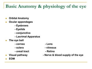
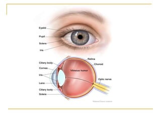
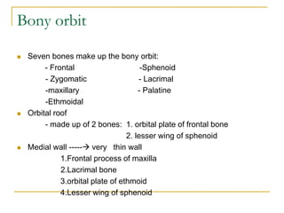
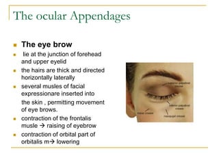
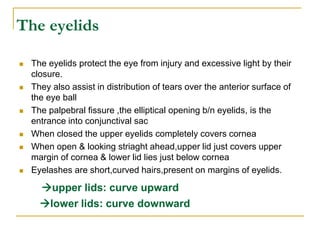
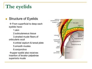
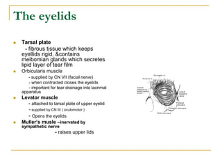
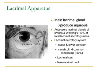
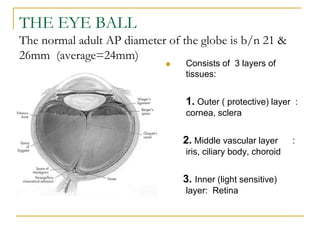
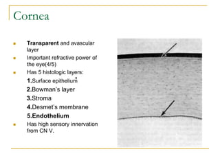
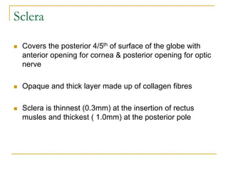
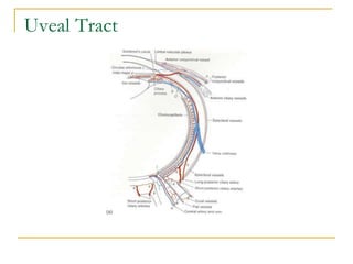
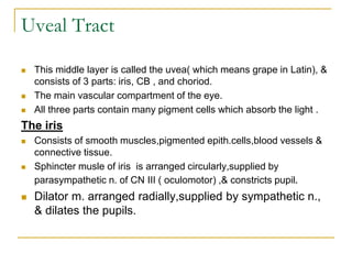
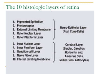
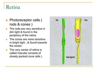
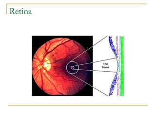
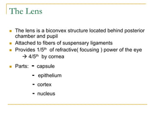
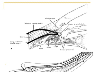
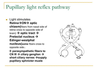
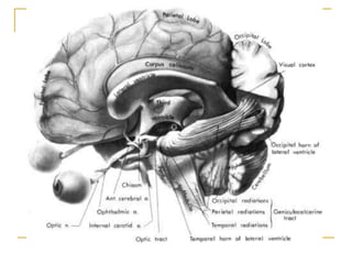
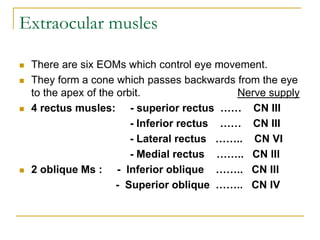
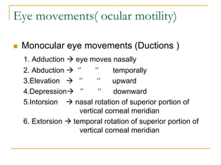
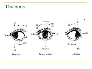
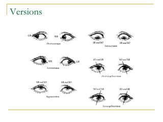
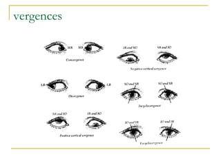
SlideShare a Scribd company logo
OPHTHALMOLOGY FOR
MEDICAl STUDENTS
Demoze Delelegn, MD
Ast. Professor of Ophthalmology
Course outline
I. Basic anatomy & physiology of the eye
II. Examination of the eye ---History taking & physical
examination
III. Diagnosis and management of ocular disorders
3.1. Diseases of the orbit
3.2. Diseases of eye lids and lacrimal Apparatus
3.3. Diseases of the conjunctiva
3.4. Diseases of the cornea
3.5. Diseases of the sclera
3.6. Diseases of uveal tract
3.7. Diseases of the lens
Course outline
 Diagnosis & mgt of ocular disorders
3.8. Diseases of retina and optic nerve
3.9. Squint
3.10. Glaucoma
3.11. Vitamin A deficiency
3.12. ocular manifestation HIV/AIDS
3.13. Ocular trauma
3.14. Refractive errors
IV. Preventive ophthalmology
Basic Anatomy & physiology of the eye
 Orbital Anatomy
 Ocular appendages
- Eyebrows
- Eyelids
- conjunctiva
- Lacrimal Apparatus
 The eye ball
- cornea - Lens
- sclera - vitreous
- uveal tract - Retina
 Visual pathway - Nerve & blood supply of the eye
 EOM
ORBITAL CAVITY
 The orbital cavities are a pair of
large bony sockets that contain
eyeball, EOMs, nerves, vessels
& orbital fat, and most of the
lacrimal apparatus.
 Each cavity is pear-shaped and
its apex is directed
posteriorly,medially, and slightly
upward; the stalk of pear lying
within optic canal.
 The medial wall runs
anteroposteriorly parallel to
sagital plane.
 The lateral wall diverges at
angle of 45.
Bony orbit
 Seven bones make up the bony orbit:
- Frontal -Sphenoid
- Zygomatic - Lacrimal
-maxillary - Palatine
-Ethmoidal
 Orbital roof
- made up of 2 bones: 1. orbital plate of frontal bone
2. lesser wing of sphenoid
 Medial wall ----- very thin wall
1.Frontal process of maxilla
2.Lacrimal bone
3.orbital plate of ethmoid
4.Lesser wing of sphenoid
 Orbital floor
1.maxillary
2.palatine
3.orbital plate of zygomatic
 Lateral wall
1. zygomatic bone
2. greater wing of sphenoid
The paranasal sinuses
 The paranasal sinuses are cavities in the interior of the maxilla
,frontal , sphenoid and ethmoidal bones
 Lined with mucoperiosteum & filled with air , they communicate with
nasal cavity through small openings
 Function:
-to act as resonators to voice
- to reduce weight of the skull
 4 sinuses:-1.maxillary sinus
2.frontal sinus
3.sphenoidal sinus
4.Ethmoidsal sinus
 Sinusitis – infection of the paranasal sinuses
The ocular Appendages
 The eye brow
 lie at the junction of forehead
and upper eyelid
 the hairs are thick and directed
horizontally laterally
 several musles of facial
expressionare inserted into
the skin , permitting movement
of eye brows.
 contraction of the frontalis
musle  raising of eyebrow
 contraction of orbital part of
orbitalis m lowering
The eyelids
 The eyelids protect the eye from injury and excessive light by their
closure.
 They also assist in distribution of tears over the anterior surface of
the eye ball
 The palpebral fissure ,the elliptical opening b/n eyelids, is the
entrance into conjunctival sac
 When closed the upper eyelids completely covers cornea
 When open & looking striaght ahead,upper lid just covers upper
margin of cornea & lower lid lies just below cornea
 Eyelashes are short,curved hairs,present on margins of eyelids.
upper lids: curve upward
lower lids: curve downward
The eyelids
 Structure of Eyelids
 From superficial to deep each
eyelids have:
1.skin
2.subcutaneous tissue
3.straited musle fibers of
orbicularis oculi
4.orbital septum & tarsal plate
5.smooth musles
6.conjunctiva
upper eyelid also receives
insertion of levator palpebrae
superioris musle
The eyelids
 Tarsal plate
- fibrous tissue which keeps
eyellids rigid, &contains
meibomian glands which secretes
lipid layer of tear film
 Orbicularis muscle
- supplied by CN VII (facial nerve)
- when contracted closes the eyelids
- important for tear drainage into lacrimal
apparatus
 Levator muscle
- attached to tarsal plate of upper eyelid
- supplied by CN III ( oculomotor )
- Opens the eyelids
 Muller’s musle –inervated by
sympathetic nerve
- raises upper lids
The conjunctiva
 Thin mucous membrane that lines eyelids and eye balls
 3 parts of conjunctiva:
1. Palpebral conjunctiva firmly attached to tarsal plate
2. Bulbar Conjunctiva
lies in contact & loosely attached to eyeball
3. Conjunctival fornix
 The junction b/n tarsal and bulbar conjunctiva
Limbus the line along which fusion of conjunctiva to
cornea occurs
The conjunctiva
 The main function of conjunctiva is to protect cornea
 Supplies some of its oxygen and metabolic needs
 Lubricates cornea with tears
 Protect s exposed part of the eye from infection
 Conjunctival secretion contains lysozme,antibody
and lymphocytes
Lacrimal Apparatus
Lacrimal Apparatus
 Main lacrimal gland
produce aqueous
 Accessory lacrimal glands of
krause & Wolfring 10% of
total lacrimal secretory mass
 Lacrimal excretory system
- upper & lower punctum
- canaliculi common
canaliculus ( 90%)
- Lacrimal sac
- Nasolacrimal duct
Lacrimal Apparatus,cont’d
 Tear film
 3 layers of tear film on the cornea:
1.Lipid layer  secreted by meibomian glands &
glands of Zeis
2. Aqueous layer
 secreted by main lacrimal gland &
accessory lacrimal glands of Krause and wolfring
3.mucin layer
 coats corneal epithelium and secreted by Goblet
cell of conjunctiva
Functions of tear film
 Contribute to optical property of tear film
 Supplying oxygen to avascular corneal
epithelium
 Providing an antibacterial and antiviral defense
 Retard evaporation
 Washing away debris
 Moistens eyeball and lubrication of eyelids
 Traps exfoliated surface cells,foreign particles
and bacteria
THE EYE BALL
The normal adult AP diameter of the globe is b/n 21 &
26mm (average=24mm)
 Consists of 3 layers of
tissues:
1. Outer ( protective) layer :
cornea, sclera
2. Middle vascular layer :
iris, ciliary body, choroid
3. Inner (light sensitive)
layer: Retina
Cornea
 Transparent and avascular
layer
 Important refractive power of
the eye(4/5)
 Has 5 histologic layers:
1.Surface epithelium
2.Bowman’s layer
3.Stroma
4.Desmet’s membrane
5.Endothelium
 Has high sensory innervation
from CN V.
Sclera
 Covers the posterior 4/5th of surface of the globe with
anterior opening for cornea & posterior opening for optic
nerve
 Opaque and thick layer made up of collagen fibres
 Sclera is thinnest (0.3mm) at the insertion of rectus
musles and thickest ( 1.0mm) at the posterior pole
Uveal Tract
Uveal Tract
 This middle layer is called the uvea( which means grape in Latin), &
consists of 3 parts: iris, CB , and choriod.
 The main vascular compartment of the eye.
 All three parts contain many pigment cells which absorb the light .
The iris
 Consists of smooth muscles,pigmented epith.cells,blood vessels &
connective tissue.
 Sphincter musle of iris is arranged circularly,supplied by
parasympathetic n. of CN III ( oculomotor) ,& constricts pupil.
 Dilator m. arranged radially,supplied by sympathetic n.,
& dilates the pupils.
Uveal Tract
The ciliary body
 Consists of ciliary epithelium and ciliary musle
 2 parts: pars plana & pars plicata
 Suspensory ligament pass from ciliary process to the
equator of the lens
 Function:
- Aqueous humor formation ( by ciliary epithelium)
- Lens accommodation ( by ciliary musle )
Uveal Tract
The choroid
 Consists mainly of blood vessels and pigment cells
 Functions:
- The choroidal circulation supplies 2/3rd of O2 needs
of retina ( nourishes outer portion of retina )
- The pigment cells absorb light inside the eye & so
prevent unwanted reflections.
The inner layer of the Globe - Retina
The inner layer of the Globe
The Retina
 Light sensitive membrane at
the back of the eye.
 The cells of the retina are
specialized & have a very
complex arrangement.
 Cell connections from outer to
in side of retina:
RPE Rod & cones
Bipolar cells Ganglion
cells axons of GC forming
opic nerve
Retina
The 10 histologic layers of retina
Retina
 Photoreceptor cells (
rods & cones )
 The rods are very sensitive in
dim light & found in the
periphery of the retina
 The cones are more sensitive
in bright light , & found towards
the center.
 The very center of retina is
called macula( consists of
closely packed cone cells )
Retina
The Lens
 The lens is a biconvex structure located behind posterior
chamber and pupil
 Attached to fibers of suspensary ligaments
 Provides 1/5th of refractive( focusing ) power of the eye
 4/5th by cornea
 Parts: - capsule
- epithelium
- cortex
- nucleus
Chambers of the eye
 Anterior chamber
The vitreous humor
 The vitreous cavity occupies 4/5th of the volume of the
globe.
 Its volume is close to 4 ml.
 Contains innert ,transparent jel-like structure:
- 99% water
- hyaluronic acid
- fine collagen fibrils
- hyalocytes
 Function: maintains the shape of the globe of the eye
The visual pathway
 The visual pathway connects the optic n. with part of
brain concerned with vision ( occipital part of cerebral
cortex – visual cortex )
 Components of visual pw:
Retinaoptic nerve  optic chiasm 
optic tract lateral geniculate body  optic
radiation  visual cortex
The visual pathway
 Optic nerve  1.2 million nerve fibers
 When both optic nerves meet at chiasm, all the fibers
from nasal part of each retina cross over to the opposite
site and fibers from the temporal side pass through
chiasm to the same side without crossing.
 Every thing in the left half of the field of vision in each
eye is seen on the right side of each retina and by the
right side of brain and viceversa.
 A few fibers in the optic nerve regulate the pupil size.
Pupillary light reflex pathway
 Light stimulates
RetinaON optic
chiasm(fibers from nasal side of
retina cross to opposite side of
brain)  optic tract 
Pretectal nucleus 
Edinger-westphal
nucleus(some fibers cross to
opposite side )
 parasympathetic fibers to
CN III  ciliary genglion 
short ciliary nerves supply
pupillary sphnicter musle
Extraocular musles
 There are six EOMs which control eye movement.
 They form a cone which passes backwards from the eye
to the apex of the orbit. Nerve supply
 4 rectus musles: - superior rectus …… CN III
- Inferior rectus …… CN III
- Lateral rectus …….. CN VI
- Medial rectus …….. CN III
 2 oblique Ms : - Inferior oblique …….. CN III
- Superior oblique …….. CN IV
Extraocular musles
 Origin
The 4 rectus musles and superior oblique musle originate from apex of the orbit.
The inferior oblique m. originate from the periosteum of the maxillary bone behind
lacrimal fossa
 Insertions
 MR : 5.5 mm from medial limbus
 LR : 6.9mm from lateral limbus
 SR : 7.7mm from superior limbus
 IR : 6.5 mm from inferior limbus
 SO : posterior to equator in superotemporal quadrant
 IO : macular area (temporally )
Anterior view
Eye movements( ocular motility)
 Monocular eye movements (Ductions )
1. Adduction  eye moves nasally
2. Abduction  ‘’ ‘’ temporally
3.Elevation  ‘’ ‘’ upward
4.Depression ‘’ ‘’ downward
5.Intorsion  nasal rotation of superior portion of
vertical corneal meridian
6. Extorsion  temporal rotation of superior portion of
vertical corneal meridian
Ductions
Binocular eye movements
 Versions-when binocular eye mov’ts are conjugate and the eyes move in
the same direction
 When the eye movements are disconjugate and eyes move in the opposite
directions , such mov’ts are known as vergences ( e.g. convergence &
divergence )
 Versions:
1.Right gaze ( dextroversion )
2.Left gaze ( levoversion )
3.Elevation or up gaze( sursumversion)
4.Depression or down gaze ( deorsumversion)
5.Dextrocycloversion both eyes rotate so that superior portion of vertical
corneal meridian moves to patients right.
6.Levocycloversion  mov’t of both eyes so that superior corneal meridian
rotates to patients left.
Versions
vergences
Actions of EOMs
Defn.:
 Primary position of gaze is when the eye is directed straight ahead
and head is also straight.
 The primary action of a musle is its major effect on the position of
the eye when the musle contracts while the eye is in primary
position.
 The secondary and tertiary actions of a musle are the additional
effects on position of the eye in primary position.
Actions of EOMs
EOM Primary action Secondary action Tertiary action
LR Abduction None None
MR Adduction None None
SR Elevation Incyclotorsion Adduction
IR Depression Excyclotorsion Adduction
SO Incyclotorsion Depression Abduction
IO Excyclotorsion Elevation Abduction
Summary of Actions of EOMs
 The horizontal muscles(LR & MR) have only primary
actions( abduction or adduction)
 The vertical muscles(SR & IR) adduction
 The oblique muscles(SO & IO) abduction
 The superior muscles(SR & SO) incyclotorsion
 The inferior muscles(IR & IO)excyclotorsion
Blood and nerve supply of the eye
 Blood supply
 Arterial supply
 From anastomosing vessels from internal and external
carotid arteries
 venous drainage
 From anterior and posterior uveavortex v.orbital
vcavernous sinus
 Lymphatic drainage
 No lymphatic vessels in the globe
 From medial eye lidssubmandibular LN
 Lateral side lidspreauricular LN
 Innervation
 Motor
 CN IIIMR,SR,IR,IO
 CN IV SO
 CN VI LR
 CN VII orbicularis oculi
 Sensory
 CN V
 CN II
 Autonomic
 Sympatheticmuller m. and dilator pupilae
 Parasympathetic ciliary m and sphincter pupilae
Thank you!

More Related Content

Similar to anatomy.ppt

Orbit and eye,
Orbit and eye, Orbit and eye,
Orbit and eye, nrkanil
 
anatomy and physiology of eye.ppt.anatomy and physiology of eyex
anatomy and physiology of eye.ppt.anatomy and physiology of eyexanatomy and physiology of eye.ppt.anatomy and physiology of eyex
anatomy and physiology of eye.ppt.anatomy and physiology of eyex
AnjuAnnMani1
 
Anatomy of eye
Anatomy of eye Anatomy of eye
The eye atlas
The eye atlas The eye atlas
The eye atlas
sosojammoly
 
anatomy of eye
anatomy of eyeanatomy of eye
anatomy of eye
vanajayarrlagadda
 
Head and neck
Head and neckHead and neck
Head and neckmchibuzor
 
Functional Anatomy of Eye.pptx
Functional Anatomy of Eye.pptxFunctional Anatomy of Eye.pptx
Functional Anatomy of Eye.pptx
Noorin Bhimani
 
Anatomy and physiology of the eye
Anatomy and physiology of the eyeAnatomy and physiology of the eye
Anatomy and physiology of the eye
Bahaa Halwany
 
Lv book (1).pdf
Lv book (1).pdfLv book (1).pdf
Lv book (1).pdf
SudhanshuSahu13
 
Anatomy of eye
Anatomy of eyeAnatomy of eye
Anatomy of eye
Dubow Digale
 
anatomyandphysiologyoftheeye-140730012447-phpapp02 (1).pptx
anatomyandphysiologyoftheeye-140730012447-phpapp02 (1).pptxanatomyandphysiologyoftheeye-140730012447-phpapp02 (1).pptx
anatomyandphysiologyoftheeye-140730012447-phpapp02 (1).pptx
BilisumaTAyana
 
INTRODUCTION TO OPHTHALMOLOGY.pdf
INTRODUCTION TO OPHTHALMOLOGY.pdfINTRODUCTION TO OPHTHALMOLOGY.pdf
INTRODUCTION TO OPHTHALMOLOGY.pdf
Simon Maina
 
01- Anatomy and Physiology of the ey ppt
01- Anatomy and Physiology of the ey ppt01- Anatomy and Physiology of the ey ppt
01- Anatomy and Physiology of the ey ppt
aamrutha180
 
Anatomy and Physiology.ppt of eye uploaded
Anatomy and Physiology.ppt of eye uploadedAnatomy and Physiology.ppt of eye uploaded
Anatomy and Physiology.ppt of eye uploaded
PrabithaManjeshwar
 
Basic anatomy and physiology
Basic anatomy and physiologyBasic anatomy and physiology
Basic anatomy and physiology
Asik Pradhan
 
Unit 13 sence organ
Unit 13 sence organUnit 13 sence organ
Unit 13 sence organ
CHANDAN260
 
Uvea anatomy
Uvea anatomyUvea anatomy
Uvea anatomy
Mero Eye
 
Ocular Anatomy
Ocular AnatomyOcular Anatomy
Ocular Anatomywcbvi
 
structure of eye ball
structure of eye ballstructure of eye ball
structure of eye ball
Azizul Islam
 

Similar to anatomy.ppt (20)

Orbit and eye,
Orbit and eye, Orbit and eye,
Orbit and eye,
 
anatomy and physiology of eye.ppt.anatomy and physiology of eyex
anatomy and physiology of eye.ppt.anatomy and physiology of eyexanatomy and physiology of eye.ppt.anatomy and physiology of eyex
anatomy and physiology of eye.ppt.anatomy and physiology of eyex
 
Anatomy of eye
Anatomy of eye Anatomy of eye
Anatomy of eye
 
The eye atlas
The eye atlas The eye atlas
The eye atlas
 
anatomy of eye
anatomy of eyeanatomy of eye
anatomy of eye
 
Head and neck
Head and neckHead and neck
Head and neck
 
Functional Anatomy of Eye.pptx
Functional Anatomy of Eye.pptxFunctional Anatomy of Eye.pptx
Functional Anatomy of Eye.pptx
 
Anatomy and physiology of the eye
Anatomy and physiology of the eyeAnatomy and physiology of the eye
Anatomy and physiology of the eye
 
Lv book (1).pdf
Lv book (1).pdfLv book (1).pdf
Lv book (1).pdf
 
Anatomy of eye
Anatomy of eyeAnatomy of eye
Anatomy of eye
 
anatomyandphysiologyoftheeye-140730012447-phpapp02 (1).pptx
anatomyandphysiologyoftheeye-140730012447-phpapp02 (1).pptxanatomyandphysiologyoftheeye-140730012447-phpapp02 (1).pptx
anatomyandphysiologyoftheeye-140730012447-phpapp02 (1).pptx
 
INTRODUCTION TO OPHTHALMOLOGY.pdf
INTRODUCTION TO OPHTHALMOLOGY.pdfINTRODUCTION TO OPHTHALMOLOGY.pdf
INTRODUCTION TO OPHTHALMOLOGY.pdf
 
01- Anatomy and Physiology of the ey ppt
01- Anatomy and Physiology of the ey ppt01- Anatomy and Physiology of the ey ppt
01- Anatomy and Physiology of the ey ppt
 
Anatomy and Physiology.ppt of eye uploaded
Anatomy and Physiology.ppt of eye uploadedAnatomy and Physiology.ppt of eye uploaded
Anatomy and Physiology.ppt of eye uploaded
 
Basic anatomy and physiology
Basic anatomy and physiologyBasic anatomy and physiology
Basic anatomy and physiology
 
Unit 13 sence organ
Unit 13 sence organUnit 13 sence organ
Unit 13 sence organ
 
Uvea anatomy
Uvea anatomyUvea anatomy
Uvea anatomy
 
Ocular Anatomy
Ocular AnatomyOcular Anatomy
Ocular Anatomy
 
Ocular Anatomy
Ocular AnatomyOcular Anatomy
Ocular Anatomy
 
structure of eye ball
structure of eye ballstructure of eye ball
structure of eye ball
 

More from Zelekewoldeyohannes

Additional notes on MG.pptx
Additional notes on MG.pptxAdditional notes on MG.pptx
Additional notes on MG.pptx
Zelekewoldeyohannes
 
Hypoglycemic Encephalopathy.pptx
Hypoglycemic Encephalopathy.pptxHypoglycemic Encephalopathy.pptx
Hypoglycemic Encephalopathy.pptx
Zelekewoldeyohannes
 
Approach to Neuromuscular Disorders.pptx
Approach to Neuromuscular Disorders.pptxApproach to Neuromuscular Disorders.pptx
Approach to Neuromuscular Disorders.pptx
Zelekewoldeyohannes
 
Cranial Nerve Seven.pptx
Cranial Nerve Seven.pptxCranial Nerve Seven.pptx
Cranial Nerve Seven.pptx
Zelekewoldeyohannes
 
DizzinessanddistconcsJune2013.pptx
DizzinessanddistconcsJune2013.pptxDizzinessanddistconcsJune2013.pptx
DizzinessanddistconcsJune2013.pptx
Zelekewoldeyohannes
 
Extrapyramidal Disorders.pptx
Extrapyramidal Disorders.pptxExtrapyramidal Disorders.pptx
Extrapyramidal Disorders.pptx
Zelekewoldeyohannes
 
Joint session discussion.pptx
Joint session discussion.pptxJoint session discussion.pptx
Joint session discussion.pptx
Zelekewoldeyohannes
 
Hyperkinetic movement disorder.pptx
Hyperkinetic movement disorder.pptxHyperkinetic movement disorder.pptx
Hyperkinetic movement disorder.pptx
Zelekewoldeyohannes
 
Diseases of the eye.pdf
Diseases of the eye.pdfDiseases of the eye.pdf
Diseases of the eye.pdf
Zelekewoldeyohannes
 
Vitamin A deficinecy.pptx
Vitamin A deficinecy.pptxVitamin A deficinecy.pptx
Vitamin A deficinecy.pptx
Zelekewoldeyohannes
 

More from Zelekewoldeyohannes (20)

Cerebral edema.pptx
Cerebral edema.pptxCerebral edema.pptx
Cerebral edema.pptx
 
Additional notes on MG.pptx
Additional notes on MG.pptxAdditional notes on MG.pptx
Additional notes on MG.pptx
 
Hypoglycemic Encephalopathy.pptx
Hypoglycemic Encephalopathy.pptxHypoglycemic Encephalopathy.pptx
Hypoglycemic Encephalopathy.pptx
 
Dizziness.pptx
Dizziness.pptxDizziness.pptx
Dizziness.pptx
 
Chorea.pptx
Chorea.pptxChorea.pptx
Chorea.pptx
 
Approach to Neuromuscular Disorders.pptx
Approach to Neuromuscular Disorders.pptxApproach to Neuromuscular Disorders.pptx
Approach to Neuromuscular Disorders.pptx
 
Cranial Nerve Seven.pptx
Cranial Nerve Seven.pptxCranial Nerve Seven.pptx
Cranial Nerve Seven.pptx
 
Immune.pptx
Immune.pptxImmune.pptx
Immune.pptx
 
ATAXIA.pptx
ATAXIA.pptxATAXIA.pptx
ATAXIA.pptx
 
DizzinessanddistconcsJune2013.pptx
DizzinessanddistconcsJune2013.pptxDizzinessanddistconcsJune2013.pptx
DizzinessanddistconcsJune2013.pptx
 
Brain stem New.ppsx
Brain stem New.ppsxBrain stem New.ppsx
Brain stem New.ppsx
 
Paraplegia.pptx
Paraplegia.pptxParaplegia.pptx
Paraplegia.pptx
 
Extrapyramidal Disorders.pptx
Extrapyramidal Disorders.pptxExtrapyramidal Disorders.pptx
Extrapyramidal Disorders.pptx
 
dvt-good.ppt
dvt-good.pptdvt-good.ppt
dvt-good.ppt
 
Joint session discussion.pptx
Joint session discussion.pptxJoint session discussion.pptx
Joint session discussion.pptx
 
Hyperkinetic movement disorder.pptx
Hyperkinetic movement disorder.pptxHyperkinetic movement disorder.pptx
Hyperkinetic movement disorder.pptx
 
Diseases of the eye.pdf
Diseases of the eye.pdfDiseases of the eye.pdf
Diseases of the eye.pdf
 
Vitamin A deficinecy.pptx
Vitamin A deficinecy.pptxVitamin A deficinecy.pptx
Vitamin A deficinecy.pptx
 
Trachoma.pptx
Trachoma.pptxTrachoma.pptx
Trachoma.pptx
 
Retinoblastoma.pptx
Retinoblastoma.pptxRetinoblastoma.pptx
Retinoblastoma.pptx
 

Recently uploaded

Unit 2- Research Aptitude (UGC NET Paper I).pdf
Unit 2- Research Aptitude (UGC NET Paper I).pdfUnit 2- Research Aptitude (UGC NET Paper I).pdf
Unit 2- Research Aptitude (UGC NET Paper I).pdf
Thiyagu K
 
The Challenger.pdf DNHS Official Publication
The Challenger.pdf DNHS Official PublicationThe Challenger.pdf DNHS Official Publication
The Challenger.pdf DNHS Official Publication
Delapenabediema
 
A Strategic Approach: GenAI in Education
A Strategic Approach: GenAI in EducationA Strategic Approach: GenAI in Education
A Strategic Approach: GenAI in Education
Peter Windle
 
How to Make a Field invisible in Odoo 17
How to Make a Field invisible in Odoo 17How to Make a Field invisible in Odoo 17
How to Make a Field invisible in Odoo 17
Celine George
 
A Survey of Techniques for Maximizing LLM Performance.pptx
A Survey of Techniques for Maximizing LLM Performance.pptxA Survey of Techniques for Maximizing LLM Performance.pptx
A Survey of Techniques for Maximizing LLM Performance.pptx
thanhdowork
 
Digital Artifact 2 - Investigating Pavilion Designs
Digital Artifact 2 - Investigating Pavilion DesignsDigital Artifact 2 - Investigating Pavilion Designs
Digital Artifact 2 - Investigating Pavilion Designs
chanes7
 
June 3, 2024 Anti-Semitism Letter Sent to MIT President Kornbluth and MIT Cor...
June 3, 2024 Anti-Semitism Letter Sent to MIT President Kornbluth and MIT Cor...June 3, 2024 Anti-Semitism Letter Sent to MIT President Kornbluth and MIT Cor...
June 3, 2024 Anti-Semitism Letter Sent to MIT President Kornbluth and MIT Cor...
Levi Shapiro
 
The Diamond Necklace by Guy De Maupassant.pptx
The Diamond Necklace by Guy De Maupassant.pptxThe Diamond Necklace by Guy De Maupassant.pptx
The Diamond Necklace by Guy De Maupassant.pptx
DhatriParmar
 
Supporting (UKRI) OA monographs at Salford.pptx
Supporting (UKRI) OA monographs at Salford.pptxSupporting (UKRI) OA monographs at Salford.pptx
Supporting (UKRI) OA monographs at Salford.pptx
Jisc
 
CACJapan - GROUP Presentation 1- Wk 4.pdf
CACJapan - GROUP Presentation 1- Wk 4.pdfCACJapan - GROUP Presentation 1- Wk 4.pdf
CACJapan - GROUP Presentation 1- Wk 4.pdf
camakaiclarkmusic
 
"Protectable subject matters, Protection in biotechnology, Protection of othe...
"Protectable subject matters, Protection in biotechnology, Protection of othe..."Protectable subject matters, Protection in biotechnology, Protection of othe...
"Protectable subject matters, Protection in biotechnology, Protection of othe...
SACHIN R KONDAGURI
 
Acetabularia Information For Class 9 .docx
Acetabularia Information For Class 9  .docxAcetabularia Information For Class 9  .docx
Acetabularia Information For Class 9 .docx
vaibhavrinwa19
 
Natural birth techniques - Mrs.Akanksha Trivedi Rama University
Natural birth techniques - Mrs.Akanksha Trivedi Rama UniversityNatural birth techniques - Mrs.Akanksha Trivedi Rama University
Natural birth techniques - Mrs.Akanksha Trivedi Rama University
Akanksha trivedi rama nursing college kanpur.
 
Introduction to AI for Nonprofits with Tapp Network
Introduction to AI for Nonprofits with Tapp NetworkIntroduction to AI for Nonprofits with Tapp Network
Introduction to AI for Nonprofits with Tapp Network
TechSoup
 
special B.ed 2nd year old paper_20240531.pdf
special B.ed 2nd year old paper_20240531.pdfspecial B.ed 2nd year old paper_20240531.pdf
special B.ed 2nd year old paper_20240531.pdf
Special education needs
 
TESDA TM1 REVIEWER FOR NATIONAL ASSESSMENT WRITTEN AND ORAL QUESTIONS WITH A...
TESDA TM1 REVIEWER  FOR NATIONAL ASSESSMENT WRITTEN AND ORAL QUESTIONS WITH A...TESDA TM1 REVIEWER  FOR NATIONAL ASSESSMENT WRITTEN AND ORAL QUESTIONS WITH A...
TESDA TM1 REVIEWER FOR NATIONAL ASSESSMENT WRITTEN AND ORAL QUESTIONS WITH A...
EugeneSaldivar
 
Normal Labour/ Stages of Labour/ Mechanism of Labour
Normal Labour/ Stages of Labour/ Mechanism of LabourNormal Labour/ Stages of Labour/ Mechanism of Labour
Normal Labour/ Stages of Labour/ Mechanism of Labour
Wasim Ak
 
Model Attribute Check Company Auto Property
Model Attribute  Check Company Auto PropertyModel Attribute  Check Company Auto Property
Model Attribute Check Company Auto Property
Celine George
 
MASS MEDIA STUDIES-835-CLASS XI Resource Material.pdf
MASS MEDIA STUDIES-835-CLASS XI Resource Material.pdfMASS MEDIA STUDIES-835-CLASS XI Resource Material.pdf
MASS MEDIA STUDIES-835-CLASS XI Resource Material.pdf
goswamiyash170123
 
BÀI TẬP BỔ TRỢ TIẾNG ANH GLOBAL SUCCESS LỚP 3 - CẢ NĂM (CÓ FILE NGHE VÀ ĐÁP Á...
BÀI TẬP BỔ TRỢ TIẾNG ANH GLOBAL SUCCESS LỚP 3 - CẢ NĂM (CÓ FILE NGHE VÀ ĐÁP Á...BÀI TẬP BỔ TRỢ TIẾNG ANH GLOBAL SUCCESS LỚP 3 - CẢ NĂM (CÓ FILE NGHE VÀ ĐÁP Á...
BÀI TẬP BỔ TRỢ TIẾNG ANH GLOBAL SUCCESS LỚP 3 - CẢ NĂM (CÓ FILE NGHE VÀ ĐÁP Á...
Nguyen Thanh Tu Collection
 

Recently uploaded (20)

Unit 2- Research Aptitude (UGC NET Paper I).pdf
Unit 2- Research Aptitude (UGC NET Paper I).pdfUnit 2- Research Aptitude (UGC NET Paper I).pdf
Unit 2- Research Aptitude (UGC NET Paper I).pdf
 
The Challenger.pdf DNHS Official Publication
The Challenger.pdf DNHS Official PublicationThe Challenger.pdf DNHS Official Publication
The Challenger.pdf DNHS Official Publication
 
A Strategic Approach: GenAI in Education
A Strategic Approach: GenAI in EducationA Strategic Approach: GenAI in Education
A Strategic Approach: GenAI in Education
 
How to Make a Field invisible in Odoo 17
How to Make a Field invisible in Odoo 17How to Make a Field invisible in Odoo 17
How to Make a Field invisible in Odoo 17
 
A Survey of Techniques for Maximizing LLM Performance.pptx
A Survey of Techniques for Maximizing LLM Performance.pptxA Survey of Techniques for Maximizing LLM Performance.pptx
A Survey of Techniques for Maximizing LLM Performance.pptx
 
Digital Artifact 2 - Investigating Pavilion Designs
Digital Artifact 2 - Investigating Pavilion DesignsDigital Artifact 2 - Investigating Pavilion Designs
Digital Artifact 2 - Investigating Pavilion Designs
 
June 3, 2024 Anti-Semitism Letter Sent to MIT President Kornbluth and MIT Cor...
June 3, 2024 Anti-Semitism Letter Sent to MIT President Kornbluth and MIT Cor...June 3, 2024 Anti-Semitism Letter Sent to MIT President Kornbluth and MIT Cor...
June 3, 2024 Anti-Semitism Letter Sent to MIT President Kornbluth and MIT Cor...
 
The Diamond Necklace by Guy De Maupassant.pptx
The Diamond Necklace by Guy De Maupassant.pptxThe Diamond Necklace by Guy De Maupassant.pptx
The Diamond Necklace by Guy De Maupassant.pptx
 
Supporting (UKRI) OA monographs at Salford.pptx
Supporting (UKRI) OA monographs at Salford.pptxSupporting (UKRI) OA monographs at Salford.pptx
Supporting (UKRI) OA monographs at Salford.pptx
 
CACJapan - GROUP Presentation 1- Wk 4.pdf
CACJapan - GROUP Presentation 1- Wk 4.pdfCACJapan - GROUP Presentation 1- Wk 4.pdf
CACJapan - GROUP Presentation 1- Wk 4.pdf
 
"Protectable subject matters, Protection in biotechnology, Protection of othe...
"Protectable subject matters, Protection in biotechnology, Protection of othe..."Protectable subject matters, Protection in biotechnology, Protection of othe...
"Protectable subject matters, Protection in biotechnology, Protection of othe...
 
Acetabularia Information For Class 9 .docx
Acetabularia Information For Class 9  .docxAcetabularia Information For Class 9  .docx
Acetabularia Information For Class 9 .docx
 
Natural birth techniques - Mrs.Akanksha Trivedi Rama University
Natural birth techniques - Mrs.Akanksha Trivedi Rama UniversityNatural birth techniques - Mrs.Akanksha Trivedi Rama University
Natural birth techniques - Mrs.Akanksha Trivedi Rama University
 
Introduction to AI for Nonprofits with Tapp Network
Introduction to AI for Nonprofits with Tapp NetworkIntroduction to AI for Nonprofits with Tapp Network
Introduction to AI for Nonprofits with Tapp Network
 
special B.ed 2nd year old paper_20240531.pdf
special B.ed 2nd year old paper_20240531.pdfspecial B.ed 2nd year old paper_20240531.pdf
special B.ed 2nd year old paper_20240531.pdf
 
TESDA TM1 REVIEWER FOR NATIONAL ASSESSMENT WRITTEN AND ORAL QUESTIONS WITH A...
TESDA TM1 REVIEWER  FOR NATIONAL ASSESSMENT WRITTEN AND ORAL QUESTIONS WITH A...TESDA TM1 REVIEWER  FOR NATIONAL ASSESSMENT WRITTEN AND ORAL QUESTIONS WITH A...
TESDA TM1 REVIEWER FOR NATIONAL ASSESSMENT WRITTEN AND ORAL QUESTIONS WITH A...
 
Normal Labour/ Stages of Labour/ Mechanism of Labour
Normal Labour/ Stages of Labour/ Mechanism of LabourNormal Labour/ Stages of Labour/ Mechanism of Labour
Normal Labour/ Stages of Labour/ Mechanism of Labour
 
Model Attribute Check Company Auto Property
Model Attribute  Check Company Auto PropertyModel Attribute  Check Company Auto Property
Model Attribute Check Company Auto Property
 
MASS MEDIA STUDIES-835-CLASS XI Resource Material.pdf
MASS MEDIA STUDIES-835-CLASS XI Resource Material.pdfMASS MEDIA STUDIES-835-CLASS XI Resource Material.pdf
MASS MEDIA STUDIES-835-CLASS XI Resource Material.pdf
 
BÀI TẬP BỔ TRỢ TIẾNG ANH GLOBAL SUCCESS LỚP 3 - CẢ NĂM (CÓ FILE NGHE VÀ ĐÁP Á...
BÀI TẬP BỔ TRỢ TIẾNG ANH GLOBAL SUCCESS LỚP 3 - CẢ NĂM (CÓ FILE NGHE VÀ ĐÁP Á...BÀI TẬP BỔ TRỢ TIẾNG ANH GLOBAL SUCCESS LỚP 3 - CẢ NĂM (CÓ FILE NGHE VÀ ĐÁP Á...
BÀI TẬP BỔ TRỢ TIẾNG ANH GLOBAL SUCCESS LỚP 3 - CẢ NĂM (CÓ FILE NGHE VÀ ĐÁP Á...
 

anatomy.ppt

  • 1. OPHTHALMOLOGY FOR MEDICAl STUDENTS Demoze Delelegn, MD Ast. Professor of Ophthalmology
  • 2. Course outline I. Basic anatomy & physiology of the eye II. Examination of the eye ---History taking & physical examination III. Diagnosis and management of ocular disorders 3.1. Diseases of the orbit 3.2. Diseases of eye lids and lacrimal Apparatus 3.3. Diseases of the conjunctiva 3.4. Diseases of the cornea 3.5. Diseases of the sclera 3.6. Diseases of uveal tract 3.7. Diseases of the lens
  • 3. Course outline  Diagnosis & mgt of ocular disorders 3.8. Diseases of retina and optic nerve 3.9. Squint 3.10. Glaucoma 3.11. Vitamin A deficiency 3.12. ocular manifestation HIV/AIDS 3.13. Ocular trauma 3.14. Refractive errors IV. Preventive ophthalmology
  • 4. Basic Anatomy & physiology of the eye  Orbital Anatomy  Ocular appendages - Eyebrows - Eyelids - conjunctiva - Lacrimal Apparatus  The eye ball - cornea - Lens - sclera - vitreous - uveal tract - Retina  Visual pathway - Nerve & blood supply of the eye  EOM
  • 5.
  • 6. ORBITAL CAVITY  The orbital cavities are a pair of large bony sockets that contain eyeball, EOMs, nerves, vessels & orbital fat, and most of the lacrimal apparatus.  Each cavity is pear-shaped and its apex is directed posteriorly,medially, and slightly upward; the stalk of pear lying within optic canal.  The medial wall runs anteroposteriorly parallel to sagital plane.  The lateral wall diverges at angle of 45.
  • 7. Bony orbit  Seven bones make up the bony orbit: - Frontal -Sphenoid - Zygomatic - Lacrimal -maxillary - Palatine -Ethmoidal  Orbital roof - made up of 2 bones: 1. orbital plate of frontal bone 2. lesser wing of sphenoid  Medial wall ----- very thin wall 1.Frontal process of maxilla 2.Lacrimal bone 3.orbital plate of ethmoid 4.Lesser wing of sphenoid
  • 8.  Orbital floor 1.maxillary 2.palatine 3.orbital plate of zygomatic  Lateral wall 1. zygomatic bone 2. greater wing of sphenoid
  • 9. The paranasal sinuses  The paranasal sinuses are cavities in the interior of the maxilla ,frontal , sphenoid and ethmoidal bones  Lined with mucoperiosteum & filled with air , they communicate with nasal cavity through small openings  Function: -to act as resonators to voice - to reduce weight of the skull  4 sinuses:-1.maxillary sinus 2.frontal sinus 3.sphenoidal sinus 4.Ethmoidsal sinus  Sinusitis – infection of the paranasal sinuses
  • 10. The ocular Appendages  The eye brow  lie at the junction of forehead and upper eyelid  the hairs are thick and directed horizontally laterally  several musles of facial expressionare inserted into the skin , permitting movement of eye brows.  contraction of the frontalis musle  raising of eyebrow  contraction of orbital part of orbitalis m lowering
  • 11. The eyelids  The eyelids protect the eye from injury and excessive light by their closure.  They also assist in distribution of tears over the anterior surface of the eye ball  The palpebral fissure ,the elliptical opening b/n eyelids, is the entrance into conjunctival sac  When closed the upper eyelids completely covers cornea  When open & looking striaght ahead,upper lid just covers upper margin of cornea & lower lid lies just below cornea  Eyelashes are short,curved hairs,present on margins of eyelids. upper lids: curve upward lower lids: curve downward
  • 12. The eyelids  Structure of Eyelids  From superficial to deep each eyelids have: 1.skin 2.subcutaneous tissue 3.straited musle fibers of orbicularis oculi 4.orbital septum & tarsal plate 5.smooth musles 6.conjunctiva upper eyelid also receives insertion of levator palpebrae superioris musle
  • 13. The eyelids  Tarsal plate - fibrous tissue which keeps eyellids rigid, &contains meibomian glands which secretes lipid layer of tear film  Orbicularis muscle - supplied by CN VII (facial nerve) - when contracted closes the eyelids - important for tear drainage into lacrimal apparatus  Levator muscle - attached to tarsal plate of upper eyelid - supplied by CN III ( oculomotor ) - Opens the eyelids  Muller’s musle –inervated by sympathetic nerve - raises upper lids
  • 14. The conjunctiva  Thin mucous membrane that lines eyelids and eye balls  3 parts of conjunctiva: 1. Palpebral conjunctiva firmly attached to tarsal plate 2. Bulbar Conjunctiva lies in contact & loosely attached to eyeball 3. Conjunctival fornix  The junction b/n tarsal and bulbar conjunctiva Limbus the line along which fusion of conjunctiva to cornea occurs
  • 15. The conjunctiva  The main function of conjunctiva is to protect cornea  Supplies some of its oxygen and metabolic needs  Lubricates cornea with tears  Protect s exposed part of the eye from infection  Conjunctival secretion contains lysozme,antibody and lymphocytes
  • 17. Lacrimal Apparatus  Main lacrimal gland produce aqueous  Accessory lacrimal glands of krause & Wolfring 10% of total lacrimal secretory mass  Lacrimal excretory system - upper & lower punctum - canaliculi common canaliculus ( 90%) - Lacrimal sac - Nasolacrimal duct
  • 18. Lacrimal Apparatus,cont’d  Tear film  3 layers of tear film on the cornea: 1.Lipid layer  secreted by meibomian glands & glands of Zeis 2. Aqueous layer  secreted by main lacrimal gland & accessory lacrimal glands of Krause and wolfring 3.mucin layer  coats corneal epithelium and secreted by Goblet cell of conjunctiva
  • 19. Functions of tear film  Contribute to optical property of tear film  Supplying oxygen to avascular corneal epithelium  Providing an antibacterial and antiviral defense  Retard evaporation  Washing away debris  Moistens eyeball and lubrication of eyelids  Traps exfoliated surface cells,foreign particles and bacteria
  • 20. THE EYE BALL The normal adult AP diameter of the globe is b/n 21 & 26mm (average=24mm)  Consists of 3 layers of tissues: 1. Outer ( protective) layer : cornea, sclera 2. Middle vascular layer : iris, ciliary body, choroid 3. Inner (light sensitive) layer: Retina
  • 21. Cornea  Transparent and avascular layer  Important refractive power of the eye(4/5)  Has 5 histologic layers: 1.Surface epithelium 2.Bowman’s layer 3.Stroma 4.Desmet’s membrane 5.Endothelium  Has high sensory innervation from CN V.
  • 22. Sclera  Covers the posterior 4/5th of surface of the globe with anterior opening for cornea & posterior opening for optic nerve  Opaque and thick layer made up of collagen fibres  Sclera is thinnest (0.3mm) at the insertion of rectus musles and thickest ( 1.0mm) at the posterior pole
  • 24. Uveal Tract  This middle layer is called the uvea( which means grape in Latin), & consists of 3 parts: iris, CB , and choriod.  The main vascular compartment of the eye.  All three parts contain many pigment cells which absorb the light . The iris  Consists of smooth muscles,pigmented epith.cells,blood vessels & connective tissue.  Sphincter musle of iris is arranged circularly,supplied by parasympathetic n. of CN III ( oculomotor) ,& constricts pupil.  Dilator m. arranged radially,supplied by sympathetic n., & dilates the pupils.
  • 25. Uveal Tract The ciliary body  Consists of ciliary epithelium and ciliary musle  2 parts: pars plana & pars plicata  Suspensory ligament pass from ciliary process to the equator of the lens  Function: - Aqueous humor formation ( by ciliary epithelium) - Lens accommodation ( by ciliary musle )
  • 26. Uveal Tract The choroid  Consists mainly of blood vessels and pigment cells  Functions: - The choroidal circulation supplies 2/3rd of O2 needs of retina ( nourishes outer portion of retina ) - The pigment cells absorb light inside the eye & so prevent unwanted reflections.
  • 27. The inner layer of the Globe - Retina
  • 28. The inner layer of the Globe The Retina  Light sensitive membrane at the back of the eye.  The cells of the retina are specialized & have a very complex arrangement.  Cell connections from outer to in side of retina: RPE Rod & cones Bipolar cells Ganglion cells axons of GC forming opic nerve
  • 30. The 10 histologic layers of retina
  • 31. Retina  Photoreceptor cells ( rods & cones )  The rods are very sensitive in dim light & found in the periphery of the retina  The cones are more sensitive in bright light , & found towards the center.  The very center of retina is called macula( consists of closely packed cone cells )
  • 33. The Lens  The lens is a biconvex structure located behind posterior chamber and pupil  Attached to fibers of suspensary ligaments  Provides 1/5th of refractive( focusing ) power of the eye  4/5th by cornea  Parts: - capsule - epithelium - cortex - nucleus
  • 34. Chambers of the eye  Anterior chamber
  • 35. The vitreous humor  The vitreous cavity occupies 4/5th of the volume of the globe.  Its volume is close to 4 ml.  Contains innert ,transparent jel-like structure: - 99% water - hyaluronic acid - fine collagen fibrils - hyalocytes  Function: maintains the shape of the globe of the eye
  • 36. The visual pathway  The visual pathway connects the optic n. with part of brain concerned with vision ( occipital part of cerebral cortex – visual cortex )  Components of visual pw: Retinaoptic nerve  optic chiasm  optic tract lateral geniculate body  optic radiation  visual cortex
  • 37. The visual pathway  Optic nerve  1.2 million nerve fibers  When both optic nerves meet at chiasm, all the fibers from nasal part of each retina cross over to the opposite site and fibers from the temporal side pass through chiasm to the same side without crossing.  Every thing in the left half of the field of vision in each eye is seen on the right side of each retina and by the right side of brain and viceversa.  A few fibers in the optic nerve regulate the pupil size.
  • 38. Pupillary light reflex pathway  Light stimulates RetinaON optic chiasm(fibers from nasal side of retina cross to opposite side of brain)  optic tract  Pretectal nucleus  Edinger-westphal nucleus(some fibers cross to opposite side )  parasympathetic fibers to CN III  ciliary genglion  short ciliary nerves supply pupillary sphnicter musle
  • 39.
  • 40. Extraocular musles  There are six EOMs which control eye movement.  They form a cone which passes backwards from the eye to the apex of the orbit. Nerve supply  4 rectus musles: - superior rectus …… CN III - Inferior rectus …… CN III - Lateral rectus …….. CN VI - Medial rectus …….. CN III  2 oblique Ms : - Inferior oblique …….. CN III - Superior oblique …….. CN IV
  • 41. Extraocular musles  Origin The 4 rectus musles and superior oblique musle originate from apex of the orbit. The inferior oblique m. originate from the periosteum of the maxillary bone behind lacrimal fossa  Insertions  MR : 5.5 mm from medial limbus  LR : 6.9mm from lateral limbus  SR : 7.7mm from superior limbus  IR : 6.5 mm from inferior limbus  SO : posterior to equator in superotemporal quadrant  IO : macular area (temporally )
  • 42.
  • 44. Eye movements( ocular motility)  Monocular eye movements (Ductions ) 1. Adduction  eye moves nasally 2. Abduction  ‘’ ‘’ temporally 3.Elevation  ‘’ ‘’ upward 4.Depression ‘’ ‘’ downward 5.Intorsion  nasal rotation of superior portion of vertical corneal meridian 6. Extorsion  temporal rotation of superior portion of vertical corneal meridian
  • 46. Binocular eye movements  Versions-when binocular eye mov’ts are conjugate and the eyes move in the same direction  When the eye movements are disconjugate and eyes move in the opposite directions , such mov’ts are known as vergences ( e.g. convergence & divergence )  Versions: 1.Right gaze ( dextroversion ) 2.Left gaze ( levoversion ) 3.Elevation or up gaze( sursumversion) 4.Depression or down gaze ( deorsumversion) 5.Dextrocycloversion both eyes rotate so that superior portion of vertical corneal meridian moves to patients right. 6.Levocycloversion  mov’t of both eyes so that superior corneal meridian rotates to patients left.
  • 49. Actions of EOMs Defn.:  Primary position of gaze is when the eye is directed straight ahead and head is also straight.  The primary action of a musle is its major effect on the position of the eye when the musle contracts while the eye is in primary position.  The secondary and tertiary actions of a musle are the additional effects on position of the eye in primary position.
  • 50. Actions of EOMs EOM Primary action Secondary action Tertiary action LR Abduction None None MR Adduction None None SR Elevation Incyclotorsion Adduction IR Depression Excyclotorsion Adduction SO Incyclotorsion Depression Abduction IO Excyclotorsion Elevation Abduction
  • 51. Summary of Actions of EOMs  The horizontal muscles(LR & MR) have only primary actions( abduction or adduction)  The vertical muscles(SR & IR) adduction  The oblique muscles(SO & IO) abduction  The superior muscles(SR & SO) incyclotorsion  The inferior muscles(IR & IO)excyclotorsion
  • 52. Blood and nerve supply of the eye  Blood supply  Arterial supply  From anastomosing vessels from internal and external carotid arteries  venous drainage  From anterior and posterior uveavortex v.orbital vcavernous sinus  Lymphatic drainage  No lymphatic vessels in the globe  From medial eye lidssubmandibular LN  Lateral side lidspreauricular LN
  • 53.  Innervation  Motor  CN IIIMR,SR,IR,IO  CN IV SO  CN VI LR  CN VII orbicularis oculi  Sensory  CN V  CN II  Autonomic  Sympatheticmuller m. and dilator pupilae  Parasympathetic ciliary m and sphincter pupilae