1. Introduction:-
The human body consists of numerous tissues and organs that are entirely
different in structure and function.The nervous system integrates and
coordinates various activities of other organ systems.It controls muscle
contraction,secretion of hormones from glands,rate and depth of
respiration,cardiac activities.It is involved in modulating and regulating a
multiple of other physiological process.
The brain and spinal cord are the central nervous system.
Nervous and sensory organs make up the peripheral nervous system.
Definition:-
Highly coplex and coordinated network of nerve and impulse carrying cells that
transports information from the brain or spinal cord to various parts of the
body is ous called NERVOUS SYSTEM.
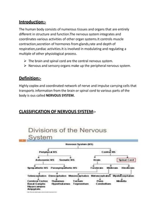
CLASSIFICATION OF NERVOUS SYSTEM:-
2. CENTRAL NERVOUS SYSTEM
The CNS consists of the brain and spinal cord with their
covering the brain is enclosed within the skull and the spinal
cord by the vertebrae that from the spinal column.
Meninges
These are 3 connective tissue membrane covering the brain
and spinal cord named from outside to inwards.
Dura Mater-outside thick membrane.
Arachnoid Mater-middle membrane separated from the
duramater by potential sublingual apace containing thin film
of fluid.
Pia Mater- thin vascular covering closely applied to the brain
and spinal cord.
Function
They protect the brain and spinal cord .
The folds of duramater prevent movement of the brain.
The epidural space allows movements of the vertebral
column without injury to the spinal cord.
The ligamentum denticulatum supports spinal cord.
Measurement
Length-45 cm
Weight-30g
3. Parts-
A cross section of spinal cord shows that it divided into 2
equal part:-
Anteriorly by short shallow median fissure ,
posteriorly by narrow septum,the posterior median
septum.
Grey mater-
Arranged like letter H around the central
canal,having 2 posterior, 2 anterior and 2 lateral
columns.
Posterior columns of grey mater-
Composed of cell bodies of sensory nerves.
Carries information from the bodu upto the
brain.
Anterior columns of grey mater-
Composed of the cell bodies of the lower motor
nervous that are stimulated by the upper motor
nervous or the interneurons linking the anterior
and posterior columns to form reflex .
4. White mater-
Arranged in 3 tracts:
Anterior
Posterior
Lateral
Tracts are formed by sensory nerve fibers ascending to
the brain ,motor nerve fibers descending from the brain
and fibers of interneurons.
BLOOD SUPPLY
Anterior spinal arteries
2 posterior spinal arteries
The radicular arteries
BRAIN:-
Lies in the cranial cavity covered by the
meninges.
Weight-1.4kg
Parts-
Cerebrum
Thalamus
Hypothalamus
Midbrain
Pons
Medulla oblongata
cerebellum
5. Cerebrum
Largest part of the brain.
Consists of two cerebral hemisphere ,partially separated
by the longitudinal fissure.
Deep within the brain,the hemispheres are connected by
a mass of white mater called corpus callosum.
Basal ganglia
These are the group of cell bodies that lie deep within
the brain and form part of the extrapyramidal tracts.
Act as relay situations with motor areas of cerebral
cortex and thalamus.
BLOOD SUPPLY-
Middle cerebral artery—superolateral surface
Anterior cerebral artery –medial surface
Posterior cerebral artery—occipital lobe
6. Diencephalon:
Meaning-between brain
Lies between cerebrum and midbrain.
Parts-
Thalamus
Hypothalamus
Metalthalamus
Epithalamus
Subthalamus
Thalamus:
Consists of 2 masses of grey and white mater situated
within the cerebral hemisphere.
Connection—thalamus conducts sensory information to the
sensory cortex.
Function—
o Acts as a major relay station whwre all specific
sensory impulses relay before terminating in the
cerebral cortex.
o Thalamus is responsible for feeling of various
emotion.
o Also provides necessary sensory information for
activity of skeletal muscle.
7. Hypothalamus –
A bilateral structure below the thalamus.
Acts as biological gland.
Weight—7g
Position –in front of the thalamus immediately above
the pituitary gland.
Function—
1.vegetative and endocrine function:
o Regulation of body temperature 37
o Regulation of water balance
o Regulation of feeding
o Regulation of cardiovascular system[increase BP
and HR]
o Endocrine function—controls secretion of pituitary
gland and release hormones of posterior pituitary.
2.Behavioural function:
o Stimulation of certain areas of hypothalamus causes
rage or fighting.
o Sexual drive is stimulated by anterior and posterior
portions of hypothalamus.
o Responsible for pleasant and unpleasant sensation.
Metathalamus—
Consists of medial and lateral geniculate bodies.
Function—
1.Medial geniculate body receives auditory impulses.
2.lateral geniculate body receives visual impulses.
8. Epithalamus—
Forms the roof of the third ventricle and
includes pineal gland.
o Has endocrine function.
Subthalamus—
Part of extrapyramidal system.
Brainstem:
o Short segment connecting the cerebrum with pons.
o The cerebral aqueduct forms the cavity of midbrain.
o Midbrain consists nuclei of 3(main and accessory iv
and v cranial nerves in periaqueductal greymater.
Pons:
o Situated infront of the cerebellum ,below the
midbrain and above the medulla oblongata.
o Joints the midbrain to the medulla oblongata .
o Contains nuclei for v,vi,vii and viii cranial nerves.
Medulla oblonata:
o Most inferior region of the brainstem.
o Joints the pons to the spinal cord.
o Length-2.5cm
o Ends at foramen magnum.
o Lower part of medulla(closed part)has central
canal,upper half(open part)forms floor of 4th
ventricle.
9. Reticular formation:
Diffuse mass of neurons and nerve fibers forming
a meshwork of reticulum in the central part of
brainstem(medulla,pons and tegmentum)is
termed as reticular formation.
Cerebellum:
o Head ganglion for proprioception.
o Largest part of midbrain.
Location:
Behind the pons and posterior portion of
cerebrum occupying the posterior cranial fossa.
Shape:
Ovoid shape.
Parts:
Consists of 2 cerebellar hemisphere joined by
vermis.
Ventricles in the brain—
The cavities inside the brain is termed as
ventricles.these are-
Right and left lateral ventricle.
Third ventricle.
Fourth ventricle.
10. CSF:
Cerebrospinal fluid covers the brain from outside as well as
inside.
Function-
o Protects the brain by covering it from inside and
outside.
o Carries nutrients to the brain and keeps brain and
spinal cord moist.
11. SPINAL CORD:-
o A cylindrical tube located in upper2/3 of
vertebral canal.
o Extends from the upper border of atlas to the
lower border of L1 vertebrae in adult.
12. PERIPHERAL NERVOUS SYSTEM---
The autonomic nervous system is a
component of the peripheral nervous
system that regulates involuntary
physiologic process including heart
rate,blood pressure,respiration,digestion
and sexual arousal.
It contains two anatomically distinct
divisions.
1.sympathetic
2.parasympathetic
The peripheral nervous system of the cranial and
spinal nerve,their ganglia,sensory receptors and
the major parts of PNS.
SPINAL NERVE:
31 pairs of spinal nerve leave the vertebral canal
by passing through the intervertebral
foramena.They are-
8 cranial nerve
12 thoracic
5 lumber
5 sacral
1 coccygeal
CRANIAL NERVE:
13. There are 12 pairs of cranial
nerve.They are—
NAME
ORIGIN
FUNCTION
Olfactory(CNI) Bipolar
cells of
olfactor
y nasal
mucosa.
Olfaction(sense
of smell)
Optic(CNII) Ganglio
n cells
of the
retina
vision
Oculomotor(CNIII) 1.Main
oculom
otor
nucleus
in mid
brain.
2.Access
ory
nucleus
of mid
brain.
Movement of
eyeball,elevatio
n of eyelid
pupillary
constriction
14. Trochlear(CNIV) Nucleas
in the
mid
brain
Superior
oblique muscle
of the eye
Trigeminal(CNV) Motor
nucleas
in the
pons
sensory
nuclei in
the
brain
stem.
Muscles of
mastication,se
n sation from
face.
Abducent(CNVI) Nucleus
on the
floor of
4th
ventricl
e
Lateral rectus
of the eye.
Facial(CNVII) Motor
nucleus
in pons
salivator
y
nucleus
Facial muscles
salivation and
lacrimation
Vestibulocochlear
(CNVIII)
Spinal
organ of
Equilibrium of
hearing.
16. NEURONS
Structural and functional unit of
nervous system.
PARTS
Cell body—perik ary on
Cell process—neurites
CELL BODY:
Also called as stroma
Variable in size and shape but too small
to be seen by naked eye.
Nucleus is large and shows nucleous.
The plasma membrane of the cell body
continues as axolemma.plasma receptor
shows receptior site.
CELL PROCESS:
Extension of the cell body.
Axon—
o Each nerve cell has only one axon.
o Single long process arising from axon hillock.
o Shape-cylindrical
o Membranes of axon called axolemma.
o Does not contain nissle granules.
o Have side branches
17. o Function-conducts impulse away from the
cell body.
Dendrons—
o Many,short,branching extremities.
o Nissle granule present
o Dendrites bear spine
o Function-conduct impulses towards
the cell body.
Neuroglia—
o The neuroglia form the connective
tissue of cns.
o Capable of division throughout life
o Can not conduct nerve impulses.
o Responsible for producing tumors of
nervous system.
18. BIBLIOGRAPHY:
Ross and Wilson “Anatomy and
physiology in Health and
Illness”(English,paperback Allison Gorant
Anne Waugh) 13th Edition,INTERNATIONAL
EDITION.Page No-
Ashalata PR,D Deepa,Text book of
ANATOMY AND PHYSIOLOGY for
Nurses,Jaypee Brothers,Medical
Publishers,The Health Science Publishers,New
Delhi,London,Ansari Road,Daryaganj,New
Delhi,110002,India,Page No-
Tp Prema KF Graicy , “Essentialls
neurological and Neurosurgical Nursing” First
Edition (2002) Jaypee Brother, Medical
Publishers, EMCA House 23/23B, Ansari Road,
Daryaganj, New Delhi 110002, India 2004
(Reprit), Page No-